

Abstract
Introduction:
The thoracodorsal artery perforator (TDAP) flap has emerged as one of the ideal perforator flaps. We, hereby, describe its versatility in indications (free/pedicled), methods of harvest (patient position and paddle orientation) and perforator consistency.
Materials and Methods:
We have performed a total of six TDAP flaps-five free and one pedicled, over a period of 1-year from March 2014 to February 2015 at a single centre. Our indications have been: Reconstruction of oral cavity, breast and upper and lower extremities.
Results:
We had neither any failures nor any re-explorations. The average perforator length is about 6 cm and the pedicle length can be extended to 12-14 cm by including the thoracodorsal artery. There is inconsistency in perforator position; however, the presence of a perforator is certain. It can be harvested in lateral, prone or supine position, thus, does not require any position change allowing a two-team approach to reconstruction. The paddle can be oriented vertically or horizontally, both healing with scars in inconspicuous locations. Apart from providing a good colour match for extremities, this flap can be thinned primarily.
Conclusion:
The versatility of TDAP has several advantages that make it a workhorse flap for most reconstructions requiring soft tissue cover. Further, the ease of harvest makes it a good perforator flap for beginners. Its use in chimerism with the underlying latissimus dorsi muscle provides reconstruction for coverage and volume replacement.
KEY WORDS: Ideal perforator flap, septocutaneous perforator, versatile, chimerism
INTRODUCTION
The goal of reconstruction is to produce the ‘ideal beautiful normal’. This most readily means replacement of like with like and this can well be done with perforator flaps, which can be thinned primarily. The attempt to reconstruct the normal should be made with the fundamental prerequisite that the donor site morbidity should be minimal. This again is well achieved with perforator flaps, which spare the underlying functional muscles.
Recently, perforator flaps, including the medial-lateral thoracic artery perforator flap, posterior tibial artery perforator flap, the propeller perforator flaps, the anterolateral thigh flap, the medial sural artery perforator flap and the chimeric fashioned thoracodorsal artery perforator (TDAP) flap, have been used widely for different anatomical areas. The TDAP flap has several established advantages, is popular in various reconstructive fields and is regarded as a satisfactory treatment option in lower extremity reconstruction as compared to the conventional fasciocutaneous/muscle flaps.[1]
The aim of this article is to highlight the versatility in the use of TDAP for various reconstructions.
MATERIALS AND METHODS
We performed the TDAP flap on six patients from March 2014 to February 2015 [Table 1].
Table 1.
TDAP flap

Surgical procedure
Markings done pre-operatively — Anatomical landmarks used:
Anterior axillary line (AAL).
Mid-axillary line (MAL).
Posterior axillary line (PAL).
Inferior angle of scapula.
Lateral border of latissimus dorsi (LD) muscle from posterior axillary fold to iliac crest.
Flap of required dimensions along the long axis of the muscle.
Handheld Doppler to localise the perforator.
For perforator localisation — Two or three perforators are marked using the following guides:
Four centimetres below the inferior angle of scapula and 1-2 cm inside of the lateral muscle border.
8-10 cm below the apex of the axilla.
An elliptical flap is marked according to the required dimensions centred around the perforator, with the anterior anterior flap border being anterior to MAL. This helps in correct identification of the interface between LD and serratus anterior muscle through which the septocutaneous perforators are given off [Figure 1].
Figure 1.

Flaps can be based on perforators from horizontal or vertical branch of thoracodorsal artery perforator. Perforator landmarks from vertical branch: 8 cm below apex of axilla and 2 cm inside the lateral border of latissimus dorsi muscle
The first incision made is the anterior incision, which also helps as an exploratory incision. The flap is elevated posteriorly above the muscle plane and perforators are looked for. The septum between LD and serratus is indicated by the presence of a fat plane. The thoracodorsal artery runs 1-2 cm behind the anterolateral border of LD on it's under surface. In two cases, when the flap was marked more posterior to the septum, the septocutaneous perforators were missed. The flap was then elevated anteriorly till the septum and perforators were identified. The flap was redesigned accordingly. The perforator was dissected till the thoracodorsal artery and the artery traced higher up till adequate length and diameter of the vessel were reached.
We encountered musculocutaneous perforator in a single case, while in all the other cases there were an average of two septocutaneous perforators. Thus, perforators were consistently present; however, they did not match with the pre-operative mapping in all the cases [Figure 2].
Figure 2.

Versatility in perforator type and in designing the flaps depending on the position of the perforator
The TDAP flap can be harvested comfortably in the lateral, prone or supine position [Figure 3].
Figure 3.

Versatility in position for flap harvest
For head and neck or lower extremity reconstructions, we have used harvested this flap in the supine position using a sand bag to elevate the side selected for flap elevation. In one case, two flaps were required for two different anatomical areas (bilateral foot injury). The exploratory incision revealed an 8 cm long perforator. The flap was harvested chimerically the upper skin paddle based on TDAP (for right medial malleolar compound fracture) and the lower skin paddle with underlying muscle based on the thoracodorsal artery (for left foot degloving injury). The TDAP flap can be designed in chimerism with the underlying LD or serratus anterior muscle for complex reconstructions [Figures 4 and 5].
Figure 4.

Versatility in flap designing — chimeric flaps of different components. Perforator length of about 6 cm
Figure 5.

Bilateral foot defect — thoracodorsal artery perforator flap for right foot and latissimus dorsi muscle flap with split thickness skin grafts for the left foot
The flap can be thinned considerably in the level of the superficial adipose layer, which is very useful for tongue reconstruction [Figure 6].
Figure 6.

Tongue reconstruction: Thoracodorsal artery perforator flap markings and harvesting in the supine position. The thinness of the flap along with good length of pedicle is seen. Final result of hemiglossectomy defect reconstruction
Primary closure was done in all cases.
RESULTS
Average perforator length: 6-8 cm.
Average length of the entire pedicle: About 12-14 cm.
The nerve was spared in all cases of TDAP.
Average time for flap harvest was 80-90 min.
DISCUSSION
The long fought battle about the preference of muscle versus fasciocutaneous flaps for the cover of bone in extremity trauma now seems to have been put at rest by the emergence of perforator flaps. These do not have the demerit of bearing fascia, hence there is no risk of shear forces hampering the reconstruction as with fasciocutaneous free flaps. Also, provision of a skin paddle abates the need for a skin graft with free muscle flap over exposed bone which makes subsequent surgeries for internal fixation of such injuries much simpler and faster[2,3,4] [Figure 7].
Figure 7.

Thoracodorsal artery perforator flap for right lower third leg defect - 2 months follow-up
The TDAP flap was first described by Angrigiani and perforator landmarks were established by him.[5]
The lateral thoracic region has three types of perforators as described by Kim:[6]
The musculocutaneous perforator from thoracodorsal artery: LD perforator flap.
The septocutaneous perforator from thoracodorsal artery: TDAP flap.
The direct cutaneous perforator from lateral thoracic artery: Lateral thoracic artery perforator flap.
TDAP flap has emerged as a workhorse flap for reconstruction of defects based on the following reasons:
It has a long vascular pedicle, approximately 18 cm. Its long pedicle helps in extremity injuries for anastomosis out of the zone of injury, good proximal vessel of larger diameter for anastomosis and ease of end-side anastomosis so as to preserve the continuity of the lower limb vessels. The latter is important in compound fractures of the leg where traumatic vessel damage may be an accompaniment and doing a free flap with end-end anastomosis raises a concern about distal vascularity. End-side anastomosis also becomes important in trauma reconstruction in patients with microangiopathy.
Allows a two-team approach.
Requires no position change as it can be harvested in supine position with sand bag/lateral tilt of the table
The thoracodorsal artery as a vascular pedicle has a lower incidence of atherosclerotic change compared to other pedicle arteries of the lower extremities.[7] This can make microvascular anastomosis easier and increases the reliability of surgical outcomes of free tissue transfer.
The TDAP flap can be harvested as a chimeric flap with LD muscle, serratus anterior muscle and / orscapular bone, thereby enabling reconstruction of complex defects using a single flap. It provides volume and cover while allowing primary closure of donor site.[8,9]
TDAP and LD muscle flap can be harvested together and utilised to cover two defects on anatomically separate areas. Thus, in a single surgery with a single donor site, two anatomically separate defects can be reconstructed.
The presence of perforators is consistent and the length of perforators in itself is good enough and can be further increased by including the main artery.
The lateral thoracic region has provision for harvest of perforator flaps based on the lateral thoracic artery and thoracodorsal artery. In case the TDAP perforator is injured during surgery, a life boat is present in the same field.
This flap is versatile in its indications as it provides cutaneous cover that can be used anywhere. It can be used as a pedicled flap for lateral neck, for pharyngeal tubing, breast, axilla and arm up to the distal third Bilateral pedicled TDAP can be used for bilateral breast reconstructions. As a free flap, its use includes intraoral and cutaneous resurfacing anywhere from head to foot including the sole [Figure 8].
Figure 8.
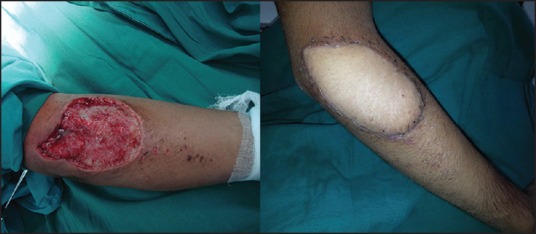
Right elbow defect resurfaced with thoracodorsal artery perforator Flap
Primary thinning of the flap makes it a good choice for reconstruction of tongue, neck contractures, extremity defects, including fingers and first web.[10] It can be used in chimerism with LD muscle to provide bulk like for breast reconstruction and maxillectomy defects. Thus, versatility in thickness and components including bone makes it a suitable choice for ideal contour and volume reconstruction.
It is versatile in the orientation of skin paddle: A vertical, oblique or transverse skin paddle can be taken with primary closure in all directions and a concealed suture line.[11] It provides an excellent tissue match for head and neck and extremity reconstruction. Also, provides durable skin as the skin of the back is the thickest.
Most other perforator flaps have a limitation in terms of the width of the flap that can be harvested permitting primary closure of the secondary defect. Lower limb perforator flaps are not a good colour match for the supraclavicular region and have a limited pedicle length. The upper limb perforator flaps also have a small pedicle length apart from leaving a conspicuous donor scar.
The biggest drawback we found was the inconsistency of perforator position and the mismatch of handheld Doppler mapping and the intra-operative finding of the perforator. But, nevertheless a perforator can be found. This flap is indeed versatile in designing of the skin flap based on perforator localisation without the fear of encroaching a different anatomical area or significantly affecting any landmark.
The maximum length that can be taken based on a single perforator needs to be assessed as this can be invaluable in treating hemicircumferential degloving injuries of upper and lower limbs and resurfacing of flexor surfaces of upper limbs as for post trauma/electrical burns functional reconstruction.
Flaps of 15-20 cm can be safely harvested on a single reliable perforator. Longer flaps can be harvested by using the various modifications to ensure perfusion as described by Hwang et al.[12]
Inclusion of more than one perforator from the same branch of thoracodorsal vessel: This is the easiest method and least morbid. The axis of flap is, thus, directed along the axis of the linking vessels which are responsible for interperforator flow.
- Inclusion of perforators from different branches of thoracodorsal artery:
- When the intervening muscle cuff between the point of origin of perforator is small, muscle is transected and sutured back after flap harvest.
- When the intervening muscle cuff is very wide, the perforator from the horizontal branch is cut 2 cm distal to the hilum and after flap elevation it is reanastomosed to the stump.
Inclusion of the intercostal artery perforator at the distal end of the flap: The intercostals perforator is noted to join the thoracodorsal artery by a connecting vessel. This perforator can be included and cut at the distal end, that is, towards its origin from the intercostals artery. Flow into this perforator from the main vessel will ensure distal flap perfusion.
CONCLUSION
TDAP has a versatile utility and surgical ease of harvest and anastomosis. It has several advantages over other perforator flaps. It may well form the work horse for most soft tissue reconstructions.
Financial support and sponsorship
Nil.
Conflicts of interest
There are no conflicts of interest.
REFERENCES
- 1.Georgescu AV. Propeller perforator flaps in distal lower leg: Evolution and clinical applications. Arch Plast Surg. 2012;39:94–105. doi: 10.5999/aps.2012.39.2.94. [DOI] [PMC free article] [PubMed] [Google Scholar]
- 2.May JW, Jr, Gallico GG, 3rd, Lukash FN. Microvascular transfer of free tissue for closure of bone wounds of the distal lower extremity. N Engl J Med. 1982;306:253–7. doi: 10.1056/NEJM198202043060501. [DOI] [PubMed] [Google Scholar]
- 3.Weinzweig N, Davies BW. Foot and ankle reconstruction using the radial forearm flap: A review of 25 cases. Plast Reconstr Surg. 1998;102:1999–2005. doi: 10.1097/00006534-199811000-00029. [DOI] [PubMed] [Google Scholar]
- 4.May JW, Jr, Rohrich RJ. Foot reconstruction using free microvascular muscle flaps with skin grafts. Clin Plast Surg. 1986;13:681–9. [PubMed] [Google Scholar]
- 5.Perignon D, Qassemyar Q, Benhaim T, Robbe M, Delay E, Sinna R. From Tansini to Angrigiani: Improvement and refinement of the thoracodorsal flap. Ann Chir Plast Esthet. 2011;56:149–55. doi: 10.1016/j.anplas.2010.09.015. [DOI] [PubMed] [Google Scholar]
- 6.Kim JT, Kim SW. Another option of perforator flap in the lateral thoracic area: Lateral thoracic perforator flap. J Reconstr Microsurg. 2014;30:443–50. doi: 10.1055/s-0034-1370358. [DOI] [PubMed] [Google Scholar]
- 7.Bartlett SP, May JW, Jr, Yaremchuk MJ. The latissimus dorsi muscle: A fresh cadaver study of the primary neurovascular pedicle. Plast Reconstr Surg. 1981;67:631–6. [PubMed] [Google Scholar]
- 8.Cavadas PC, Teran-Saavedra PP. Combined latissimus dorsi-thoracodorsal artery perforator free flap: The “razor flap”. J Reconstr Microsurg. 2002;18:29–31. doi: 10.1055/s-2002-19706. [DOI] [PubMed] [Google Scholar]
- 9.Van Landuyt K, Hamdi M, Blondeel P, Monstrey S. The compound thoracodorsal perforator flap in the treatment of combinedsoft-tissue defects of sole and dorsum of the foot. Br J Plast Surg. 2005;58:371–8. doi: 10.1016/j.bjps.2004.10.026. [DOI] [PubMed] [Google Scholar]
- 10.Jeon BJ, Lim SY, Pyon JK, Bang SI, Oh KS, Mun GH. Secondary extremity reconstruction with free perforator flaps for aesthetic purposes. J Plast Reconstr Aesthet Surg. 2011;64:1483–9. doi: 10.1016/j.bjps.2011.06.019. [DOI] [PubMed] [Google Scholar]
- 11.Lee SH, Mun GH. Transverse thoracodorsal artery perforator flaps: Experience with 31 free flaps. J Plast Reconstr Aesthet Surg. 2008;61:372–9. doi: 10.1016/j.bjps.2007.10.050. [DOI] [PubMed] [Google Scholar]
- 12.Hwang JH, Lim SY, Pyon JK, Bang SI, Oh KS, Mun GH. Reliable harvesting of a large thoracodorsal artery perforator flap with emphasis on perforator number and spacing. Plast Reconstr Surg. 2011;128:140e–50. doi: 10.1097/PRS.0b013e318221dc3e. [DOI] [PubMed] [Google Scholar]