Sir,
Giant lipomas are occasional mesenchymal tumours that are usually located deep in the body. The lipoma cells are believed to arise from primordial mesenchymal fatty tissue cells; thus, they are not of adult fat cell origin. They tend to increase in size with body weight gain, but interestingly, weight loss usually does not decrease their sizes. They do not cause any symptoms other than those determined by the space occupying mass. They are described as ‘giant’ beyond 1 kg of weight and 10 cm of diameter.
In literature, giant lipomas of the back were reported as exceptional findings, with variable weight and dimensions.[1,2,3] Giant lipomas are usually surgically treated by direct excision, even though their treatment through liposuction is described.[4]
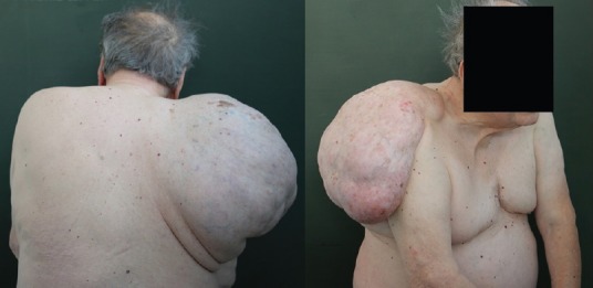
An 84-year-old man was admitted to our hospital because of a subcutaneous mass in his upper back. Thirty years earlier, he had noticed a firm, small, painless subcutaneous mass on the upper back [Figure 1]. The mass had progressively enlarged over the years. He never consulted a doctor before coming to our attention. The patient was in a state of good health before admission.
Figure 1.

Macroscopic appearance
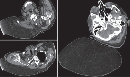
On physical examination, a well-circumscribed, freely movable giant subcutaneous mass, approximately 40 cm in diameter, was palpated in the upper back. Computed tomography showed a huge heterogeneous hypodense mass located in the subcutaneous fat tissue [Figure 2]. There was no evidence of calcification or necrosis in the tumour. Magnetic resonance imaging showed a mass of mixed heterogeneous high intensity, identical to that of the subcutaneous adipose tissue. The patient underwent complete surgical excision under general anaesthesia.
Figure 2.

Preoperative chest computed tomography scan
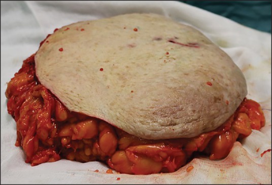
The mass had dimensions of 36 cm × 40 cm × 24 cm and weighed 5.75 kg after removal [Figure 3]. To the best of our knowledge, this could be the largest lipoma ever described in literature.
Figure 3.

Surgical specimen
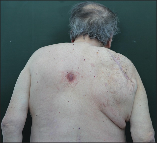
Histopathologic examination showed a benign neoplasm composed of normal adipose cells with strands of connective tissue. The patient had a postoperative course without complications or recurrences. After 6 months of follow-up, the patient was very pleased with the result [Figure 4].
Figure 4.

Six months after surgical excision
Financial support and sponsorship
Nil.
Conflicts of interest
There are no conflicts of interest.
REFERENCES
- 1.Terzioglu A, Tuncali D, Yuksel A, Bingul F, Aslan G. Giant lipomas: A series of 12 consecutive cases and a giant liposarcoma of the thigh. Dermatol Surg. 2004;30:463–7. doi: 10.1111/j.1524-4725.2004.30022.x. [DOI] [PubMed] [Google Scholar]
- 2.Pachani AB, Shojai AR, Gautam R, Topno M, Panchal A, Sharma V, et al. A giant congenital lipoma over the back. Indian J Surg. 2010;72:361–2. doi: 10.1007/s12262-010-0111-7. [DOI] [PMC free article] [PubMed] [Google Scholar]
- 3.Hakim E, Kolander Y, Meller Y, Moses M, Sagi A. Gigantic lipomas. Plast Reconstr Surg. 1994;94:369–71. doi: 10.1097/00006534-199408000-00025. [DOI] [PubMed] [Google Scholar]
- 4.Silistreli OK, Durmus EU, Ulusal BG, Oztan Y, Görgü M. What should be the treatment modality in giant cutaneous lipomas?. Review of the literature and report of 4 cases. Br J PlastSurg. 2005;58:394–8. doi: 10.1016/j.bjps.2004.09.005. [DOI] [PubMed] [Google Scholar]


