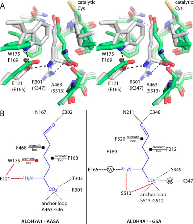
Figure 7.
Comparison of substrate recognition in ALDH7A1 and ALDH4A1. (A) Superposition of ALDH7A1 complexed with AA (white) and ALDH4A1 complexed with Glu (green). Black dashes indicate interactions that are unique to ALDH7A1. Magenta dashes denote those unique to ALDH4A1. (B) Substrate interaction diagrams for ALDH7A1 and ALDH4A1 inferred from the enzyme–product complex structures. Dashes denote hydrogen bonds and ion pairs. Squares denote van der Waals interactions between aromatic box residues and the aliphatic chain of the substrate. Interactions unique to either enzyme are colored red. W in a circle represents water-mediated interactions.

