

Abstract
The effect of resveratrol on the damage induced by methotrexate (MTX) in rat duodenum and jejunum tissue was investigated and evaluated in comparison with famotidine. The rats were divided into four groups as healthy group (HG), resveratrol+MTX (RMTX) group, famotidine+MTX (FMTX) group and the control group which received MTX (MTXC). RMTX group was given resveratrol 25 mg/kg and FMTX group famotidin 25 mg/kg, while MTXC and HG groups were orally administered distilled water once a day for 30 days. The rats in RMTX, FMTX and MTXC groups were given MTX of 5 mg/kg dose by the same way for 30 days. At the end of this period, amount of MDA, 8-OH/Gua and tGSH, and MPO gene expression were measured in the duodenal and jejunal tissues and the results were histopathologically evaluated. Resveratrol and famotidine were found to significantly prevent elevation of the MDA, 8-OH/Gua and MPO parameters with MTX and decrease of the levels of tGSH in the duodenal and jejunal tissues. Both drugs prevented severe damage to the villus and crypt epithelium in the duodenum and jejunum, congestion and hemorrhage, inflammatory cell infiltration and necrosis in the mucosa and submucosa due to MTX administration. Resveratrol could be considered in the clinical practice for treatment of the tissue damage in the intestines due to use of MTX, in comparison with famotidine. Resveratrol may be more advantageous than famotidine in long-term use against MTX toxicity since it does not inhibit gastric acid secretion.
Keywords: Resveratrol, oxidative stress, small intestine
Introduction
Methotrexate (MTX) which is known as a chemotherapeutic agent is also widely used in treatment of inflammatory diseases such as psoriasis, dermatomyositis, sarcoidosis and rheumatoid arthritis [1]. However, MTX may cause severe damage to the hematopoietic system, bone marrow and gastrointestinal system (GIS), because its effect on the above mentioned pathologies is not selective [2]. One of the major toxic effects related to MTX is GIS injury [3]. The most obvious side effect of MTX on GIS is seen in the small intestine. MTX has been reported to produce mucositis in the intestines [4]. It has also been shown that, mucositis caused by MTX in the rodent model is similar to the intestinal mucosa injury seen in humans [5]. Oxidative stress and especially neutrophil infiltration were found to be predominant in MTX-induced small intestinal injury [6]. Again, it has been reported that MTX decreases the levels of glutathione (tGSH) in the intestinal and gastric tissues, significantly increases the activity of myeloperoxidase (MPO) which is indicator of inflammatory response and increases amount of malondialdehyde (MDA) [7]. Demiryilmaz et al. reported that, MTX raises the levels of MDA which is one of the end products of lipid peroxidation, decreases amount of GSH which is an endogenous antioxidant and increases amount of 8-OH/Gua which is a DNA damage product [8]. It is understood from the literature that, oxidative stress is an important factor in the GIS toxicity caused by MTX. Although there are numerous studies about the prevention of toxicity in the GIS induced by MTX, a definitive solution is yet to be found. Therefore, scientific studies about the treatment of GIS toxicity due to MTX are being continued. Resveratrol which we tested in our study against the small intestinal toxicity caused by MTX, is a biologically active phenol and it is a natural stilbene (3,5,4-trihydroxy stilbene) [9]. Resveratrol is found in many fruits. It is found more abundantly in grape skins [10]. Resveratrol is a potent in vivo antioxidant with a high collector effect on the free oxygen radicals and the other oxidants. Resveratrol has been experimentally shown to decrease ischemia-reperfusion induced oxidative stress in heart, brain and kidney tissues [11,12]. This information obtained suggests that, resveratrol could be helpful in prevention of GIS toxicity caused by MTX. There was not any information found in the literature screening regarding prevention of methotrexate (MTX) induced damage to the duodenum and jejunum with resveratrol. Therefore, objective of this study was to investigate the effect of resveratrol on the oxidative damage to the duodenum and jejunum induced by MTX in rats. Also we aimed to compare resveratrol with famotidine which is used in the treatment of gastrointestinal ulcers.
Materials & methods
Animals
Experimental animal were supplied from the Recep Tayyip Erdoğan University, Medical Experimental and Research Center. Twenty-four Wistar albino male rats with weights differed between 240 g and 255 g were randomly selected to be used in the experiment. Four groups were created with six in each group before the experiment and the rats were housed and fed in the laboratory at the room temperature (22°C). Animal experiments were performed in accordance with the National Guidelines for the Use and Care of Laboratory Animals and were approved by the local animal ethics committee of Recep Tayyip Erdogan University, Rize, Turkey (Ethics Committee Number: 2014/64, Dated: 30.10.2014).
Chemical agents
Of the agents used in the experiment; resveratrol was supplied from SIGMA, thiopental sodium from I.E. Ulagay-Turkey, Methotrexate from Med-Drug-Turkey and famotidine from Fako Drugs-Istanbul.
Experimental groups
Experimental animals were divided into four groups as healthy (HG), resveratrol+MTX (RMTX), famotidine+MTX (FMTX) and the controls which received MTX (MTXC).
Performing of the test
RMTX group was given resveratrol 25 mg/kg (n=6) and FMTX group was given famotidin 25 mg/kg (n=6), while MTXC and HG groups were orally administered distilled water as the solvent by gavage. The rats in RMTX, FMTX and MTXC groups were given MTX of 5 mg/kg dose by the same way after one hour of the drugs and distilled water administration. This dose of MTX may cause gastric damage [8]. This procedure was repeated for 30 days. At the end of this period, all animals were sacrified with high doses of anesthesia. Then, amount of MDA, 8-OH/Gua and tGSH, and MPO gene expression were measured in the removed duodenal and jejunal tissues and the results were histopathologically evaluated. The results obtained from the RMTX and FMTX groups were compared with those obtained from the HG and MTCX groups.
Preparing the samples
The 25 mg of the tissue was homogenized using a solution of 1.15% KCl (Merck, Germany). The homogenate were centrifuged at 4000 rpm for 30 minutes at +4°C. Supernatants were then used for NO and MDA measurements. Tissues (25 mg) taken for tGSH analysis were washed with isotonic sodium chloride (İ.E. ULAGAY, Turkey) and subsequently brought to 2 mL total volume with phosphate buffer solution [0.213 gr NaH2PO4·2H2O (Merck, Germany) + 1.563 gr Na2HPO4·2H2O (Merck, Germany) + 0.038 gr EDTA (Sigma-Aldrich, Germany) + 100 mL dH2O, pH=7,4] and then were homogenized in an icy environment. After that, the tissues were centrifuged at 1000 rpm for 15 minutes at a temperature of +4°C. The supernatant was used as the sample for analysis. The protein concentration of the supernatant was measured using the method described by Bradford [13].
MDA analysis
According to the method defined by Ohkawa H et al., MDA forms a pink complex with thiobarbituric acid (TBA) at 95°C, which can be measured using spectrophotometry at a wavelength of 532 nm [14]. The 0.1 mL homogenat was added to a solution containing 0.1 mL of 8.1% sodium dodesil sulphate (SDS), 1.5 mL of 20% acetic acid (Merck, Germany) 1.5 mL of 0.9% TBA (Sigma-Aldrich, Germany), and 0.3 mL dH2O. The mixture was incubated at 95°C for 1 h. Upon cooling, 5 mL of n-butanol: pyridine (v/v, 15:1, Merck, Germany) was added. The mixture was vortexed for 1 min and centrifuged for 30 min at 4000 rpm. The absorbance of the 0.15 mL supernatant was measured at 532 nm by spectrophotometry. The Standard curve was obtained by using 1,1,3,3-tetramethoxypropane (Sigma-Aldrich, Germany).
tGSH analysis
According to the method defined by Sedlak J et al., DTNB [5,5’-dithiobis (2-nitrobenzoic acid)] disulfite is chromogenic in the medium, and DTNB is reduced easily by sulfhydryl groups. The yellow colour produced during the reduction is measured by spectrophotometry at 412 nm [15]. For measurement, cocktail solution (5.85 mL 100 mM Na-Fosfat buffer, 2.8 mL 1 mM DTNB (Sigma-Aldrich, Germany), 3.75 mL 1 mM NADPH (Sigma-Aldrich, Germany) and 80 µL 625 U/L Glutatyon redüktaz (Sigma-Aldrich, Germany)) was prepared. Before measurement, 0.1 mL meta-phosphoric acid (Sigma-Aldrich, Germany) was added onto 0.1 mL homogenate and centrifuged for 2 min at 2000 rpm for deproteinization. 0.15 mL cocktail solution was added onto 50 µL of supernatant. The Standard curve was obtained by using GSSG (Sigma-Aldrich, Germany).
DNA oxidation analysis
Tissue preparation
The 50 mg tissue was homogenized at +4°C using 1 mL of homogenization buffer solution [30 mM Tris pH 8, 10 mM EDTA, 10 mM 2-mercapto ethanol and 0.5% (v/v) Triton X-I00 (Sigma-Aldrich, Germany)]. The mixture was centrifuged for 10 minutes at 1000 g and the supernatant was discarded. The pellet was resuspended using 1 mL of extraction buffer (0.1 M Tris pH 8, 0.1 M NaCl, 20 mM EDTA) and was then homogenized using a vortex for 30 seconds. After that, it was centrifuged at 1000 g for 2 minutes. The pellet was again resuspended using the extraction buffer solution. The suspension was mixed well using the vortex. 400 μL of phenol (Sigma-Aldrich, Germany) was added to the mixture and it was mixed thoroughly for 1 minute using the vortex. After 10 minutes of waiting for the phases to separate, we removed the upper phase and transferred it to a clean tube. Then, 400 μL of chloroform-isopropanol (Sigma-Aldrich, Germany) was added to the clean tube (24:1) and it was centrifuged at 10000× g for 10 minutes. The upper phase was again transferred into a new tube. Next, 40 μL of 3 M sodium acetate (Merck, Germany), pH=5 and 800 μL of ice-cold ethanol (Merck, Germany), were added to the mixture obtained from the last centrifuge and it was shaken to ensure the mixing of the fluids. The mixture was centrifuged at 10000× g for 15 minutes and the upper part was removed completely. 1 mL of 70% ethanol was added to the lower part [16]. Then, 0.5 mL of 60% formic acid (Sigma-Aldrich, Germany) was added to 1 mL of the mixture obtained and it was left for 60 minutes at 150°C. The tubes were held at room temperature in order to eliminate the formic acid and approximately 1 mL of the mixture was stored at -20°C until the day of the analysis [17].
HPLC analysis of 8-hydroxiguanin
The levels of 8-OHdG and deoxyguanine (dG) were measured using different wavelengths and using HPLC-UV and HPLC-ECD electrochemical detectors. Before HPLC analysis, hydrolysed DNA samples were dissolved again using HPLC eluent. The final volume was 1 mL. Then, 20 μL of the final hydrolysate was injected into the HPLC-ECD (HP, HP 1049A ECD detector, Agilent 1100 modular systems, HP 1049A ECD detector, Germany). A reverse phase C18 (RP-C18) analytical column (250 mm x 4.6 mm x 4.0 µm, Phenomenex, CA) was used. The mobile phase consisted of acetonitrile (Merck, Germany) (97:3, v/v) containing 0.05 M potassium phosphate (Merck, Germany), (pH 5.5) buffer solution with a flow rate of 1 mL per minute. The absorbance of the dG concentrate was measured at 245 nm and 8-OHdG was read electrochemically (600 mV). The amounts of dG and 8-OHdG were determined using dG and 8-OHdG (Sigma-Aldrich, Germany) standards. 8-OHdG7 dG106 was interpreted as a sign of DNA damage [16,18,19].
Gene expression of MPO
RNA isolation: RNA was isolated from the homogenizated tissue samples using the Roche Magna Pure Compact LC device (Meinheim-Germany) with MagNA MagNA Pure LC RNA Kit (Roche Diagnostics). The quantity and quality of the isolated RNA was assessed with a nucleic acid measurement device (Maestro, Nano). 50 μl of RNA samples were stored at -80°C.
cDNA synthesis: cDNA was synthesized from the isolated RNA samples using the Transcriptor First Strand cDNA synthesis kit (Roche Diagnostics). For each subject, 1 μl ddH2O, 10 μL RNA and 2 μl Random Primer were combined and incubated in Thermal Cycler for 10 min at 65°C. After incubation, 4 μl Reaction Buffer, 0.5 μl RNAase, 2 μl Deoxynucleotide Mix, and 0.5 μl Reverse Transciptase were added and the reactions were incubated at 10 min at 25°C, 30 min at 55°C, 5 min at 85°C, then held at 4°C.
Quantitative gene expression evaluation by real-time polymerase chain reaction (RT-qPCR): For each cDNA sample, gene expression of MPO and the reference gene (G6PD) was analyzed using the Roche LightCycler 480 II Real-Time PCR instrument (Meinheim-Germany). PCR reactions in a final volume of 20 μl: 5 μl cDNA, 3 μl distiled water, 10 μl LightCycler 480 Probes Master (Roche Diagnostics) and 2 μl primer-probe set (Real-Time Ready single assay-Roche). Cycle conditions of the relative quantitative PCR (qPCR) were preincubation at 95°C for 10 min, followed by 45 amplification cycles of 95°C for 10 s, 6°C for 30 s, 72°C for 1 s, followed by cooling at 40°C for 30 s. qPCR analysis and calculation of quantification cycle (Cq) values for Relative Quantification were performed with by the LightCycler 480 Software, Version 1.5 (Roche Diagnostics). Relative quantitative amounts were calculated by dividing the target genes by the expression level of the reference gene. Reference gene was used for normalization of target gene expression.
Histopathological examination
Duodenal and jejunal tissues removed from the rats were fixed in 10% formalin solution for 24 hours. Sections of 4 micron thickness were obtained from the paraffin blocks that were obtained following the routine tissue monitoring and stained with hematoxylin & eosin. All sections were evaluated under light microscopy (Olympus BX 52, Tokyo, Japan) by a pathologist who was not aware of the treatment protocols.
Statistical analysis
Statistical analyses were carried out using the Statistical Package for Social Sciences, Windows version 18.0 (SPSS, Chicago, IL, USA). Descriptive statistics for each variable were determined. Normality of the data distribution was assessed with the Kolmogorov-Smirnov test. Results for continuous variables were demonstrated as mean ± standard error of the mean (mean ± SEM). Significance of differences between the groups was determined using one-way ANOVA test followed by Fisher’s post-hoc LSD (least significant differences) analysis. A P value less than 0.05 was considered significant.
Results
Biochemical results
As is seen in the Figure 1, amount of MDA was found to be higher and tGSH lower in the duodenal tissue of the rat group administered MTX. Again, it is seen in this chart that, resveratrol prevented elevation of the amount of MDA and decrease of tGSH with MTX significantly compared to famotidine. However, resveratrol and famotidine decreased the amount of increase of 8-OH/Gua with MTX almost by the same rate. That is the difference between the RMTX and FMTX groups in terms of 8-OH/Gua was not statistically significant. Likewise, MTX caused an increase in the amounts of MDA ve 8-OH/Gua and decrease in the amount of tGSH in the jejunum tissue (Figure 2).
Figure 1.
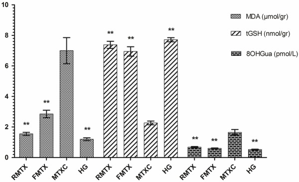
The effects of Resveratrol and Famotidin on MDA, tGSH and 8-OH/Gua levels in the duodenal tissues of rats given methotrexate. Bars are mean ± SEM. RMTX, FMTX and HG groups are compared with MTXC group. **: P<0.001.
Figure 2.

The effects of Resveratrol and Famotidin on MDA, tGSH and 8-OH/Gua levels in the jejunal tissue of rats given methotrexate. Bars are mean ± SEM. RMTX, FMTX and HG groups are compared with MTXC group. **: P<0.001.
It was found that resveratrol and famotidine affected the levels of these oxidant and antioxidant parameters in the jejunum almost by the same rate. That is the difference between the RMTX, FMTX and HG in terms of the amount of tGSH was not statistically significant.
Results of the MPO gene expression
MPO gene expression in the duodenal and jejunal tissues of the groups which were given MTX significantly increased compared to the RMTX, FMTX and HG groups. Inhibitory effects of the MPO gene expression on the resveratrol and famotidine were found rather close (Figure 3).
Figure 3.
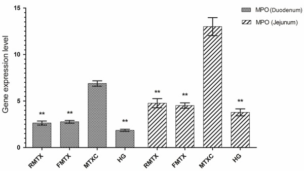
The effects of Resveratrol and Famotidin on MPO gene expression in the duodenal and jejunal tissues of rats given MTX. RMTX, FMTX and HG groups are compared with MTXC group. **: P<0.001.
Histopathological results
Duodenal tissue
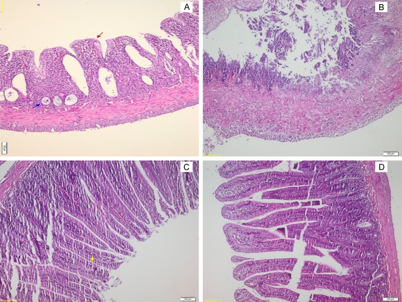
As is seen from Figure 4A, normal mucosa and structures are monitored on the full-thickness histopathological sections of duodenal tissue of the HG rats. Whereas on the superficial histopathological sections of duodenal tissue of the MTXC group; we observed villus epithelial damage in the mucosa (red arrow), congestion and hemorrhage (yellow arrow) in the lamina propria and mixed inflammatory cells infiltration (blue arrow) containing PMNL and eosinophil leukocytes (Figure 5A). In addition to the mixed inflammatory cells infiltration exceeding the muscularis propria and reaching to the serosa (blue arrow, Figure 5B), full thickness necrosis in the mucosa and submucosa (Figure 5C) and crypt epithelial damage is monitored in the MTXC group (Figure 5D). Mild villus superfacial epithelial damage (red arrow), mixed inflammatory cells infiltration and mostly protected crypt structures (blue arrow) are seen in the duodenal tissue of the RMTX group (Figure 5E). Again, we observed mild villus epithelial damage (red arrow), mixed inflammatory cells infiltration and mostly protected crypt structures (blue arrow) in the FMTX group rats (Figure 5F)
Figure 4.
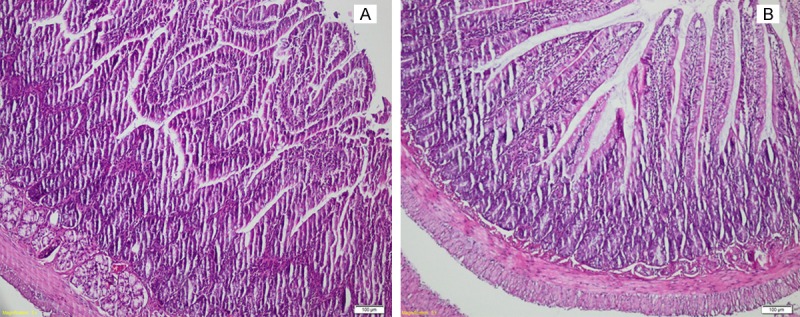
A. The histo-pathological examination of the duodenum tissue of healthy group (HG), (H&E ×100), B. The histo-pathological examination of the jejunum tissue of healthy group (HG), (H&E ×100).
Figure 5.
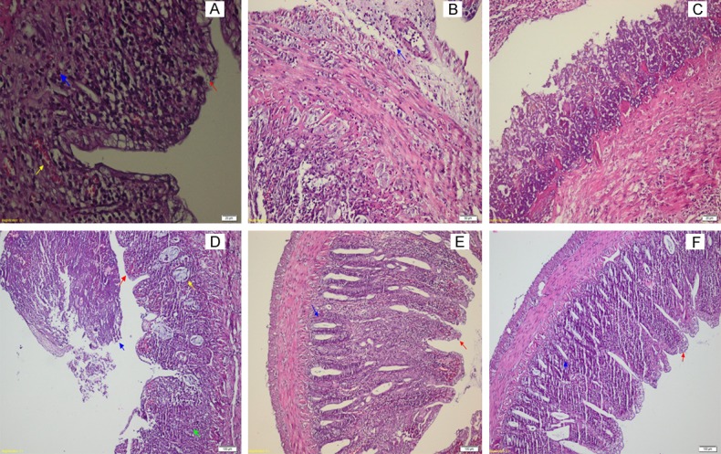
A. Section of the duodenum tissue of MTXC group (H&E ×400), B. Section of the duodenum tissue of MTXC group (H&E ×200), C. Section of the duodenum tissue of MTXC group (H&E ×400), D. Section of the duodenum tissue of MTXC group (H&E ×100), E. Section of the duodenum tissue of RMTX group (H&E ×100), F. Section of the duodenum tissue of FMTX group (H&E ×100).
Jejunal tissue
Normal jejunal tissue is seen on Figure 4B in the histopathological sections of HG group rats. Whereaes, villus epithelial (red arrow) and diffuse crypt epithelial damage are seen in the jejunal tissue of the MTXC group (blue arrow, Figure 6A). In addition, diffuse necrosis is observed in the mucosa and submucosa of this group (Figure 6B). Mild mixed inflammatory cells infiltration (yellow), mostly protected villus superficial epithelial and crypt epithelial damage are monitored in the jejunal tissue of the RMTX group which received resveratrol (Figure 6C). Almost all protected villus superficial and crypt epithelia are monitored in the FMTX group (Figure 6D).
Figure 6.
A. Section of the jejunum tissue of MTXC group (H&E ×400), B. Section of the jejunum tissue of MTXC group (H&E ×400), C. Section of the jejunum tissue of RMTX group (H&E ×100), D. Section of the jejunum tissue of FMTX group (H&E ×100).
Discussion
In this study, effect of resveratrol on the oxidative duodenal and jejunal damage developing due to the MTX administration was investigated and evaluated in comparison with famotidine. As is known, MTX leads to biochemical and morphological abnormalities in the intestinal mucosa [20,21]. It is reported that, levels of MDA which is an indicator of lipid peroxidation increase and levels of GSH decrease in the intestinal tissues of rats administered MTX [22]. It has been reported that DNA damage due to purine and pyrimidine synthesis plays a role in the pathogenesis of MTX toxicity [23]. It has been shown that, neutrophil infiltration which is an indicator of the inflammatory response plays a role in the intestinal oxidative damage caused by MTX [6]. MTX is known to activates the neutrophils in the intestinal tissue, increasing MPO production, while excessive production of MPO plays a crucial role in the production of oxidative agents [8,24]. This information from the literature is consistent with biochemical results obtained in our study from the duodenal and jejunal tissues of rats administered MTX. This in turn demonstrates that antioxidant therapy is of importance in the prevention of tissue damage due to MTX. Likewise, it is argued in numerous studies that antioxidants have been used against tissue damage due to MTX, with successful results [8,25,26]. In our study, resveratrol and famotidine prevented increase of MDA and decrease of tGSH in the duodenum and jejunum with MTX administration almost at the same level. That is resveratrol and famotidine reversed change of the oxidant/antioxidant balance by MTX in favor of oxidants in the intestinal tissue. Resveratrol and famotidine are known to have antioxidant features [11,27]. There was not any information found in the literature screening regarding the protective effect of resveratrol on the methotrexate (MTX) induced damage to the duodenum and jejunum, but there were several studies in the literature demonstrating that resveratrol suppresses oxidative stress in the various tissues caused by MTX [28]. Again, it has been reported that, resveratrol prevents increase of the amount of MDA and decrease the levels of GSH in the intestinal (ileum) tissue in which ischemia-reperfusion is administered [29]. Also there was not any study in the literature regarding effects of famotidine against MTX-induced damage, but famotidine has been reported to decrease the amount of damaged gastric tissue, creating protective effect by raising the levels of GSH [30].
Amount of 8-OH/Gua in the duodenal and jejunal tissues of the groups which received resveratrol and famotidine was found to be decreased compared to the MTXC group, which indicates that resveratrol and famotidine protect the intestinal tissue against harmful effects of the free radicals. As is known, free oxygen radicals can react with macromolecules in the cells, leading to significant cellular damage such as DNA oxidation [31]. Following the free radical reactions, base changes occur in nucleic acids and chain breaks occur in DNA. If this change is not restored, DNA becomes mutated. 8-OH/Gua has been accepted as a mutagenic form of DNA [32]. Resveratrol has been reported to reduce DNA breaking with hydroxyl radical scavenging effect [33]. In a study by Alturfan AA et al. on rats, resveratrol was reported to reduce the amount of 8-OH/d Gua which increases during the renal damage [34]. Kurt A et. al. demonstrated that, famotidine has protective effect on DNA and suppresses the formation of 8-OH/d in the damaged tissues [35]. This information from the literature supports the protective effect of resveratrol and famotidine on DNA.
As it was mentioned above, MTX increased MPO gene expression in the duodenal and jejunal tissues. The levels of gene expression in the intestinal tissues of the groups administered resveratrol and famotidine was closer to that of the healthy group. It is known that, MPO which is secreted by the neutrophil infiltration and activated neutrophil in MTX-induced intestinal damage plays a role in the oxidant production [24], suggesting that resveratrol and famotidine prevent neutrophil activation and increase of the MPO production. Evidence from the literature supports our this opinion [27,36,37].
In addition, in this study we encountered serious pathological findings in the duodenal and jejunal tissues of the MTXC group, in which oxidant parameters was elevated. Excessive production of the reactive oxidant products are accused in the pathogenesis of MTX-induced intestinal pathology [6,38]. As it is understood from the experimental outcomes; severe villus and crypt damage, congestion and hemorrhage, PMNL and mixed inflammatory cells infiltration and, full thickness necrosis in the mucosa and submucosa are seen in the MTXC group in which the amounts of MDA, 8-OH/Gua and MPO were higher and tGSH was lower than the other groups. In a study conducted long before by Taminiau JA et al., MTX was reported to cause villus and crypt epithelial cellular damage [5]. Again Kesik V et al. reported that, besides villus and crypt epithelial cellular loss, MTX creates congestion and hemorrhage in the mucosa [39]. It has been argued in that study as well as the other similar studies that, MTX induced damage in the intestine might be resulted from PNL infiltration and marked inflammation [40]. Because increased PNL infiltration leads to increase of reactive oxygen radicals which cause oxidative stress [41]. These pathological changes in the intestinal tissue may leads epithelial cells until necrosis [42]. Information from the literature is in line with our results. Protective effects of resveratrol and famotidine on the duodenal and jejunal mucosa as well as villus and crypt are seen to be almost the same. In a study by Southcott E et al., MTX was reported to cause more damage in the jejenum than in the duodenum [43]. However, in our study there was not a histologically significant difference between the duodenal and jejunal tissues.
MTX produced a significant damage in the duodenal and jejunal tissues. Resveratrol and famotidine prevented this damage caused by MTX. Resveratrol could be considered in the clinical practice for treatment of the tissue damage in the intestines due to use of MTX, in comparison with famotidine. Resveratrol may be more advantageous than famotidine in long-term use against MTX toxicity since it does not inhibit gastric acid secretion.
Disclosure of conflict of interest
None.
References
- 1.Cetinkaya A, Bulbuloglu E, Kurutas EB, Kantarceken B. N-acetylcysteine ameliorates methotrexate-induced oxidative liver damage in rats. Med Sci Monit. 2006;12:BR274–278. [PubMed] [Google Scholar]
- 2.Jolivet J, Cowan KH, Curt GA, Clendeninn NJ, Chabner BA. The pharmacology and clinical use of methotrexate. N Engl J Med. 1983;309:1094. doi: 10.1056/NEJM198311033091805. [DOI] [PubMed] [Google Scholar]
- 3.Nagakubo J, Tomimatsu T, Kitajima M, Takayama H, Aimi N, Horie T. Characteristics of transport of fluoresceinated methotrexate in rat small intestine. Life Sci. 2001;69:739–747. doi: 10.1016/s0024-3205(01)01162-6. [DOI] [PubMed] [Google Scholar]
- 4.Loehry C, Creamer B. Three-demensional structure of the rat small intestinal mucosa related to mucosal dynamics. II. Mucosal structure and dynamics in the lactating rat. Gut. 1969;10:116–118. doi: 10.1136/gut.10.2.116. [DOI] [PMC free article] [PubMed] [Google Scholar]
- 5.Taminiau J, Gall D, Hamilton J. Response of the rat small-intestine epithelium to methotrexate. Gut. 1980;21:486–492. doi: 10.1136/gut.21.6.486. [DOI] [PMC free article] [PubMed] [Google Scholar]
- 6.Miyazono Y, Gao F, Horie T. Oxidative stress contributes to methotrexate-induced small intestinal toxicity in rats. Scand J Gastroenterol. 2004;39:1119–1127. doi: 10.1080/00365520410003605. [DOI] [PubMed] [Google Scholar]
- 7.Jahovic N, Sener G, Cevik H, Ersoy Y, Arbak S, Yegen BC. Amelioration of methotrexate-induced enteritis by melatonin in rats. Cell Biochem Funct. 2004;22:169–178. doi: 10.1002/cbf.1071. [DOI] [PubMed] [Google Scholar]
- 8.Demiryilmaz I, Uzkeser H, Cetin N, Hacimuftuoglu A, Bakan E, Altuner D. Effect of mirtazapine on gastric oxidative stress and DNA injury created with methotrexate in rats. Asian Jf Chem. 2013;25:2047–2050. [Google Scholar]
- 9.Mazza G. Anthocyanins in grapes and grape products. Crit Rev Food Sci Nutr. 1995;35:341–371. doi: 10.1080/10408399509527704. [DOI] [PubMed] [Google Scholar]
- 10.Juan ME, Vinardell MP, Planas JM. The daily oral administration of high doses of trans-resveratrol to rats for 28 days is not harmful. J Nutr. 2002;132:257–260. doi: 10.1093/jn/132.2.257. [DOI] [PubMed] [Google Scholar]
- 11.Aggarwal BB, Shishodia S. Resveratrol in health and disease. CRC Press; 2005. [Google Scholar]
- 12.de la Lastra CA, Villegas I. Resveratrol as an anti-inflammatory and anti-aging agent: mechanisms and clinical implications. Mol Nutr Food Res. 2005;49:405–430. doi: 10.1002/mnfr.200500022. [DOI] [PubMed] [Google Scholar]
- 13.Bradford MM. A rapid and sensitive method for the quantitation of microgram quantities of protein utilizing the principle of protein-dye binding. Anal Biochem. 1976;72:248–254. doi: 10.1016/0003-2697(76)90527-3. [DOI] [PubMed] [Google Scholar]
- 14.Ohkawa H, Ohishi N, Yagi K. Assay for lipid peroxides in animal tissues by thiobarbituric acid reaction. Anal Biochem. 1979;95:351–358. doi: 10.1016/0003-2697(79)90738-3. [DOI] [PubMed] [Google Scholar]
- 15.Sedlak J, Lindsay RH. Estimation of total, protein-bound, and nonprotein sulfhydryl groups in tissue with Ellman’s reagent. Anal Biochem. 1968;25:192–205. doi: 10.1016/0003-2697(68)90092-4. [DOI] [PubMed] [Google Scholar]
- 16.Shigenaga MK, Aboujaoude EN, Chen Q, Ames BN. Assays of oxidative DNA damage biomarkers 8-oxo-2’-deoxyguanosine and 8-oxoguanine in nuclear DNA and biological fluids by high-performance liquid chromatography with electrochemical detection. Methods Enzymol. 1994;234:16–33. doi: 10.1016/0076-6879(94)34073-0. [DOI] [PubMed] [Google Scholar]
- 17.Kaur H, Halliwell B. Measurement of oxidized and methylated DNA bases by HPLC with electrochemical detection. Biochem J. 1996;318:21–23. doi: 10.1042/bj3180021. [DOI] [PMC free article] [PubMed] [Google Scholar]
- 18.Floyd RA, Watson JJ, Wong PK, Altmiller DH, Rickard RC. Hydroxyl free radical adduct of deoxyguanosine: sensitive detection and mechanisms of formation. Free Radic Res Commun. 1986;1:163–172. doi: 10.3109/10715768609083148. [DOI] [PubMed] [Google Scholar]
- 19.Asami S, Hirano T, Yamaguchi R, Tomioka Y, Itoh H, Kasai H. Increase of a type of oxidative DNA damage, 8-hydroxyguanine, and its repair activity in human leukocytes by cigarette smoking. Cancer Res. 1996;56:2546–2549. [PubMed] [Google Scholar]
- 20.Takeuchi H, Kosakai Y, Tsurui K, Hasegawa K, Horie T, Awazu S. Change in small intestinal brush border membranes of rats following methotrexate administration. Pharmacol Toxicol. 1989;65:269–273. doi: 10.1111/j.1600-0773.1989.tb01171.x. [DOI] [PubMed] [Google Scholar]
- 21.Tsurui K, Kosakai Y, Horie T, Awazu S. Vitamin A protects the small intestine from methotrexate-induced damage in rats. J Pharmacol Exp Ther. 1990;253:1278–1284. [PubMed] [Google Scholar]
- 22.Jahovic N, Cevik H, Sehirli AO, Yegen BC, Sener G. Melatonin prevents methotrexate-induced hepatorenal oxidative injury in rats. J Pineal Res. 2003;34:282–287. [PubMed] [Google Scholar]
- 23.Uraz S, Tahan V, Aygun C, Eren F, Unluguzel G, Yuksel M, Senturk O, Avsar E, Haklar G, Celikel C, Hulagu S, Tozun N. Role of ursodeoxycholic acid in prevention of methotrexate-induced liver toxicity. Dig Dis Sci. 2008;53:1071–1077. doi: 10.1007/s10620-007-9949-3. [DOI] [PubMed] [Google Scholar]
- 24.Zimmerman BJ, Granger DN. Reperfusion injury. Surg Clin North Am. 1992;72:65–83. doi: 10.1016/s0039-6109(16)45628-8. [DOI] [PubMed] [Google Scholar]
- 25.Yapcaa OE, Borekcib B, Turanc MI, Gulapoglud M, Salmane S. The effect of mirtazapine on methotrexate-induced oxidative damage and infertility in rats. Scienceasia. 2014;40:152–156. [Google Scholar]
- 26.Kolli VK, Abraham P, Isaac B, Kasthuri N. Preclinical efficacy of melatonin to reduce methotrexate-induced oxidative stress and small intestinal damage in rats. Dig Dis Sci. 2013;58:959–969. doi: 10.1007/s10620-012-2437-4. [DOI] [PubMed] [Google Scholar]
- 27.Suleyman H, Cadirci E, Albayrak A, Polat B, Halici Z, Koc F, Hacimuftuoglu A, Bayir Y. Comparative study on the gastroprotective potential of some antidepressants in indomethacin-induced ulcer in rats. Chem Biol Interact. 2009;180:318–324. doi: 10.1016/j.cbi.2009.03.002. [DOI] [PubMed] [Google Scholar]
- 28.Dalaklioglu S, Genc GE, Aksoy NH, Akcit F, Gumuslu S. Resveratrol ameliorates methotrexate-induced hepatotoxicity in rats via inhibition of lipid peroxidation. Hum Exp Toxicol. 2013;32:662–671. doi: 10.1177/0960327112468178. [DOI] [PubMed] [Google Scholar]
- 29.Karabulut AB, Kirimlioglu V, Kirimlioglu H, Yilmaz S, Isik B, Isikgil O. Protective effects of resveratrol on spleen and ileum in rats subjected to ischemia-reperfusion. Transplant Proc. 2006;38:375–377. doi: 10.1016/j.transproceed.2006.01.017. [DOI] [PubMed] [Google Scholar]
- 30.Polat B, Albayrak Y, Suleyman B, Dursun H, Odabasoglu F, Yigiter M, Halici Z, Suleyman H. Antiulcerative effect of dexmedetomidine on indomethacin-induced gastric ulcer in rats. Pharmacol Rep. 2011;63:518–526. doi: 10.1016/s1734-1140(11)70518-7. [DOI] [PubMed] [Google Scholar]
- 31.Kisaoglu A, Borekci B, Yapca OE, Bilen H, Suleyman H. Tissue damage and oxidant/antioxidant balance. Eurasian J Med. 2013;45:47. doi: 10.5152/eajm.2013.08. [DOI] [PMC free article] [PubMed] [Google Scholar]
- 32.Yapca OE, Borekci B, Suleyman H. Ischemia-Reperfusion Damage. Eurasian J Med. 2013;45:126. doi: 10.5152/eajm.2013.24. [DOI] [PMC free article] [PubMed] [Google Scholar]
- 33.Burkitt MJ, Duncan J. Effects of trans-resveratrol on copper-dependent hydroxyl-radical formation and DNA damage: evidence for hydroxyl-radical scavenging and a novel, glutathione-sparing mechanism of action. Arch Biochem Biophys. 2000;381:253–263. doi: 10.1006/abbi.2000.1973. [DOI] [PubMed] [Google Scholar]
- 34.Alturfan AA, Tozan-Beceren A, Şehirli AÖ, Demiralp E, Şener G, Omurtag GZ. Resveratrol ameliorates oxidative DNA damage and protects against acrylamide-induced oxidative stress in rats. Mol Biol Rep. 2012;39:4589–4596. doi: 10.1007/s11033-011-1249-5. [DOI] [PubMed] [Google Scholar]
- 35.Kurt A, Isaoglu U, Yilmaz M, Calik M, Polat B, Hakan H, Ingec M, Suleyman H. Biochemical and histological investigation of famotidine effect on postischemic reperfusion injury in the rat ovary. J Pediatr Surg. 2011;46:1817–1823. doi: 10.1016/j.jpedsurg.2011.04.092. [DOI] [PubMed] [Google Scholar]
- 36.Guha P, Dey A, Chatterjee A, Chattopadhyay S, Bandyopadhyay SK. Pro-ulcer effects of resveratrol in mice with indomethacin-induced gastric ulcers are reversed by L-arginine. Br J Pharmacol. 2010;159:726–734. doi: 10.1111/j.1476-5381.2009.00572.x. [DOI] [PMC free article] [PubMed] [Google Scholar]
- 37.Tunali-Akbay T, Sehirli O, Ercan F, Sener G. Resveratrol protects against methotrexate-induced hepatic injury in rats. J Pharm Pharm Sci. 2010;13:303–310. doi: 10.18433/j30k5q. [DOI] [PubMed] [Google Scholar]
- 38.Maeda T, Miyazono Y, Ito K, Hamada K, Sekine S, Horie T. Oxidative stress and enhanced paracellular permeability in the small intestine of methotrexate-treated rats. Cancer Chemother Pharmacol. 2010;65:1117–1123. doi: 10.1007/s00280-009-1119-1. [DOI] [PubMed] [Google Scholar]
- 39.Kesik V, Uysal B, Kurt B, Kismet E, Koseoglu V. Ozone ameliorates methotrexate-induced intestinal injury in rats. Cancer Biol Ther. 2009;8:1623–1628. doi: 10.4161/cbt.8.17.9203. [DOI] [PubMed] [Google Scholar]
- 40.Acipayam C, Bayram I, Daglioglu K, Doran F, Yilmaz S, Sezgin G, Totan Ates B, Ozkan A, Tanyeli A. The protective effect of hesperidin on methotrexate-induced intestinal epithelial damage in rats: an experimental study. Med Princ Pract. 2014;23:45–52. doi: 10.1159/000355900. [DOI] [PMC free article] [PubMed] [Google Scholar]
- 41.Hamada K, Kakigawa N, Sekine S, Shitara Y, Horie T. Disruption of ZO-1/claudin-4 interaction in relation to inflammatory responses in methotrexate-induced intestinal mucositis. Cancer Chemother Pharmacol. 2013;72:757–765. doi: 10.1007/s00280-013-2238-2. [DOI] [PubMed] [Google Scholar]
- 42.Moghadam AR, Mohajeri D, Namvaran-Abbas-Abad A, Manafi H, Shahi D, Mazani M. Protective effect of turmeric extract on ethotrexate-induced intestinal damage and oxidative stress. Chin J Nat Med. 2013;11:477–483. doi: 10.1016/S1875-5364(13)60087-4. [DOI] [PubMed] [Google Scholar]
- 43.Southcott E, Tooley KL, Howarth GS, Davidson GP, Butler RN. Yoghurts containing probiotics reduce disruption of the small intestinal barrier in methotrexate-treated rats. Dig Dis Sci. 2008;53:1837–1841. doi: 10.1007/s10620-008-0275-1. [DOI] [PubMed] [Google Scholar]