Abstract
Endoscopic ultrasound (EUS)-guided biliary drainage is accepted as a less invasive, alternative treatment for patients in whom endoscopic retrograde cholangiopancreatography has failed. Most patients with malignant hilar obstruction undergo EUS-guided hepaticogastrostomy. The authors present the case of a 77-year-old man with advanced hilar cholangiocarcinoma who had undergone a roux-en-Y hepaticojejunostomy several months prior. He developed progressive jaundice and a low-grade fever that persisted for one week. The enteroscopic-assisted endoscopic retrograde cholangiopancreatography failed, thus the patient was scheduled for EUS-guided biliary drainage. In order to obtain adequate drainage, both intrahepatic systems were drained. This report describes the technique used for bilateral drainage via a transgastric approach. Currently, only a few different techniques for EUS-guided right system drainage have been reported in the literature. This case demonstrates that bilateral EUS-guided biliary drainage is feasible and effective in patients with hilar cholangiocarcinoma, and thus can be used as an alternative to percutaneous biliary drainage.
Keywords: Biliary drainage, Endoscopic ultrasound-guided, Bilateral systems, Transmural drainage
Core tip: Endoscopic ultrasound-guided left system drainage via hepaticogastrostomy can be performed with > 90% technical and clinical success in patients with obstructive jaundice. A transgastric approach for endoscopic ultrasound-guided hepaticogastrostomy to drain both intrahepatic systems was successfully performed by manipulating the guidewire until it passed across the stricture point; the two systems were then bridged with a metal stent. The authors propose that this technique is feasible and effective for bilateral biliary drainage.
INTRODUCTION
Endoscopic ultrasound (EUS)-guided biliary drainage was first reported in 2001 by Giovannini et al[1]. A subsequent case series described by Burmester et al[2] in 2003 demonstrated the feasibility of this technique using two platforms depending on the route of approach: EUS-guided transpapillary and transgastric. Since then, this therapeutic intervention has become an alternative treatment for patients with malignant bile duct obstruction in whom endoscopic retrograde cholangiopancreatography (ERCP) has failed, and in patients who prefer internal rather than percutaneous biliary drainage. When performed by experienced endoscopists, this technique has technical and clinical success rates of 75%-100% and 65%-92%, respectively[3-9]. As a result, EUS-guided biliary drainage has become more popular.
EUS-guided rendezvous with a transpapillary approach or antegrade stent insertion or EUS-guided transgastric drainage (EUS-guided hepaticogastrostomy) may be useful for patients with malignant hilar obstruction in whom ERCP has failed[10]. The case reported here describes a 77-year-old man with advanced hilar cholangiocarcinoma who underwent surgical bypass and developed obstructive jaundice thought to be due to tumor recurrence at the anastomosis site. EUS-guided bilateral biliary drainage was performed after enteroscopic-assisted ERCP failed.
CASE REPORT
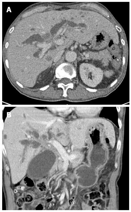
A 77-year-old man with hilar cholangiocarcinoma had undergone roux-en-Y hepaticojejunostomy several months prior. He subsequently developed progressive jaundice and a low-grade fever that persisted for a week. Liver function analyses showed hyperbilirubinemia with total bilirubin of 12.7 mg/dL (reference range: 0-1.5 mg/dL), a direct bilirubin level of 11.3 mg/dL (reference range: 0-1.2 mg/dL), and an elevated alkaline phosphatase level (395 U/L; reference range: 40-125 U/L). A computed tomography (CT) scan revealed a large, ill-defined, enhanced lesion in the hilar region that had invaded hepatic segment IV and caused bilateral intrahepatic duct dilation (Figure 1). Enteroscopic-assisted ERCP failed, and the patient was therefore scheduled for EUS-guided biliary drainage.
Figure 1.

Computed tomography examination. A: Axial; B: Coronal computed tomography images showing an ill-defined mass in the hilar region causing bilateral intrahepatic duct dilation.
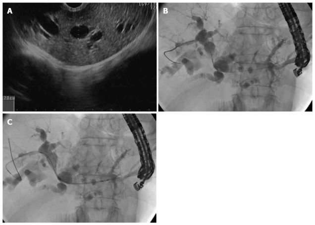
To obtain adequate drainage, the endoscopist aimed to drain both intrahepatic systems. After obtaining informed consent for this procedure, the patient was placed in a supine position under total intravenous anesthesia. A 19-gauge needle was used to puncture through the gastric cardia into hepatic segment II, and a purulent discharge was aspirated. A 0.025 ViZiguide (Terumo Medical Corp., Tokyo, Japan) was then inserted using a tapered-tip sphincterotome (PR-V234Q; Terumo Medical Corp.), and the guidewire was manipulated until it passed from the left through to the right intrahepatic duct (Figure 2). The neo-tract dilation was performed using 7.0, 8.5 and 10.0 Fr Soehendra dilating catheters. A 10 mm × 80 mm uncovered self-expanding metal stent was then inserted and deployed to connect the right and left systems (Figure 2B). Finally, a 10 mm × 100 mm covered self-expanding metal stent was deployed to form the bilo-enteric tract (Figure 3).
Figure 2.

Guidewire insertion. A: Echoview demonstrating dilated of left system B: Cholangiogram showing the tapered-tip sphincterotome; C: The guidewire was passed to the right system from the gastric site and an uncovered self-expanding metal stent was deployed.
Figure 3.

Dilation. A: Cholangiogram showing deployment of the covered self-expanding metal stent; B: After the guidewire was removed.
There were no immediate postoperative complications, and the patient was transferred to a regular ward. Nil per os was maintained for five days, and in the absence of eventful conditions, the patient was discharged after an additional three days. At the 6-wk follow-up, the patient was doing well with a normal total biliary level (1.2 mg/dL), but elevated alkaline phosphatase (247 U/L).
DISCUSSION
EUS-guided left system drainage via hepaticogastrostomy is a common procedure with > 90% technical and clinical success rates for patients with obstructive jaundice[11]. Typically, a transmural approach via the stomach is utilized, either by cauterization, non-cauterization/balloon dilation, or graded dilation, followed by stent insertion. In the case presented here, the right and left systems were completely separated by tumor invasion. Therefore, in order to drain the right system, a second stent was bridged across the stricture point at the common hepatic duct. In 2013, Park et al[12] reported a drainage procedure using direct access to the right intrahepatic duct via the duodenal bulb. More recently, Ogura et al[13] reported a case series of 11 patients who underwent right system drainage via a transgastric approach similar to the one reported here, though with the use of different instruments. The results of the present case provide further evidence that EUS-guided bilateral biliary drainage is feasible and effective for patients with hilar cholangiocarcinoma, and can be used as an alternative to percutaneous biliary drainage.
COMMENTS
Case characteristics
A 77-year-old male patient with type IV malignant hilar obstruction who presented with jaundice and fever.
Clinical diagnosis
Malignant tumor obstructing the hilar area causing bilateral biliary system blockage.
Differential diagnosis
Cholangiocarcinoma, hepatocellular carcinoma, or mixed cholangio-hepatocellular carcinoma.
Laboratory diagnosis
Hyperbilirubinemia with a total bilirubin level of 12.7 mg/dL and direct bilirubin of 11.3 mg/dL, and an elevated alkaline phosphatase level (395 U/L).
Imaging diagnosis
Computed tomography revealed a soft tissue mass involving the common hepatic duct and portions of the left intrahepatic duct resulting in upstream dilation of both systems.
Treatment
Endoscopic ultrasound (EUS)-guided bilateral biliary drainage via a transgastric approach.
Related reports
EUS-guided hepaticogastrostomy is commonly performed to drain the left system. In contrast, there are few reports demonstrating techniques to drain both intrahepatic systems via a single puncture site.
Term explanation
EUS-guided hepaticogastrostomy is a novel alternative treatment for biliary drainage in patients for whom endoscopic retrograde cholangiopancreatography has failed and who prefer internal rather than percutaneous biliary drainage or surgical bypass procedures.
Experiences and lessons
EUS-guided hepaticogastrostomy for bilateral drainage of both hepatic systems is feasible if the guidewire is successfully manipulated to pass the stricture point from the left system across to the right.
Peer-review
This case report describes the successful use of EUS-guided bilateral biliary drainage in a patient with malignant hilar obstruction.
Footnotes
Institutional review board statement: Approval was obtained from the ethical committee of the Siriraj Internal Review Board.
Informed consent statement: Informed consent was obtained from the patient.
Conflict-of-interest statement: The authors have no conflicts of interest related to this article.
Open-Access: This article is an open-access article which was selected by an in-house editor and fully peer-reviewed by external reviewers. It is distributed in accordance with the Creative Commons Attribution Non Commercial (CC BY-NC 4.0) license, which permits others to distribute, remix, adapt, build upon this work non-commercially, and license their derivative works on different terms, provided the original work is properly cited and the use is non-commercial. See: http://creativecommons.org/licenses/by-nc/4.0/
Peer-review started: March 7, 2015
First decision: April 13, 2015
Article in press: June 26, 2015
P- Reviewer: Galasso D, Steinbruck K, Xu Y S- Editor: Yu J L- Editor: A E- Editor: Ma S
References
- 1.Giovannini M, Moutardier V, Pesenti C, Bories E, Lelong B, Delpero JR. Endoscopic ultrasound-guided bilioduodenal anastomosis: a new technique for biliary drainage. Endoscopy. 2001;33:898–900. doi: 10.1055/s-2001-17324. [DOI] [PubMed] [Google Scholar]
- 2.Burmester E, Niehaus J, Leineweber T, Huetteroth T. EUS-cholangio-drainage of the bile duct: report of 4 cases. Gastrointest Endosc. 2003;57:246–251. doi: 10.1067/mge.2003.85. [DOI] [PubMed] [Google Scholar]
- 3.Artifon EL, Aparicio D, Paione JB, Lo SK, Bordini A, Rabello C, Otoch JP, Gupta K. Biliary drainage in patients with unresectable, malignant obstruction where ERCP fails: endoscopic ultrasonography-guided choledochoduodenostomy versus percutaneous drainage. J Clin Gastroenterol. 2012;46:768–774. doi: 10.1097/MCG.0b013e31825f264c. [DOI] [PubMed] [Google Scholar]
- 4.Khashab MA, Valeshabad AK, Afghani E, Singh VK, Kumbhari V, Messallam A, Saxena P, El Zein M, Lennon AM, Canto MI, et al. A comparative evaluation of EUS-guided biliary drainage and percutaneous drainage in patients with distal malignant biliary obstruction and failed ERCP. Dig Dis Sci. 2015;60:557–565. doi: 10.1007/s10620-014-3300-6. [DOI] [PubMed] [Google Scholar]
- 5.Dhir V, Artifon EL, Gupta K, Vila JJ, Maselli R, Frazao M, Maydeo A. Multicenter study on endoscopic ultrasound-guided expandable biliary metal stent placement: choice of access route, direction of stent insertion, and drainage route. Dig Endosc. 2014;26:430–435. doi: 10.1111/den.12153. [DOI] [PubMed] [Google Scholar]
- 6.Gupta K, Perez-Miranda M, Kahaleh M, Artifon EL, Itoi T, Freeman ML, de-Serna C, Sauer B, Giovannini M. Endoscopic ultrasound-assisted bile duct access and drainage: multicenter, long-term analysis of approach, outcomes, and complications of a technique in evolution. J Clin Gastroenterol. 2014;48:80–87. doi: 10.1097/MCG.0b013e31828c6822. [DOI] [PubMed] [Google Scholar]
- 7.Iqbal S, Friedel DM, Grendell JH, Stavropoulos SN. Outcomes of endoscopic-ultrasound-guided cholangiopancreatography: a literature review. Gastroenterol Res Pract. 2013;2013:869214. doi: 10.1155/2013/869214. [DOI] [PMC free article] [PubMed] [Google Scholar]
- 8.Artifon EL, Ferreira FC, Sakai P. Endoscopic ultrasound-guided biliary drainage. Korean J Radiol. 2012;13 Suppl 1:S74–S82. doi: 10.3348/kjr.2012.13.S1.S74. [DOI] [PMC free article] [PubMed] [Google Scholar]
- 9.Song TJ, Lee SS, Park do H, Seo DW, Lee SK, Kim MH. Preliminary report on a new hybrid metal stent for EUS-guided biliary drainage (with videos) Gastrointest Endosc. 2014;80:707–711. doi: 10.1016/j.gie.2014.05.327. [DOI] [PubMed] [Google Scholar]
- 10.Prachayakul V, Aswakul P. Endoscopic ultrasound-guided biliary drainage as an alternative to percutaneous drainage and surgical bypass. World J Gastrointest Endosc. 2015;7:37–44. doi: 10.4253/wjge.v7.i1.37. [DOI] [PMC free article] [PubMed] [Google Scholar]
- 11.Fabbri C, Luigiano C, Lisotti A, Cennamo V, Virgilio C, Caletti G, Fusaroli P. Endoscopic ultrasound-guided treatments: are we getting evidence based--a systematic review. World J Gastroenterol. 2014;20:8424–8448. doi: 10.3748/wjg.v20.i26.8424. [DOI] [PMC free article] [PubMed] [Google Scholar]
- 12.Park SJ, Choi JH, Park do H, Choi JH, Lee SS, Seo DW, Lee SK, Kim MH. Expanding indication: EUS-guided hepaticoduodenostomy for isolated right intrahepatic duct obstruction (with video) Gastrointest Endosc. 2013;78:374–380. doi: 10.1016/j.gie.2013.04.183. [DOI] [PubMed] [Google Scholar]
- 13.Ogura T, Sano T, Onda S, Imoto A, Masuda D, Yamamoto K, Kitano M, Takeuchi T, Inoue T, Higuchi K. Endoscopic ultrasound-guided biliary drainage for right hepatic bile duct obstruction: novel technical tips. Endoscopy. 2015;47:72–75. doi: 10.1055/s-0034-1378111. [DOI] [PubMed] [Google Scholar]


