Abstract
Objectives
The purpose of this study was to investigate the efficacy of tylvalosin (Aivlosin Water Soluble Granules, ECO Animal Health) in drinking water for control of Mycoplasma hyopneumoniae (M hyo) on a farm with chronic enzootic pneumonia (EP) problems and high prevalence of mycoplasma-like lesions at slaughter.
Design
On a 4000-sow farm in the southeast of Spain, 1500 animals of same age were randomly divided into two groups: 900 pigs in the treated group (TG) and 600 pigs in the non-treated control group (CG). TG was medicated for seven days with tylvalosin in drinking water (2.5 mg tylvalosin/kg bodyweight (BW)) at weaning (from 21st to 28th day of life) and a second treatment when moved to finisher barn (from 63rd to 70th day of life).
Results
In the TG, there was a significant reduction in the severity (P<0.001) and number of animals with lung lesions (P<0.001) compared with CG. TG had an increased average daily gain and decreased average number of days in finishing. TG had a lower average carcase weight, but improved homogeneity. M hyo was not detected by q-PCR in samples, taken from lungs with characteristic EP lesions in the TG (0/9), in contrast to the CG (8/9 positive).
Conclusions
A strategic medication with Aivlosin at 2.5 mg tylvalosin/kg BW in drinking water for seven days at weaning and when moved to finisher barn significantly reduces mycoplasma-like lung lesions and improves productivity parameters.
Keywords: Mycoplasmas, Pigs, Treatment, Tylvalosin
Introduction
Mycoplasma hyopneumoniae (M hyo) is the principal aetiological agent for enzootic pneumonia (EP), a chronic respiratory disease considered to be one of the most widespread and economically damaging diseases on pig operations (Madec and others 1992). The lung lesions are characterised by well-defined, greyish-red depressed cranioventral areas of consolidation (Kobisch and others 1993).
The tools used to control EP are focused on the prevention of other opportunistic agents, which may aggravate EP, improvement of management, antibiotic treatment and prophylaxis by vaccination. The most frequently used antibiotics against M hyo include tetracyclines and macrolides (Maes and others 2008). Any protocol evaluating the efficacy of treatment against EP must involve an evaluation of the main clinical parameters and an examination of the lungs at sacrifice in the slaughterhouse.
Tylvalosin, the active ingredient of Aivlosin 625 mg/g Water Soluble Granules (ECO Animal Health, UK), is a modern macrolide that has shown its effectiveness in the control of porcine proliferative enteropathy, EP and swine dysentery (Marco and others 2008, Pommier and others 2008, Guedes and others 2009). Aivlosin 42.5 mg/g Premix is registered in the EU for the treatment and prevention of M hyo at 2.125 mg/kg bodyweight (BW) in finished feed for seven days.
The objective of this field trial was to evaluate the effect of Aivlosin 625 mg/g Water Soluble Granules formulation on productivity parameters, mycoplasma-like lesions in lungs at slaughter and the prevalence of M hyo by q-PCR in bronchoalveolar lavage fluid (BALF) and lungs on a farm with a chronic problem of EP.
Materials and methods
Farm and study design
The trial was performed on a 4000-sow farm located in the southeast of Spain with a history of EP problems. This farm had tested positive for reproductive and respiratory syndrome virus (PRRSv), porcine circovirus type 2 (PCV2), Actinobacillus pleuropneumoniae and atrophic rhinitis, demonstrated by serology and PCR. The sows were vaccinated against PRRSv with a mixed protocol using live attenuated and killed commercial vaccines.
Historically M hyo vaccines had been used; however, this was discontinued two years prior to the trial. There was an increase of 30 per cent in the prevalence of mycoplasma-like lesions at slaughter over the eight months before the start of the trial with the prevalence rising from 50 per cent to 80 per cent. The periodical serology tests performed in the year prior to the trial showed that 70 per cent of the animals tested positive for M hyo during the finishing period. Due to this problem, it was decided to test the efficacy of tylvalosin.
In total, 1500 pigs were randomly divided into treated group (TG, 900 pigs) and control group (CG, 600 pigs) and monitored from the first day of life to slaughter. The TG was medicated via water (2.5 mg tylvalosin/kg BW) for seven days at weaning (three weeks of age) and then again when moved to finisher barn (nine weeks of age). CG was the non-medicated control. Both groups were housed in two buildings of the same farm, fed mechanically with the same feed and managed by the same person.
Productivity parameters
The productivity parameters calculated at finishing in both groups were the percentage of mortality, average days in finishing (ADF), feed conversion ratio (FCR), average feed intake (AFI), initial weight (IW), weight at slaughter (WS), average daily weight gain (ADG), cost per kilogram gained in finishing in dollars (CKF) and cost of medicines per pig in dollars (CMP), excluding the cost of the drug tested. The productivity parameters were calculated as previously described (Pallarés and others 2001). As regards mortality, postmortem examination was performed on all pigs that died during the trial period in order to determine the cause of death and evaluate the mortality. Carcase weights for each group were recorded at slaughterhouse.
Lung study at slaughterhouse
In total, 311 lungs from the TG and 204 lungs from the CG were examined at the slaughterhouse to document the presence of mycoplasma-like lesions characterised by well-defined, greyish-red depressed areas of consolidation, mainly situated in the apical and cardiac lobes, the anterior portion of the diaphragmatic lobes and intermediate lobe (Kobisch and others 1993). The lesions were scored according to the system described by Madec and Derrien (1981) with a scoring between 0 and 28 points. Briefly, each one of the seven lung lobes are scored from 0 to 4 (0, absence of lesion; 1, point of lesion lesser than a coin of 2€; 2, focal point of lesion higher that a coin of 2€ but does not take up half of the lobe surface; 3, more important lesion but there is functional parenchyma in the lobe; and 4, the lobe is totally affected). The final score is the sum of the scores of each one of the seven lobes. The percentage of damaged lungs was calculated, and the number of lungs included in each lesion score from 0 to 28 was recorded for each group.
Sample collection
BALF samples from three randomly selected animals in each group were collected two weeks after weaning and the first treatment with tylvalosin as previously described (van Leengoed and Kamp 1989, Abiven and Pommier 1993). Briefly, a 40 cm intratracheal catheter was inserted into the airways, with the piglet fixated. Once inserted towards the end of bronchiolar way the catheter was moved in order to collect BALFs. The tip of the catheter was cut off and deposited in an Eppendorf container with 1.5 ml of PBS. The samples were immediately transported to the laboratory. The samples were centrifuged and 1.2 ml of PBS was discarded. The DNA isolation was done on the 300 µl residing at the bottom of the Eppendorf. On the extracted DNA, a q-PCR technique (Marois and others 2010) against M hyo was performed using SYBR-Green chemistry and by means of an ABI 7300 thermalcycler in mode plus/minus. The specificity of the reaction was assessed adding a dissociation cycle at the end of the q-PCR to determine the melting temperature of amplicons.
Blood samples were taken by cervical venipuncture using BD Vacutainer needles (1.2×38) and tubes from 10 randomly selected pigs in each group at the start and 12 at the end of the finishing period. Tubes were centrifuged and serum was transferred to Eppendorf containers and saved at −80°C until analysis. The percentage of animals positive to M hyo was calculated using an Ingezim Mhyo Compac kit (Ingenasa, Spain).
One sample of 1 cm3 from each one of the nine randomly selected lungs with mycoplasma-like lesions per group was taken and preserved in 10 per cent neutral buffered formalin for histopathology or kept fresh for q-PCR for M hyo, PRRSv, PCV2 and A. pleuropneumoniae. The formalin-fixed samples of lung tissue were embedded in paraffin wax and sectioned at 4 μm using a microtome and stained with haematoxylin and eosin. Microscopic lesions in all samples were evaluated. DNA and RNA were isolated from 20 mg of tissue including airways.
Statistical analysis
All data were included in a SPSS (V.15.0) database. The mean values and yearly sds of productivity parameters available (IW, WS, ADF, FCR and ADG) were calculated for all pigs slaughtered from the same farm in the previous year (2012) and compared with TG and CG. It was considered a significant difference for productive parameters when the difference between TG and CG values was higher than the yearly sd.
The differences in the frequencies for lungs with mycoplasma-like lesions, number of lungs with each lesion score, presence of interstitial lesions and suppurative bronchopneumonia, seropositive blood samples for M hyo and positive lung samples by q-PCR for M hyo were analysed by means of a contingency table using the χ2 test. The comparison for carcase weights was carried out using an analysis of variance means comparison test. For all analyses, P<0.05 was considered significant.
Results
Productivity parameters
Table 1 contains data on the productivity parameters during the finishing period. Differences were significant for WS, ADG and ADF. The ADF was 10.9 days shorter with 0.640 kg higher IW and 2.55 kg lower WS in TG compared with CG.
TABLE 1:
Mean values of performance parameters obtained during the finishing phase in both groups
| Group | n | ADF | Mortality % (n) | FCR | AFI (kg) | IW (kg) | WS (kg) | ADG (kg) | CKF ($) | CMP ($) |
|---|---|---|---|---|---|---|---|---|---|---|
| TG | 902 | 133.4 | 3.88 (35)* | 2.636 | 1.81 | 18.54 | 106.56 | 0.66 | 2.19 | 1.74 |
| CG | 600 | 144.3 | 1.5 (9) | 2.710 | 1.74 | 17.90 | 109.01 | 0.631 | 2.18 | 3.03 |
| Difference | 10.9 | 2.38 | −0.074 | 0.07 | −0.64 | −2.45 | 0.029 | 0.01 | 1.29 | |
| Yearly population sd | 7.4 | 0.093 | 2.03 | 1.86 | 0.028 |
*There was a colibacilar diarrhoea outbreak in the TG between the second and third week of finishing with a mortality of 20 pigs (2.21%) due to this problem
ADF, average days in finishing; ADG, average daily weight gain; AFI, average feed intake; CG, control group; CKF, cost per kilogram gained in finishing in dollars; CMP, cost of medicines per pig in dollars; FCR, feed conversion ratio; IW, initial weight; TG, treated group; WS, weight at slaughter
There was a higher percentage of mortality in TG compared with CG (3.88 v 1.5); however, this was due to a colibacilar diarrhoea problem between the second and third week of finishing. There were no differences in mortality due to respiratory problems between groups.
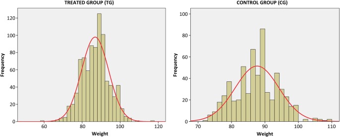
There was an average carcase weight of 86.81±0.24 and 87.65±0.27 kg for respectively TG and CG (P=0.023). The distribution of carcase weights in both groups is shown in Fig 1.
FIG 1:

Distribution of carcase weights obtained for both groups. The red line indicates a normal distribution for these frequencies
Lung study at slaughterhouse
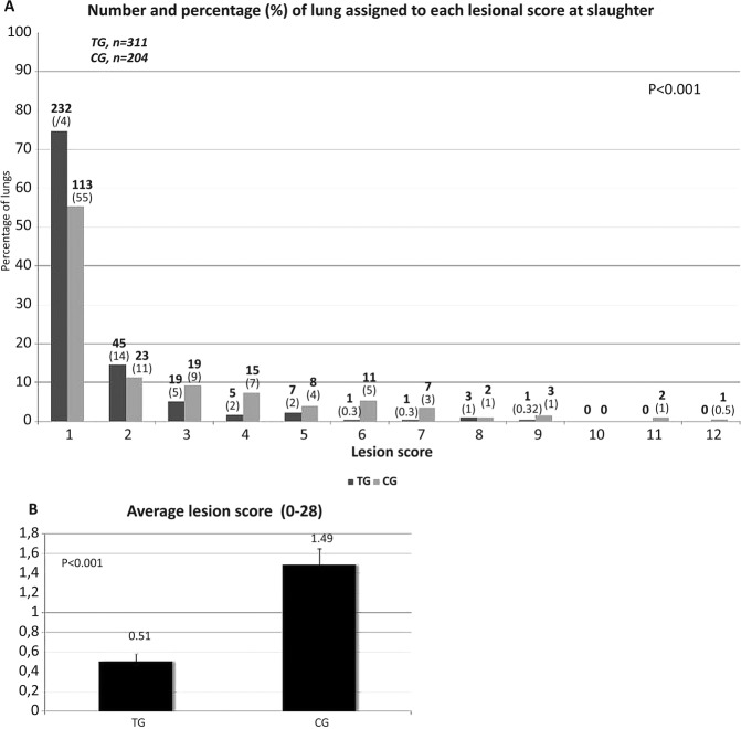
There were significant differences (P<0.001) for the average score between the TG (0.51±0.07) and CG (1.49±0.16), as well as the percentage of lung damaged (25.4 per cent for TG v 44.6 per cent for CG, P<0.001) and the number of lungs with each lesion score (Fig 2).
FIG 2:
(A) Number (bold) and percentage (in parentheses) of lungs assigned into each lesion score. (B) Average lesion score for each group (mean±standard error of the mean). CG, control group; TG, treated group
Serology, histopathology and q-PCR
From the six BALFs collected at weaning, all three samples from the TG were negative, while 2/3 samples in the CG were positive for M hyo.
At the start of the finishing period, 20 per cent (TG) and 70 per cent (CG) of the samples collected (10 samples in each group) for M hyo serology were positive, while at the end of the finishing period (12 samples in each group) this was 25 per cent (TG) and 75 per cent (CG). In both cases, the difference between groups was significant (P=0.025 and 0.014 for each time point, respectively).
The type of microscopic lung lesions observed, the number of lung samples showing each type of microscopic lesion and the number of positive lung samples to different swine pathogens are summarised in Table 2. Microscopic lung lesions indicative for M hyo infection were characterised by peribronchiolar and perivascular hyperplasia of the lymphoid tissues with the presence of inflammatory cells in the alveolar septa. Microscopic lesions of interstitial pneumonia were characterised by septal infiltration with mononuclear cells, type 2 pneumocyte hypertrophy and hyperplasia, and increased amounts of alveolar exudate consisting of mixed inflammatory cells and necrotic debris. Microscopic lesions of suppurative bronchopneumonia were characterised by the presence of abundant neutrophils, macrophages and cellular debris within the lumen of alveoli, bronchioles and bronchi. There were no differences for interstitial pneumonia and suppurative bronchopneumonia frequency among TG and CG (P=0.342 and 0.157, respectively).
TABLE 2:
Type of microscopic lung lesions observed, number of lung samples showing each type of microscopic lesion and number of positive lung samples to different swine pathogens in both groups
| Histopathology |
PCR |
||||||
|---|---|---|---|---|---|---|---|
| Mycoplasma-like lesions | Interstitial pneumonia | Suppurative bronchopneumonia | Mycoplasma hyopneumoniae | PRRSv | PCV2 | Actinobacillus pleuropneumoniae | |
| TG | 9/9 | 4/9 | 3/9 | 0/9 | 2/9 | 5/9 | 0/9 |
| CG | 9/9 | 6/9 | 6/9 | 8/9 | 2/9 | 6/9 | 0/9 |
| P | – | 0.342 | 0.157 | <0.001 | – | 0.629 | – |
CG, control group; PCV2, porcine circovirus type 2; PRRSv, reproductive and respiratory syndrome virus; TG, treated group.
Regarding the q-PCR for M hyo, there was significant difference comparing TG and CG (P<0.001) while there were no differences in the frequency of positive samples for PRRSv or PCV2. No sample was positive for A. pleuropneumoniae.
Discussion
Vaccines have demonstrated their effectiveness against M hyo in reducing clinical signs and lung lesions and improving productivity parameters but have not been able to eliminate the organism from the respiratory tract of the pig (Maes and others 2008). Other researchers reported that vaccines do not prevent M hyo colonisation (Thacker and others 1998) and that with the use of antimicrobials there was a reduction in the number of M hyo organisms (Vicca and others 2005). The effectiveness of tylvalosin against M hyo has been previously demonstrated in an eradication programme using tylvalosin (Marco and others 2008). In the present trial, it was demonstrated that the strategic use of tylvalosin administered in drinking water at two key time points of the pigs’ life reduces the pathological changes and negative impact on productivity caused by M hyo. The bacteria were not found by q-PCR in any of the samples of lung tissue of TG taken at slaughter, which may indicate that tylvalosin eliminated the organism from the lung. This is supported by the finding that all BALF samples taken in the nursery only two weeks after the first treatment were also negative.
The humoral immune response was different in each group on the basis of the blood samples tested. The kinetic in CG was the same observed in the serological studies performed before the trial with a percentage of seropositive pigs of 75 per cent at the end of the finishing period; however, in the group medicated with tylvalosin, only 25 per cent of the animals tested were seropositive at the end of the finishing period. This is an important reduction in the level of specific antibodies, which also has been reported in treatment with other antibiotics such as florfenicol (Ciprián and others 2012).
A previous study showed that for every 10 per cent increase of affected lung surface caused by pneumonia there was a reduction in the ADG of 37.4 g (Straw and others 1989). Compared with CG, we found a significant reduction of the lung lesion average score and the number of animals with mycoplasma-like lesions in the TG. The ADG (+0.028 kg) was significantly improved in the TG. In some studies, significant differences in lung lesions and ADG were found in animals treated in-feed with chlortetracycline (Del Pozo Sacristán and others 2012) or florfenicol (Ciprián and others 2012), or a single intramuscular administration of tulathromycin (Mckelvie and others 2005) compared with non-treated animals.
An increase in mean lung score was associated with a decrease of the carcase quality in a previous study (Ostanello and others 2007). In our case, the decrease of lung lesions score and of the number of lungs with mycoplasma-like lesions in the TG resulted in improved homogeneity and quality of the carcases, resulting in a better financial result for the farmer and the meat company.
The average carcase WS in the TG was significantly lower, but the animals in this group were sent to the slaughterhouse almost 11 days before and the IW in finishing was 2.45 kg lower than the CG. A lower ADF improves the FCR because less maintenance feed is needed. Also, the occupation rate of the units will be improved.
Administration in water gives a more accurate treatment compared with in-feed administration regarding start and duration of the treatment. In feed, it is more difficult to medicate for a precise number of treatment days due to feed bin size and management. Furthermore, initiation of the treatment at onset of clinical symptoms is easy with water administration in contrast to in-feed.
The medication cost per pig (excluding the cost of the tested drug) was reduced by $1.29 in the TG. The estimated cost of the treatment with tylvalosin per pig in this trial was $0.18, so the reduction of medication cost per pig including the treatment was $1.11. No deaths related to respiratory problems were diagnosed in the TG in contrast to the CG where two animals died because of pneumonia and two other pigs were wasted as a consequence of respiratory disease. Since tylvalosin is also registered for porcine proliferative enteropathy and swine dysentery and has shown its effectiveness (Pommier and others 2008, Guedes and others 2009), in case of concurrence of these diseases with EP, we may have an additional positive effect. In our case, there were no differences between groups in the presence of viral pathogens (PRRSv and PCV2) in the samples of lungs with mycoplasma-like lesions, but there was a reduction of microscopic lesions of suppurative bronchopneumonia indicative of bacterial pathogens in the TG, so the reduction of the presence of M hyo resulted in a decrease of the action of secondary bacterial pathogens.
A strategic medication with Aivlosin at 2.5 mg tylvalosin/kg BW in drinking water for seven days at weaning and when moved to finisher barn significantly reduced mycoplasma-like lung lesions and improved productivity parameters. The decreased presence of M hyo reduced clinical signs of secondary respiratory pathogens. In consequence, this antimicrobial could be a useful tool for both prevention and treatment of EP.
Acknowledgments
The authors thank Piensos Garla S.A. for allowing them to carry out this study and Germán Díaz and Denis Hernández for their assistance during the study.
Footnotes
Contributors: FJP, CL, MR and GR designed the study, analysed the data and drafted the manuscript.
Funding: Esteve Veterinaria, Laboratorios Dr. Esteve S.A.
Competing interests: CL, works for Esteve Veterinaria, Laboratorios Dr. Esteve S.A., distributor of Aivlosin in Spain and MR works for ECO Animal Health.
Provenance and peer review: Not commissioned; externally peer reviewed.
Data sharing statement: The main results of this research are presented in this paper.
References
- Abiven P., Pommier P. (1993) Technique de lavage tracheobronchique par voie transnasale pour la detection de Mycoplasma hyopneumoniae chez le porc vivant non anesthesie. Veterinary Research 24, 515–522 [PubMed] [Google Scholar]
- Ciprián A., Palacios J. M., Quintanar D., Batista L., Colmenares G., Cruz T., Romero A., Schnitzlein W., Mendoza S. (2012) Florfenicol feed supplemented decrease the clinical effects of Mycoplasma hyopneumoniae experimental infection in swine in México. Research in Veterinary Science 92, 191–196 doi:10.1016/j.rvsc.2011.01.010 [DOI] [PubMed] [Google Scholar]
- Del Pozo Sacristán R., Rodríguez A. L., Sierens A., Vranckx K., Boyen F., Dereu A., Haesebrouck F., Maes D. G. (2012) Efficacy of in-feed medication with chlortetracycline in a farrow-to-finish herd against a clinical outbreak of respiratory disease in fattening pigs. Veterinary Record 171, 645 doi:10.1136/vr.100976 [DOI] [PubMed] [Google Scholar]
- Guedes R. M. C., França S. A., Machado G. S., Blumer M. A., da Costa Cruz E. C. Jr. (2009) Use of tylvalosin-medicated feed to control porcine proliferative enteropathy. Veterinary Record 165, 342–345 doi:10.1136/vr.165.12.342 [DOI] [PubMed] [Google Scholar]
- Kobisch M., Blanchard B., Le Potier M. F. (1993) Mycoplasma hyopneumoniae infection in pigs: duration of the disease and resistance to reinfection. Veterinary Research 24, 67–77 [PubMed] [Google Scholar]
- Madec F., Derrien H. (1981) Fréquence, intensité et localisation des lésions pulmonaires chez le porc charcutier. Journées de la Recherche Porcine en France 13, 231–236 [Google Scholar]
- Madec F., Fourichon C., Morvan P., Labbé A. (1992) Économie et santé en production porcine. Inra Prodution Animals 5, 149–161. [Google Scholar]
- Maes D., Segales J., Meyns T., Sibila M., Pieters M., Haesebrouck F. (2008) Control of Mycoplasma hyopneumoniae infections in pigs. Veterinary Microbiology 126, 297–309 doi:10.1016/j.vetmic.2007.09.008 [DOI] [PMC free article] [PubMed] [Google Scholar]
- Marco E., Quiroga M., Menjón R., Bollo J. M., Calco E., Donadeu M., Cia C., Duran O. (2008) Eradication of Mycoplasma hyopneumoniae in a head using Aivlosin. Proceedings of an International Symposium: International Porcine Veterinary of Swine Congress Durban, 22 to 26 June, 2008. p 197 [Google Scholar]
- Marois C., Dory D., Fablet C., Madec F., Kobisch M. (2010) Development of a quantitative Real-Time TaqMan PCR assay for determination of the minimal dose of Mycoplasma hyopneumoniae strain 116 required to induce pneumonia in SPF pigs. Journal of Applied Microbiology 108, 1523–1533 doi:10.1111/j.1365-2672.2009.04556.x [DOI] [PubMed] [Google Scholar]
- Mckelvie J., Morgan J. H., Nanjiani I. A., Sherington J., Rowan T. G., Sunderland S. J. (2005) Evaluation of tulathromycin for the treatment of pneumonia following experimental infection of swine with Mycoplasma hyopneumoniae. Veterinary Therapeutics 6, 197–202 [PubMed] [Google Scholar]
- Ostanello F., Dottori M., Gusmara C., Leotti G., Sala V. (2007) Pneumonia disease assessment using a slaughterhouse lung-scoring method. Journal of Veterinary Medicine. A, Physiology, Pathology, Clinical Medicine 54, 70–75 doi:10.1111/j.1439-0442.2007.00920.x [DOI] [PubMed] [Google Scholar]
- Pallarés F. J., Gómez S., Muñoz A. (2001) Evaluation of the zootechnical parameters of vaccinating against swine enzootic pneumonia under field conditions. Veterinary Record 148, 104–107 doi:10.1136/vr.148.4.104 [DOI] [PubMed] [Google Scholar]
- Pommier P., Keita A., Pagot E., Duran O., Cloet P. R. (2008) Comparison of tylvalosin with tylosin for the control of subclinical ileitis in swine. Revue de Médecine Vétérinaire 159, 579–582 [Google Scholar]
- Straw B. E., Tuovinen V. K., Bigras-Poulin M. (1989) Estimation of the cost of pneumonia in swine herds. Journal of the American Veterinary Medical Association 195, 1702–1706 [PubMed] [Google Scholar]
- Thacker E., Thacker B., Boetteher T., Jayappa H. (1998) Comparison of antibody production, lymphocyte stimulation, and protection induced by four commercial Mycoplasma hyopneumoniae bacterins. Journal of Swine Health and Production 6, 107–112 [Google Scholar]
- van Leengoed L. A., Kamp E. M. (1989) A method for bronchoalveolar lavage in live pigs. Veterinary Quarterly 11, 65–72 doi:10.1080/01652176.1989.9694201 [DOI] [PubMed] [Google Scholar]
- Vicca J., Maes D., Jonker L., de Kruif A., Haesebrouck F. (2005) Efficacy of in-feed medication with tylosin for the treatment and control of Mycoplasma hyopneumoniae infections. Veterinary Record 156, 606–610 doi:10.1136/vr.156.19.606 [DOI] [PubMed] [Google Scholar]


