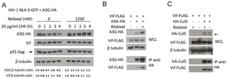
Figure 5. Redoxal augments A3G protein stability without disrupting Vif interaction with A3G, or Cul5.

A. Redoxal augments A3G protein stability in cells expressing HIV-1 Vif. 293T cells were co-transfected with 100 ng A3G-3xHA and 1000 ng pNL4-3GFPΔEnv HIV-1 plasmids. At 5 h post transfection, the media was replaced with media supplemented with 1250 nM redoxal. After 36 h of redoxal treatment, CHX was added. At the time intervals indicated after CHX treatment cells were lysed and A3G, Vif, p55Gag, and β-tubulin protein levels were analyzed. Values shown below the blots represent relative A3G or Vif protein levels in treated cells determined by densitometry using ImageJ software and normalization to A3G or Vif protein levels in untreated cells (DMSO control). B. Redoxal does not disrupt Vif-A3G interaction in 293T cells. 293T cells were co-transfected with 2000 ng pNLA1.Vif-FLAG and 300 ng A3G-3xHA or pCDNA3.1 (empty vector). The cells were then treated with 1250 nM redoxal for 40 h. At 48 h post transfection, cells were lysed and subjected to protein analysis. Western blot of cell lysates or anti-HA co-immunoprecipitated proteins were probed using anti-Vif and anti-HA antibodies. C. Redoxal does not affect Vif-Cul5 interaction in co-immunoprecipitation assays. 293T cells were co-transfected with 2000 ng pNLA1.Vif-FLAG and either 4000 ng HA-Cullin 5 or pCDNA3.1 (empty vector) expression constructs. Transfected cells were treated with DMSO or 1250 nM redoxal for 40 h. Cells were lysed 48 h after transfection. Cell lysates or anti-HA co-immunoprecipitated proteins were analyzed using anti-Vif and anti-HA antibodies.
