

Abstract
Intrabony cross arch migration (transmigration) of impacted teeth is a very rare dental anomaly. Transmigration is commonly seen in permanent dentition of the lower jaw. The tooth most commonly involved is the mandibular canine. The prevalence of transmigrated canine has been found to be only 0.14–0.31%. Transmigration of the canine most frequently occurs in a mesial direction resulting in migration across the mandibular symphysis to the opposite side of the dental arch. We report a case series (4 cases) of rare transmigration of mandibular canines and emphasise the need to use panoramic radiography in cases presenting with either over retained deciduous canines and/or missing permanent canines. We also propose a newer modification to the present classification of transmigration for one of these cases as the present classification does not include all the entities.
Background
Transmigration is a term used to describe movement of an unerupted tooth across the midline without the influence of any pathological entity (Tarsitano et al)1–3 is a rare phenomenon with prevalence as low as 0.14–0.31%. Transmigration is more often seen in the mandibular arch than in the maxillary arch.3 The tooth commonly involved is the lower mandibular canine. In the mandible, the tooth moves in a medial direction crossing the mandibular midline to land at variable regions in the opposite mandibular arch. The intraosseous migration of a tooth apparently starts during the early mixed dentition stage and may continue over a period of many years. Transmigration of canines does not usually cause major problems; the process is asymptomatic and usually discovered only during routine examination. Symptoms, if present, are mainly due to loss of aesthetics owing to absence of the transmigrated tooth.
Retained primary mandibular canines and/or absence of permanent mandibular canines should always be viewed with suspicion by the practitioner. Early diagnosis with timely orthodontic or surgical intervention can help dentists preserve these canines, as they play an important role aesthetically as well as functionally in human dentition.4 Timely diagnosis not only limits the amount of disability but also helps in repositioning the canine with as little trauma as possible. It is especially important, therefore, to have access to routine radiographs taken during childhood and adolescence to assess growth and development of the child; these can be very valuable in preventing and intercepting this phenomenon.
In the present article we report a case series (4 cases) of mandibular canine transmigration to highlight the importance of early detection by panoramic radiographical examination. Among these cases, there is a report of an unusual case of reverse oblique migration of a mandibular right canine crossing the jaw midline and piercing the mid-ramus region of the left mandible, which is a rather unique condition, and never before cited in the literature; we propose a modification to the existing classification for this case.
Case presentation
Case 1
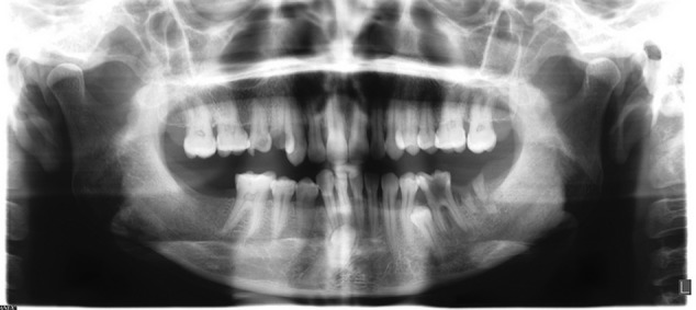
A 26-year-old man presented with multiple missing teeth (figure 1). His dental history revealed multiple missing teeth that were extracted due to caries and/or periodontal issues. Routine radiographs such as intraoral peri apical (IOPA) view and orthopantomograph (OPG) were advised to aid in replacement of missing teeth allowing proper rehabilitation of aesthetics and function to the patient. A panoramic radiograph showed multiple missing teeth and restorations in the maxilla and mandible. Importantly, a rare finding, presence of a tooth in a longitudinal direction, was seen in the mid-ramus region of the left jaw. Since the patient had undergone extraction of several teeth, and transmigration is usually symptomless, the patient was not aware of this phenomenon. The anatomy of the tooth confirmed it to be the right mandibular canine transmigrated to the ramus on the left side (the crown dictates the migration of tooth and history of extraction with 33 from the case record of the patient).
Figure 1.

Reverse oblique transmigration of right mandibular canine to left mid-ramus region.
Although several dental treatments (extractions and restorations) were performed earlier for the patient, it was only now that the presence of such pathology was found. This emphasises the importance of using OPG in conjunction with IOPAs in diagnosing missing teeth before restorative work begins. Overall scouting of the jaw becomes all the more necessary if teeth are reported to be missing from the beginning. Although several extractions and restorative work were performed by previous dentists, inability to take proper diagnostic aids is the reason for non-diagnosing of the transmigration.
This presentation is also unique in that there is no mention of similar cases (regarding position of the migrated tooth) in the literature. Therefore, it was difficult to classify it, which was not a problem in the following three cases. We wish to add to the pre-existing and commonly used classification system (Mupparapu's classification for transmigration) to type 6 (namely, canine impacted either horizontally or vertically beyond the body of the mandible on the contralateral side).
Case 2
A 28-year-old man presented with malaligned lower front teeth (figure 2). A complete clinical examination revealed over-retained 83 and missing 43, 31, 37 and 47. The patient had no history of prior extractions. Owing to absence of 43, 31, 37 and 47, the adjacent teeth had either drifted to the adjacent space or supra-eruption of opposing teeth had formed malalignment and derangement of occlusion. To move the teeth to their correct positions and to rehabilitate occlusion to a normal state, routine radiographs were advised before contemplating a treatment plan. A panoramic radiograph revealed intrabony longitudinal transmigration of 43 to periapical region of 36 at the level of inferior border of mandible (Mupparapu type 4 category of transmigration).
Figure 2.

Transmigration of right mandibular canine below apices of left mandibular molar near inferior border of mandible.
Case 3
A 26-year-old man presented with pain in the lower left rear tooth region and mobility in the lower right front teeth (figure 3). Clinical examination revealed carious destruction with 36, Grade 2 mobility with 83 and missing 16 and 43. A panoramic radiograph was advised as part of routine diagnostic aid, which revealed intrabony longitudinal migration of 43 to the periapical region of 33 crossing the midline (Mupparapu type 2 category of transmigration). Since transmigration (impacted tooth) rarely produces symptoms, the patient was given the diagnosis and advised periodical check-ups to look for pathological changes.
Figure 3.

Transmigration of right mandibular canine crossing the midline up to the apex of left mandibular canine.
Case 4
A 30-year-old woman presented with pain in the lower left rear tooth region (figure 4). Clinical examination revealed carious destruction of 37's crown and deep occlusal caries at 36 with a missing 43. A panoramic radiograph was taken to evaluate for missing tooth 43, which revealed intrabony vertical transmigration of 43 beneath the perapical region of 31, 32 and 41 present in midline. Thus a diagnosis of transmigration was made (Mupparapu type 5 category of transmigration).
Figure 4.
Vertical transmigration of right mandibular canine in the midline near the apices of incisors.
Discussion
The term transmigration was coined by Ando et al5 Transmigration should be considered when half the length of the crown of an unerupted tooth crosses the midline.4 Studies have suggested that transmigration of canines is a rare phenomenon with an incidence as low as 0.31%.5 Mandibular canines are the commonly involved teeth. Mupparapu reported transmigration of canines to be more frequently associated with females than males with a ratio of 1.6:1,4 but we found it to be more prevalent in males. Mandibular left side is affected more than the right1–5 but in three out of four cases, the right side was more affected, which contradicts the literature.
Maxillary canines are more commonly impacted teeth in comparison with mandibular canines; however, the phenomenon of transmigration of maxillary canines is lower in comparison to transmigration of mandibular canines in the literature until now. The mandibular canine is the only tooth in which migration and crossing of the midline has been documented. The reason cited for mandibular canine transmigration is due to the larger cross-sectional area of the anterior mandible compared with that of the anterior maxilla.4 Also, transmigration of maxillary canines may not be common because of the closeness between the roots of the maxillary incisors and floor of the nasal fossa. Other reasons mentioned include restriction in the path of maxillary tooth movement by the roots of adjacent teeth, maxillary sinus and mid-palatal suture, all of which can resist free movement of the tooth.4
The aetiology of transmigration is unknown; several propositions have been put forth, such as:
Abnormal displacement of dental lamina during embryonic life.
Non-eruption of canines due to deviation during development arising from obstacles such as root fragments or odontomes, the most commonly accepted explanation.4
Agenesis of the adjacent tooth, in particular the lateral incisor, may favour the retention of the primary canine at a more medial position than normal. Such a position of primary canine cannot provide a guide/pathway for eruption of permanent canine. Thus, an unerupted canine has the possibility of deviating from its normal developmental site, moving to a more horizontal position and migrating through the symphyseal bone, but only if enough space is available in front of the mandibular incisors.5
Also, the conical crown shape and long root of the mandibular canine aid in transmigration.
The greatest amount of movement or transmigration takes place during early stages of tooth development. Movement of a transmigrated canine is more rapid before the formation of its root.6 One case observed that about 3–4 mm of movement of a transmigratory canine occurred during a period of 1 year.7
Impacted teeth may migrate mesially or distally in the bone. The movement of teeth always occurs in the direction of the crown, which explains the reason for transmigration of canines to the opposite arch. The migratory passage of a canine through the mandible is in the direction of its long axis with the tip of the crown leading the way.8 The mandibular permanent lateral incisor usually migrates distally. In our first case we revealed transmigration of a mandibular canine to the mid-ramus (rarely reported) region of the left jaw crossing the midline, which is rare.
It is noted that movement takes place along the paths of least resistance.3–5 In our first case, perhaps it is because the multiple missing teeth in the mandible allowed intrabony migration of the right mandibular canine that it shifted to the mid-ramus region on the left side, which is a very rare phenomenon.
Mupparapu (2007)9 proposed a classification for mandibular canine transmigration depending on its path of deviation into five types (table 1 and figure 5).1–6 10
Table 1.
Classification based on position of unerupted mandibular canine
| Type-1 | Canine positioned mesioangularly across the midline within the jaw bone, labial or lingual to anterior teeth and the crown portion of the tooth crossing the midline (45.6%) |
| Type-2 | Canine horizontally impacted near the inferior border of the mandible below the apices of the incisors (20%) |
| Type-3 | Canine erupting either mesial or distal to the opposite canine (14%) |
| Type-4 | Canine horizontally impacted near the inferior border of the mandible below the apices of either premolars or molars on the opposite side (17%) |
| Type-5 | Canine positioned vertically in the midline (the long axis of the tooth crossing the midline) irrespective of eruption status (1.5%) |
Figure 5.

Diagrammatic representation of Mupparapu's classification.
In our first case, the right mandibular canine migrated to the left mid-ramus portion of the mandible, which, so far, is a unique occurrence not yet classified. We propose a modification to the present classification by Mupparapu and suggest a type 6, namely, a canine impacted either horizontally or vertically beyond the body of the mandible on the contralateral side. Our case 2 was of type 4 variety, while case 3 was type 2 and case 4, type 5 variant.
In cases of missing or over retained deciduous mandibular canines where periapical radiographs fail to detect abnormality, transmigration of canines should be a strong suspicion. These canines lie horizontally below the inferior alveolar canal or migrate toward the midline and, as a result, may not be visible in periapical radiographs. This emphasises the need for panoramic radiography.3–7 In cases 1 and 2, transmigration were only detected following panoramic radiography. Failure to use panoramic radiography would be the reason for non-detection after several treatments, as seen in case 1.
Various treatment modalities such as surgical extraction of transmigrated canines, transplantation, exposure and orthodontic alignment have been suggested.3–5 7 8 11 12 Surgical extraction is the most favoured treatment. Several pathologies are associated with impacted teeth, such as cyst and malignant transformation from its overlying lining. Since transmigration is a type of impacted tooth, periodic check-ups are necessary. Treatment is given only if abnormalities such as development of an apical cyst, neuralgia, resorption of adjacent tooth root or displacement of teeth occur. If the patient is asymptomatic, the transmigrated canine can be left in place; however, regular follow-up with radiographs is required to monitor the movement of these teeth. In all our four cases, the patients were asymptomatic so were advised regular follow-up.
Even though the teeth have transmigrated to the contralateral side, they maintain their nerve supply from the originating side, so it is necessary to anaesthetise the nerve bilaterally before surgical extraction with local anaesthesia.4 5 13 This may be performed so as to anaesthetise the nerve supplying the tooth (contralateral side) and buccal and lingual anaesthesia (ipsilateral side).
Conclusion
Transmigration of a mandibular canine is a rare event. It may be associated with genetic inherited anomalies or a defect in dental follicle metabolism. It is the duty of the clinician to suspect intrabony migration of a tooth in every case of missing or over-retained teeth followed by proper radiographic investigation. Early detection of transmigration can aid in treatment of such teeth maintaining cross arch stability.
Learning points.
Transmigration is usually found during routine radiographic examination, since most cases usually remain dormant.
Transmigrant teeth can give rise to complications to adjacent teeth (resorption, displacement) and adjacent vital structures (eg, nerve paraesthesia, cystic transformation).
It is the duty of every clinician to detect transmigration if a clinically missing permanent canine or over-retained deciduous canine is noted.
Transmigrant canines should be carefully followed regularly for any signs of ensuing pathologies and should be treated only when associated with the problems mentioned above.
Footnotes
Competing interests: None.
Patient consent: Obtained.
Provenance and peer review: Not commissioned; externally peer reviewed.
References
- 1.Tarsitano JJ, Wooten JW, Burditt JT. Transmigration of nonerupted mandibular canines: report of cases. J Am Dent Assoc 1971;82:1395–7. [DOI] [PubMed] [Google Scholar]
- 2.Joshi M, Shetye S. Transmigration of mandibular canines: a review of the literature and report of two cases. Quintessence Int 1994;25:291–4. [PubMed] [Google Scholar]
- 3.Umashree N, Kumar A, Nagaraj T. Transmigration of mandibular canine. Case Rep Dent 2013;2013:697671 http://dx.doi.org/10.1155/2013/697671 [DOI] [PMC free article] [PubMed] [Google Scholar]
- 4.Auluck A, Nagpal A, Setty S et al. Transmigration of impacted mandibular canines—report of 4 cases. J Can Dent Assoc 2006;72:249–52. [PubMed] [Google Scholar]
- 5.Kumar JS. Transmigration of mandibular canine: a case report with review of literature. J Indian Aca Oral Med Radiol 2012;24:88–90. doi:10.5005/jp-journals-10011-1267 [Google Scholar]
- 6.Aktan A, Kara S, Akgunlu F et al. Unusual cases of the transmigrated mandibular canines: report of 4 cases. Eur J Dent 2008;2:122–6. [PMC free article] [PubMed] [Google Scholar]
- 7.Joshi MR. Transmigrant mandibular canines: a record of 28 cases and a retrospective review of the literature. Angle Orthod 2001;71:12–22. [DOI] [PubMed] [Google Scholar]
- 8.Shapira Y, Kuftinec M. Intrabony migration of impacted teeth. Angle Orthod 2003;73:738–43. [DOI] [PubMed] [Google Scholar]
- 9.Mupparapu M, Auluck A, Suhaz S et al. Patterns of intraosseous transmigration and ectopic eruption of bilaterally transmigrating mandibular canines: radiographic study and proposed classification. Quintessence Int 2007;38:821–8. [PubMed] [Google Scholar]
- 10.Nagaraj T, Umashree N. Transmigration of mandibular canine. E J Dent 2011;1:72–4. [DOI] [PMC free article] [PubMed] [Google Scholar]
- 11.Torres-Lagares D, Flores-Ruiz R, Infante-Cossío P et al. Transmigration of impacted lower canine. Case report and review of literature. Med Oral Patol Oral Cir Bucal 2006;11:E171–4. [PubMed] [Google Scholar]
- 12.Qaradaghi IF. Bilateral transmigration of impacted mandibular canines: report of two cases and review. Rev Clin Pesq Odontol 2010;6:271–5. [Google Scholar]
- 13.Camilleri S, RCS M, Scerri E. Transmigration of mandibular canines—a review of literature and a report of five cases. Angle Orthod 2003;73:753–62. [DOI] [PubMed] [Google Scholar]