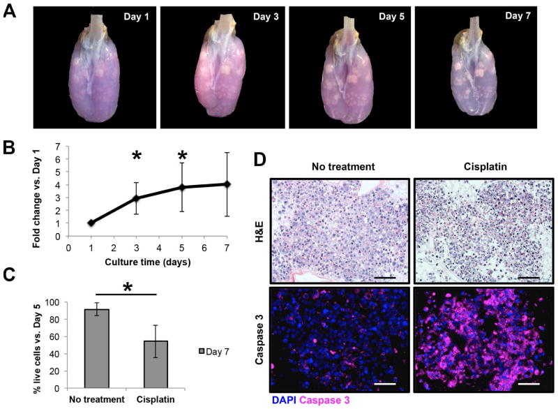
Fig. 4.
The resazurin reduction perfusion assay in a three-dimensional culture model for non-small cell lung cancer within decellularized scaffolds. (A) Time-lapse photography of a decellularized scaffold seeded with H358 adenocarcinoma cells after the performance of the resazurin reduction perfusion assay. The resazurin reduction perfusion assay on day 1 facilitated the localization of engrafted cells within the decellularized lung scaffold by providing a blue background that contrasted with cells, but also by highlighting the specific sites with higher resazurin reduction activity in pink. The evolution of the tumor nodules in terms of size and density can be appreciated over the culture period. (B) Proliferation curve of H358 cells under three-dimensional culture conditions within decellularized lung scaffolds, based on the resazurin reduction perfusion assay, demonstrating increasing cancer cell numbers over the culture period. Cell number increase was compared to day 1 measurements to account for variations in cell engraftment between experimental replicates (n=6 for days 1, 3 and 5; n=3 for day 7). There was a significant increase in the number of live cells on day 3 (2.9±1.2-fold, p=0.012) and day 5 (3.8±1.9-fold, p=0.015) when compared to day 1. (C) Use of the resazurin reduction perfusion assay to assess the cytotoxic effect of a single dose of the chemotherapeutic agent cisplatin. Relative to pre-treatment results (day 5), the number of live cancer cells within the decellularized lung scaffolds decreased to 54.2%±18.8% after cisplatin treatment, as compared to a small decrease to 91.5%±7.6% in non-treated controls (p=0.03; n=3 for each experimental arm). (D) Histologic evaluation of H358 tumor nodules cultured within decellularized lung scaffolds. The H&E staining for control scaffolds showed tumor nodules with viable cancer cells, while for cisplatin-treated scaffolds there were several dead cells characterized by nuclear pyknosis and fragmentation. This observation correlated with activated caspase-3 staining thus confirming the cytotoxic effect of cisplatin. Data are presented as means with standard deviations. *p<0.05.

