. 2015 Sep 22;4:e08022. doi: 10.7554/eLife.08022

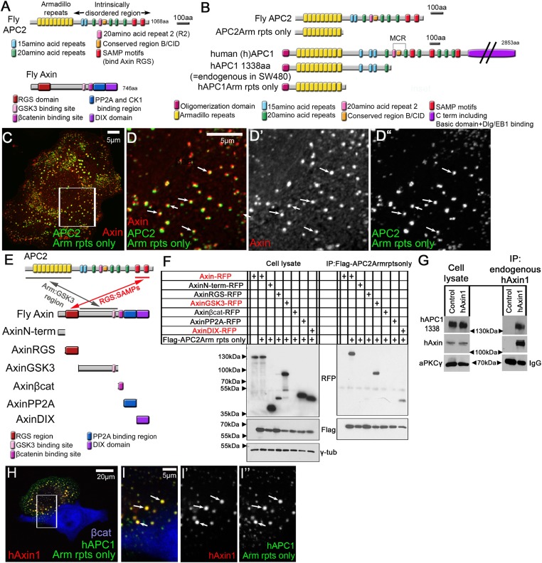
Figure 1. APC2's Arm rpts provide a second means of interacting with the Axin complex.

(A) Fly APC2 and Axin. (B) Constructs used. hAPC1-1338 = the endogenous truncated hAPC1 in SW480 cells. (C and D) SW480 cells coexpressing GFP-APC2Arm rpts only and Axin-RFP, which localize adjacent to one another (arrows). (D) Insets = box in (C). (E) Known and novel APC:Axin interaction sites (top) and Axin constructs (bottom). (F and G) IPs from SW480 cells. (F) APC's Arm rpts coIP with Axin's middle region that contains the GSK3 binding site. Axin's DIX domain was weakly detected. The βcat binding site fragment was not detected in either immunoblots or immunofluorescence (not shown), suggesting rapid degradation. (G) IP of endogenous hAxin1 or control Ig. Truncated endogenous hAPC1-1338 coIPs with hAxin1 at endogenous levels. (H) GFP-hAPC1Arm rpts only and hAxin1-RFP colocalize in puncta (arrows). (I) Insets = box in (H).

DOI: http://dx.doi.org/10.7554/eLife.08022.003

Figure 1.

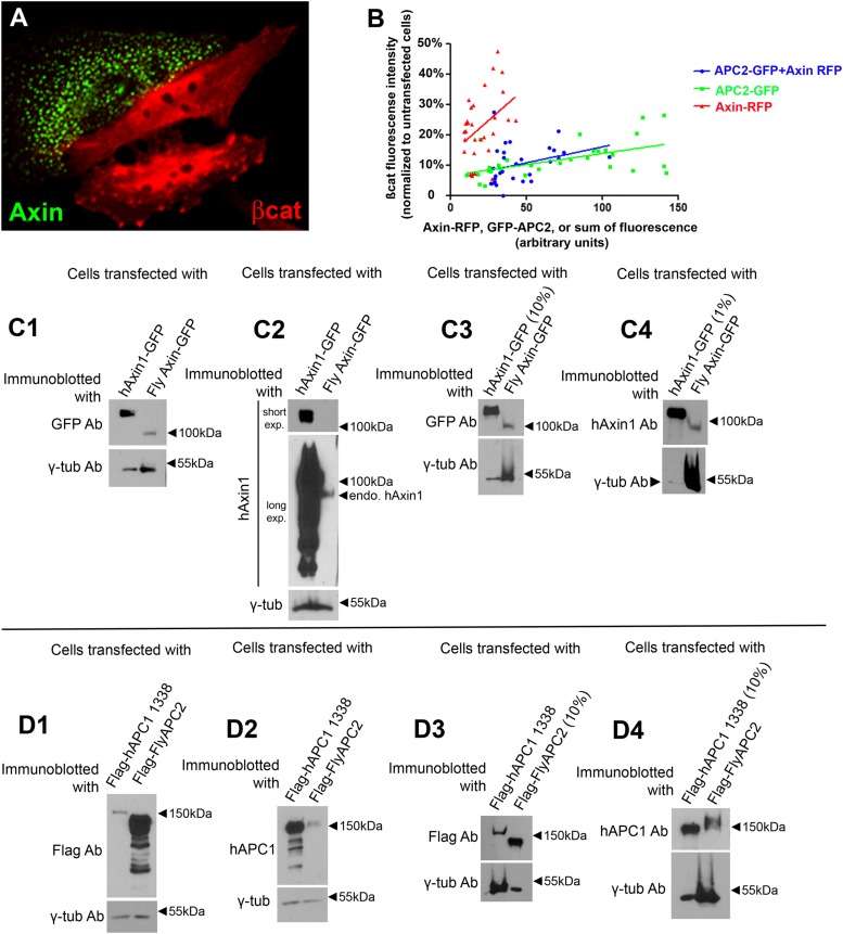
Figure 1—figure supplement 1. Assessing levels of over-expression of Axin and APC.

Figure 1—figure supplement 1.

(A) When overexpressed in SW480 cells, fly Axin forms puncta. (B) Plot of immunofluorescence intensities in SW480 cells transfected with GFP-APC2, or Axin-RFP, or GFP-APC2 + Axin–RFP, and stained for βcat via antibody. βcat intensities of transfected cells were normalized to adjacent untransfected cells and plotted against the GFP, or RFP, or sum of GFP and RFP intensities. 10 cells were measured each time in 3 independent experiments. (C) Measuring the levels of Axin overexpression in SW480 cells. Immunoblot analysis of cells transfected with human Axin1-GFP (hAxin1-GFP) or fly Axin-GFP with the indicated antibodies. γ-tubulin was used as a loading control. (C1,C2) Equal volumes of cell lysate were loaded. (C3) 10% the amount of hAxin1-GFP lysate was loaded. (C4) 1% of the amount of hAxin1-GFP lysate was loaded. One representative immunoblot of 3 independent experiments. Details of calculations used are in the Results and Materials and methods—full data is in Table 1. (D) Measuring the levels of APC2 overexpression in SW480 cells. Immunoblot analysis of cells transfected with Flag-tagged truncated human APC1-1338 (see Figure 1B) or fly APC2 with the indicated antibodies. γ-Tubulin was used as a loading control. (D1,D2) Equal volumes of cell lysate were loaded. (D3) 10% the amount of Flag-flyAPC2 lysate loaded. (D4) 10% the amount of Flag hAPC1-1338 lysate was loaded. One representative immunoblot of 3 independent experiments.

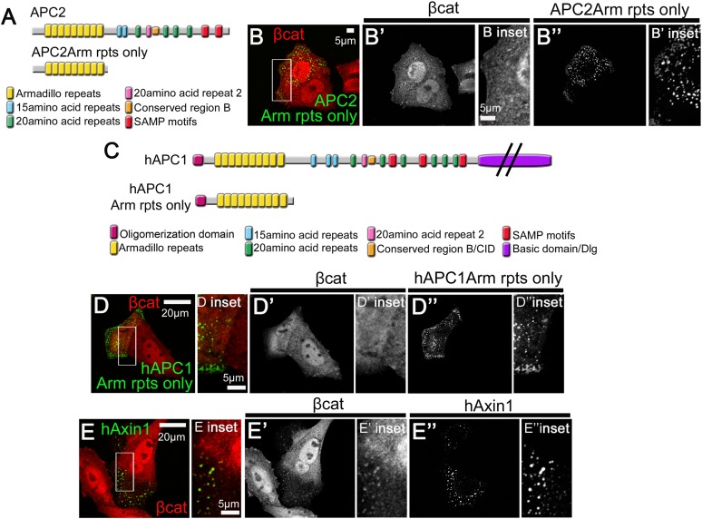
Figure 1—figure supplement 2. Human APC's Arm repeat domain colocalizes with Axin.

Figure 1—figure supplement 2.

(A) Diagrams of fly APC2 and APC2Arm rpts only. (B) SW480 cells expressing GFP-APC2Arm rpts only. APC2Arm rpts only forms cytoplasmic puncta and is unable to reduce βcat levels (insets = box in B). (C) Diagrams of hAPC1 and hAPC1Arm rpts only mutant. (D) GFP-hAPC1Arm rpts only expressed in SW480 cells. hAPC1Arm rpts also forms cytoplasmic puncta and is unable to reduce βcat levels (insets = box in D). (E) GFP-hAxin1 expressed in SW480 cells forms cytoplasmic puncta, and does reduce βcat levels, but βcat accumulates in the puncta (insets = box in D).