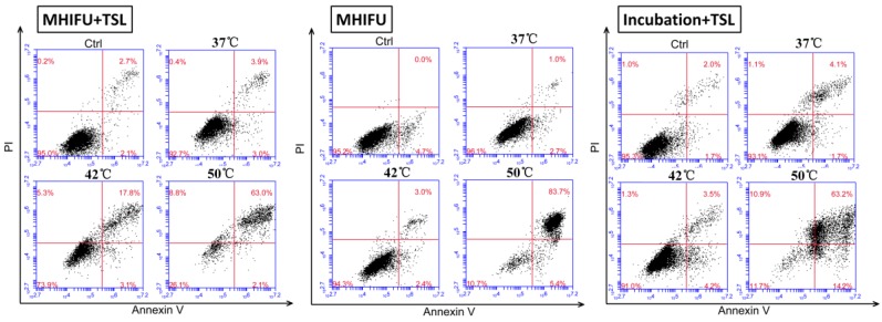
Figure 5.
Quantitative analysis of apoptosis by flow cytometry. The cells were divided four groups: MHIFU-mediated drug delivery; MHIFU irradiation solely; water incubation-mediated drug delivery and control group. It can be seen that the cell did not show significant change in viability at 37 °C for each group. For 42 °C, the late stage apoptotic cells were detected only in the first group, indicating MHIFU irradiation solely would not exert an adverse effect on the viability while the third group did not show obvious apoptosis. When the temperature increased to 50 °C, apoptosis in each group increased dramatically because of high-hyperthermia.

