PRESENTATION
We present multi-modal imaging of two phakic patients who developed lens nucleus dislocation following multiple complicated vitrectomy surgeries. We feel vitreoretinal physicians should be aware of this phenomenon and its presentation. To the best of our knowledge, high-quality multi-modal imaging of this presentation has not been published.
Two phakic patients with moderate nuclear sclerosis underwent 25-gauge pars plana vitrectomy (PPV). The first case underwent six PPV procedures; an initial macular hole repair was later complicated by rhegmatogenous retinal detachment, proliferative vitreoretinopathy, tractional retinal detachment and suprachoroidal hemorrhage. The patient noted a sudden change in vision on day 15 after the last surgery and posterior dislocation of the lens nucleus was detected.
The second patient underwent PPV for traumatic vitreous hemorrhage, during which a retinal tear was found and repaired. This patient noted a sudden change in vision 3 months after the surgery.
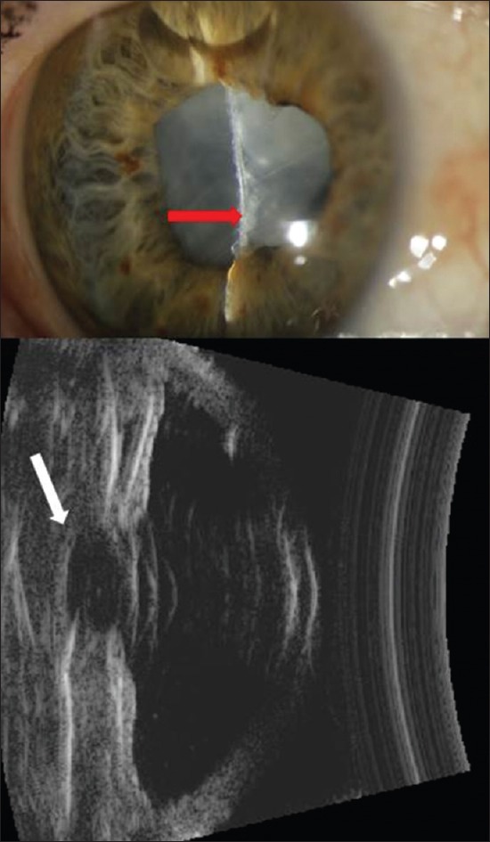
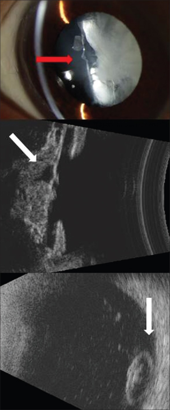
In both patients, slit lamp biomicroscopy demonstrated cataractous cortical material within the lens capsule with an odd appearance. In the first patient, ophthalmoscopy allowed a clear view to the posterior segment, and the nucleus appeared to be resting on the retina inferiorly, still under silicone oil tamponade. Slit-lamp photography and ultrasound biomicroscopy documented these findings [Figure 1]. B-scan ultrasonography did not yield high quality images due to the presence of silicone oil. Ultrasound biomicroscopy in the second patient revealed absence of the lens nucleus within the lens capsule. B-scan ultrasonography showed a shifting hyper reflective mass resting on the retina, representing a dislocated lens nucleus [Figure 2].
Figure 1.

Top: Slit-lamp photograph shows residual cortical material within the lens capsule with absent lens nucleus and lack of typical anterior capsule convexity (red arrow). Bottom: Ultrasound biomicroscopic image showing a transpupillary section; hyperreflective cortical material within the lens capsule is visible with a hyporeflective space (white arrow) representing the cavity left after the dislocation of the nucleus.
Figure 2.

Top: Slit-lamp image showing residual cortical material within the lens capsule with absent lens nucleus and lack of typical anterior convexity (red arrow). Middle: Ultrasound biomicroscopic image showing a transpupillary section; hyperreflective cortical material within the lens capsule is visible with a hyporeflective space (oblique white arrow) representing the cavity left after the dislocation of the nucleus. Bottom: B-scan ultrasound image of the posterior segment showing a hyperreflective mass consistent with dislocated lens nucleus (vertical white arrow).
The second patient underwent a PPV with lens fragmentation without complication. A large rent was observed in the posterior capsule, however an intraocular lens was successfully implanted into the ciliary sulcus.
DISCUSSION
Late spontaneous dislocation of the lens nucleus following PPV is a rarely reported occurrence with limited photographic documentation. Posterior capsular rupture and spontaneous lens dislocation has been shown to uncommonly occur in eyes with posterior polar cataracts[1,2] and posterior lenticonus[3] wherein weakness of the posterior capsular bag combined with an increasing nucleus size from nuclear sclerosis may cause the posterior capsule to burst leading to posterior dislocation of the nucleus into the vitreous. Spontaneous dislocation has also been reported in children likely due to congenital anomalies.[3,4,5] Hypermature cataracts have been reported spontaneously dislocate.[6] Another such case was reported in conjunction with pseudoexfoliation syndrome[7] and another with synchysis scintillans.[8]
Dislocation may also result from iatrogenic trauma to the posterior lens capsule during PPV. We believe this is the likely mechanism of nucleus dislocation in the presented cases, possibly combined with the initial trauma (in the second patient). Only one such case has been previously reported, to the best of our knowledge, and photographic documentation in that case was limited.[9] Published in 1978, Sigelman et al noted no predisposing factors or congenital anomalies in their patient undergoing PPV.[9]
In the first case presented herein, lens dislocation occurred 2 weeks after the latest of several PPV surgeries, while in the second case, dislocation occurred 3 months after a single uncomplicated PPV surgery. Neither of these patients had known lens capsule pathology predisposing to nuclear dislocation. In the first case, extensive vitreous base shaving and repeated surgeries gave ample opportunity for iatrogenic lens capsule trauma. In the second case, the antecedent trauma may have weakened the posterior capsule. Both cases were successfully managed with PPV, nucleus fragmentation and removal, and implementation of an intraocular lens in the ciliary sulcus with good visual outcome.
Herein, we present high quality multi-modal photographic documentation for two these cases of lens nucleus dislocation into the posterior segment following PPV surgery. While delayed presentation as in our cases is uncommon, we feel vitreoretinal surgeons should be aware of this phenomenon, its predisposing factors, and appearance. Furthermore, we believe that management of this condition may be successful using standard techniques of PPV, nucleus fragmentation and removal, and intraocular lens implantation in the ciliary sulcus.
Financial Support and Sponsorship
Nil.
Conflicts of Interest
There are no conflicts of interest.
REFERENCES
- 1.Ashraf H, Khalili MR, Salouti R. Bilateral spontaneous rupture of posterior capsule in posterior polar cataract. Clin Experiment Ophthalmol. 2008;36:798–800. doi: 10.1111/j.1442-9071.2008.01889.x. [DOI] [PubMed] [Google Scholar]
- 2.Ho SF, Ahmed S, Zaman AG. Spontaneous dislocation of posterior polar cataract. J Cataract Refract Surg. 2007;33:1471–1473. doi: 10.1016/j.jcrs.2007.05.007. [DOI] [PubMed] [Google Scholar]
- 3.Vajpayee RB, Sandramouli S. Bilateral congenital posterior-capsular defects: A case report. Ophthalmic Surg. 1992;23:295–296. [PubMed] [Google Scholar]
- 4.Vajpayee RB, Sharma N, Dada T, Gupta V, Kumar A, Dada VK. Management of posterior capsule tears. Surv Ophthalmol. 2001;45:473–488. doi: 10.1016/s0039-6257(01)00195-3. [DOI] [PubMed] [Google Scholar]
- 5.Kanigowska K, Gralek M, Trzebicka A, Chipczynska B. Spontaneous bilateral posterior capsule rupture in child – Case report. Klin Oczna. 2007;109:337–339. [PubMed] [Google Scholar]
- 6.Hemalatha C, Norhafizah H, Shatriah I. Bilateral spontaneous rupture of anterior lens capsules in a middle-aged woman. Clin Ophthalmol. 2012;6:1955–1957. doi: 10.2147/OPTH.S37276. [DOI] [PMC free article] [PubMed] [Google Scholar]
- 7.Takkar B, Mahajan D, Azad S, Sharma Y, Azad R. Spontaneous posterior capsular rupture with lens dislocation in pseudoexfoliation syndrome. Semin Ophthalmol. 2013;28:236–238. doi: 10.3109/08820538.2012.760622. [DOI] [PubMed] [Google Scholar]
- 8.Kumar V, Goel N, Piplani B, Raina UK, Ghosh B. Spontaneous posterior dislocation of nucleus with synchysis scintillans. Cont Lens Anterior Eye. 2011;34:144–146. doi: 10.1016/j.clae.2011.02.009. [DOI] [PubMed] [Google Scholar]
- 9.Sigelman J, Ketover BP, Coleman DJ. Spontaneous lens rupture following pars plana vitrectomy. Ann Ophthalmol. 1978;10:65–71. [PubMed] [Google Scholar]


