Abstract
The viscoelastic properties of blood clot have been studied most commonly using thrombelastography (TEG®) and thromboelastometry (ROTEM®). ROTEM®-based bleeding treatment algorithms recommend administering platelets to patients with low EXTEM clot strength (e.g., clot amplitude at 10 minutes [A10] <40 mm) once clot strength of the ROTEM® fibrin-based test (FIBTEM) is corrected. Algorithms based on TEG® typically use a low value of maximum amplitude (e.g., <50 mm) as a trigger for administering platelets. However, this parameter reflects the contributions of various blood components to the clot, including platelets and fibrin/fibrinogen. The platelet component of clot strength may provide a more sensitive indication of platelet deficiency than clot amplitude from a whole blood TEG® or ROTEM® assay. The platelet component of the formed clot is derived from the results of TEG®/ROTEM® tests performed with and without platelet inhibition. In this article, we review the basis for why this calculation should be based on clot elasticity (e.g., the E parameter with TEG® and the CE parameter with ROTEM®) as opposed to clot amplitude (e.g., the A parameter with TEG® or ROTEM®). This is because clot elasticity, unlike clot amplitude, reflects the force with which the blood clot resists rotation within the device, and the relationship between clot amplitude (variable X) and clot elasticity (variable Y) is nonlinear. A specific increment of X (ΔX) will be associated with different increments of Y (ΔY), depending on the initial value of X. When calculated correctly, using clot elasticity data, the platelet component of the clot can provide a valuable insight into platelet deficiency in emergency bleeding.
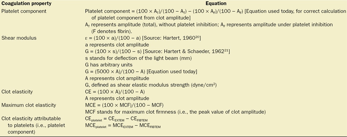
Clot amplitude is a standard parameter derived from viscoelastic methods of coagulation monitoring with thrombelastography (TEG®; Haemonetics Corp, Braintree, MA) or thromboelastometry (ROTEM®; Tem International GmbH, Munich, Germany). This variable is interpreted as a measure of clot strength. Although red blood cells make up over 90% of blood clot volume,1 clot strength is derived from the interaction of the fibrin network and platelets.2,3 Fibrin-based clot strength is dependent mainly on factor XIII and fibrinogen,4 whereas platelets contribute to overall clot strength by binding and tightening fibrin fibers.3 The platelet component of clot strength can be inhibited pharmacologically with, for example, a glycoprotein IIb/IIIa receptor antagonist or cytochalasin D. The platelet component of clot strength is defined as the difference in shear modulus measured with and without platelet inhibition.5–7 The calculation is shown in Table 1. The platelet component of clot strength is usually expressed either dimensionless in the same way as “clot elasticity” (CE) or in units of dyne/cm2 (e.g., G, which is numerically 50 times the TEG® parameter E or the equivalent ROTEM® parameter CE). Dyne is a unit of force that, although superseded by the SI system (1 dyne/cm2 = 0.1 N/m² = 0.1 Pa), is still used in the scientific literature pertaining to viscoelastic coagulation assessment. It is important to note that the assessment of the platelet component to clot strength may lead to misleading results if the calculation is performed using clot amplitude instead of CE. In this article, we explore parameters used for defining platelet deficiency with TEG® and ROTEM®.
Table 1.
Equations for Calculating Parameters of Interest
Viscoelastic Coagulation Monitoring
TEG® and ROTEM® are photokymographic8 devices designed to measure coagulation under conditions with oscillation but without blood flow.9 This reflects in vivo conditions of trauma and surgery, where blood vessels are cut or disrupted; blood flow is interrupted and the clot functions to close the vessel (hemostatic clot). It should also be considered that blood is unlikely to be fully static in vivo. The oscillations that characterize the TEG® and ROTEM® devices, which reduce clot strength compared with quiescent conditions,10 mimic the “nonflow”/“sluggish flow” conditions of surgery and trauma.
In common with many other biological tissues, blood clots have viscosity and elasticity properties. Application of stress to a blood clot results in a molecular rearrangement known as “creep,” characterizing viscosity. However, when the stress is removed, the clot’s elasticity returns to its original form. The Maxwell model takes into account a material’s viscoelastic properties in relation to stress, strain, and changes in these parameters over time. Early consideration of clot properties suggested that a blood clot could be considered to behave as a Maxwell body.11 However, recent porcine studies indicate that the Zener model may be more appropriate.12,13 The Zener model (also referred to as “the standard linear solid model”) is an alternative method of modeling the behavior of a viscoelastic material which, unlike the Maxwell model, includes a description of creep.
In addition to factor XIII,4 platelets and fibrin/fibrinogen are recognized as the key determinants of whole blood clot strength.14,15 After platelets have bound to fibrin via the glycoprotein IIb/IIIa receptor, the clot contracts through the action of cytoplasmic motility proteins inside platelets, such that fluid (serum) is expelled.16 With TEG® and ROTEM® devices, the clot is attached to both the pin and the cup, and clot retraction is therefore hindered. However, the clot contractile forces may contribute to clot stiffness, which translates into increased clot amplitude. It has been proposed that the relative contributions of platelets and fibrin to the clot amplitude (strength) are approximately 80% and 20%, respectively.17,18 Two points should be raised regarding this proposition. First, the estimation of the platelet and fibrin contribution to clot strength should, theoretically, be based on CE and not on clot amplitude. For example, using rabbit whole blood samples, Nielsen et al.7 calculated that platelets contribute 87% of CE in the absence of tissue factor and 94% upon exposure to tissue factor. Second, the extent to which platelets contribute to hemostasis probably differs from their contribution to CE: there is no evidence that platelets make an 80% to 95% contribution to hemostasis.
In 1948, Hartert19 introduced a viscoelastic device for measuring the shear modulus of a blood clot. Whole blood was added to a cup, and a plunger (pin) was immersed in the blood. The apparatus was designed so that rotation of the plunger was recorded via deflection of a light beam, with a 100-mm deflection representing the maximal rotation of 4°45′ (the scale of 0–100 mm was chosen arbitrarily). Hartert used the symbol ε to denote CE.19 In 1960, he used the same symbol to denote shear modulus of the clot and defined its relationship with clot amplitude as shown by the equation in Table 1.20 The equation was written slightly differently by the same author in 1962 (Table 1).21 Importantly, the equation shows that the relationship between deflection of the light beam (subsequently defined as clot amplitude) and shear modulus is not linear. The symbol ε was used synonymously with G in the 1962 publication. This is confusing, because within the same publication Hartert calculated the shear modulus of a clot with amplitude 2.5 cm to be 5000 dyne/cm2.14 This is the basis for today’s calculation of G (defined as shear elastic modulus strength in units dyne/cm2) from clot amplitude (A) (Table 1).22,23 Numerically, G (dyne/cm2) has a value 50 times that of Hartert’s parameter G with arbitrary units.
In a discussion of Hartert’s work, Copley stated that the name Hartert gave for his method— thromboelastography—was ill-chosen and even misleading, because the term “thrombus” is reserved for intravascular clotting, whereas blood clot in Hartert’s device is formed in vitro.20 Copley suggested the term “coaguloelastograph” or “blood clot elastograph” for Hartert’s apparatus20; these suggestions were reiterated by Evans et al.24 in 2006.
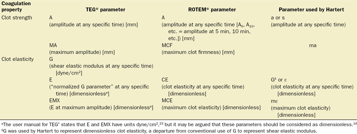
Today, the principles of using either TEG® or ROTEM® remain similar to the early work of Hartert. Whole blood or plasma is placed into a cup, although unlike in Hartert’s experiments reagents, such as celite or kaolin, are added to stimulate coagulation. Similarities between the current TEG® apparatus and that designed by Hartert are that the angle of rotation is the same for both devices (4°45′) and the TEG® oscillation period is 10 seconds (6 full oscillations per minute) compared with 9 seconds (6.7 oscillations per minute) with Hartert’s apparatus.21,23 The principles of the ROTEM® device are similar to those of TEG®, although with ROTEM® the oscillation period is 12 seconds (5 full oscillations per minute) and the central pin, instead of the cup, is rotated so that resistance to its rotation is measured (resistance increases as the clot forms). With both TEG® and ROTEM®, the principal measurement is clot amplitude, which shows the extent to which rotation is either triggered (TEG®) or resisted (ROTEM®) by clot formation. The scale for clot amplitude with both TEG® and ROTEM® generally ranges from 0 to 100 mm, with the maximal value chosen arbitrarily. Both elasticity and viscosity of the forming blood clot contribute to the clot amplitude.24
Standard parameters used to characterize the coagulation process using TEG® or ROTEM® are summarized in Table 2. The lower section of the table is described as relating to the elasticity of the clot. This is not strictly true because both viscosity and elasticity contribute to clot amplitude,24 and it is not possible with TEG® or ROTEM® to ascertain the contributions that each of these properties of blood clots contribute to the amplitude. As a result, the true elasticity of a blood clot cannot be calculated from TEG®/ROTEM® data. However, the elasticity parameters are directly related to the force with which the blood clot resists rotation within the device. In addition, it is believed that viscosity makes only a small contribution to clot amplitude.20 We will therefore continue to use the term CE within this article, in relation to the parameters G, E, EMX, CE, and maximum clot elasticity (MCE).
Table 2.
Major Parameters Associated with Thrombelastography and Thromboelastometry
As with Hartert’s original device, clot amplitude has a nonlinear relationship with elasticity (Fig. 1). Although the scale for elasticity ranges between zero and infinity, it would have been possible to configure the TEG® or ROTEM® device to display elasticity as the primary reading instead of amplitude. Had this approach been adopted (Fig. 2), calculation of the platelet component from TEG® or ROTEM® results would have been more straightforward from the beginning. Such adjustment could now be implemented by modifying the device software. Thus, future presentation of the platelet component as a primary TEG®/ROTEM® parameter, as shown in Figure 2, is conceivable.
Figure 1.
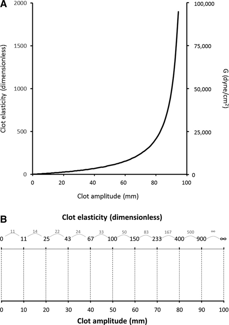
Relationships of clot amplitude (e.g., A) with clot elasticity (e.g., E for TEG®; CE for ROTEM®) and shear modulus (G). Because a specific increment of clot amplitude is associated with different increments of clot elasticity or shear modulus, depending on the initial value of clot amplitude, the relationship between amplitude and elasticity or shear modulus is nonlinear. In A, the single curvilinear line can show relationships of clot amplitude with both G and clot elasticity because G is 50 times clot elasticity. With respect to TEG® parameters, E = (100 × A)/(100 − A) while G = (5000 × A)/(100 − A). The conversion scale in B illustrates in a different way how clot amplitude is converted to clot elasticity; 10-mm increments in clot amplitude are associated with variable increments in clot elasticity. Configuration of a viscoelastic device so that the primary output is clot elasticity would enable the platelet component to be calculated by subtracting the primary FIBTEM reading from the primary EXTEM reading. A = Clot amplitude at any specific time (TEG® or ROTEM® notation); E = clot elasticity at any specific time (TEG® notation); CE = clot elasticity at any specific time (ROTEM® notation); G = shear modulus at any specific time.
Figure 2.
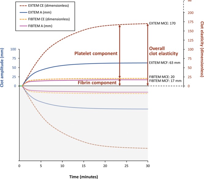
Derivation of the platelet component from viscoelastic assays performed in whole blood, in the presence and absence of platelet inhibition. The graph represents data obtained from a healthy volunteer with coagulation parameters in the normal range (roTEG®05 device; software version 2.95–2.99, December 2001; readings taken every 10 s are represented by curves of best fit). The platelet component is defined as the difference in clot elasticity between values obtained from assays with and without platelet inhibition. Conversion of clot amplitude to clot elasticity is therefore needed for calculation of the platelet component. As shown with ROTEM®, calculation of the platelet component requires data from the EXTEM and FIBTEM assays. With TEG®, the RapidTEG and Functional Fibrinogen assays could be used for this purpose; the procedure for calculating the platelet component would be the same. A = clot amplitude; CE = clot elasticity; EXTEM = ROTEM® extrinsically activated test; FIBTEM = ROTEM® test designed to assess fibrin-based clotting; MCE = maximum clot elasticity; MCF = maximum clot firmness.
With the TEG® device, the cup rotates both clockwise and counterclockwise, with movement that can be described as oscillatory. Amplitude is derived from rotation of the plunger, occurring as the blood forms a bond between these 2 parts of the apparatus. The clockwise and counterclockwise rotation angles of the plunger are recorded, and the central point at which the plunger remains before the clot is formed represents no rotation. With the ROTEM® device, it is the plunger that rotates (oscillates); the cup remains stationary and the rotation angle is decreased as the clot forms. As with TEG®, clot amplitude is calculated from the maximal rotation angle of the plunger. There is a question with both devices whether clot amplitude represents the difference between the central point and the full rotation in one direction or the difference between full rotation in one direction versus full rotation in the opposite direction. With Hartert’s device, the maximal rotation was 4°45′, representing 2°22.5′ clockwise from the resting point and the same rotation counterclockwise. Therefore, the 100-mm maximal deflection represented 50 mm in each direction from the resting point. The ROTEM® device provides measurements from a single point in the rotation cycle (i.e., maximal rotation in one direction). The measured deflection from the resting point is doubled to obtain clot amplitude (A), and the generation of traces showing both positive and negative deflection is artificial. With TEG®, readings are taken more frequently, so that values are obtained for rotation in both directions. Consequently, Figure 2 could be represented differently with TEG®; the curves above and below the x-axis would have equal weighting, and the y-axis scale could go downward to −50 and upward to +50. However, clot amplitude with TEG® (A) represents both positive and negative deflection, meaning that, in practice, TEG® clot amplitude values correspond to those of ROTEM® (although differences in cup size/geometry and in assay components mean that values are not directly comparable between the 2 devices).
PARAMETERS FOR ASSESSING PLATELET CONTRIBUTION TO CLOT STRENGTH
ROTEM®
Results from 2 ROTEM® tests are used to guide platelet administration: EXTEM and FIBTEM. The EXTEM test provides a measure of clot strength with extrinsic activation of whole blood coagulation via tissue factor. Both fibrin and platelets contribute to EXTEM clot strength, meaning that EXTEM alone does not provide a specific measure of the platelet contribution to clot strength. The FIBTEM test is the same as EXTEM but with the addition of cytochalasin D to prevent platelets from contributing to the clot strength. By comparing results from the EXTEM and FIBTEM tests, a specific assessment of the contribution of platelets to clot strength (platelet component) can be obtained.
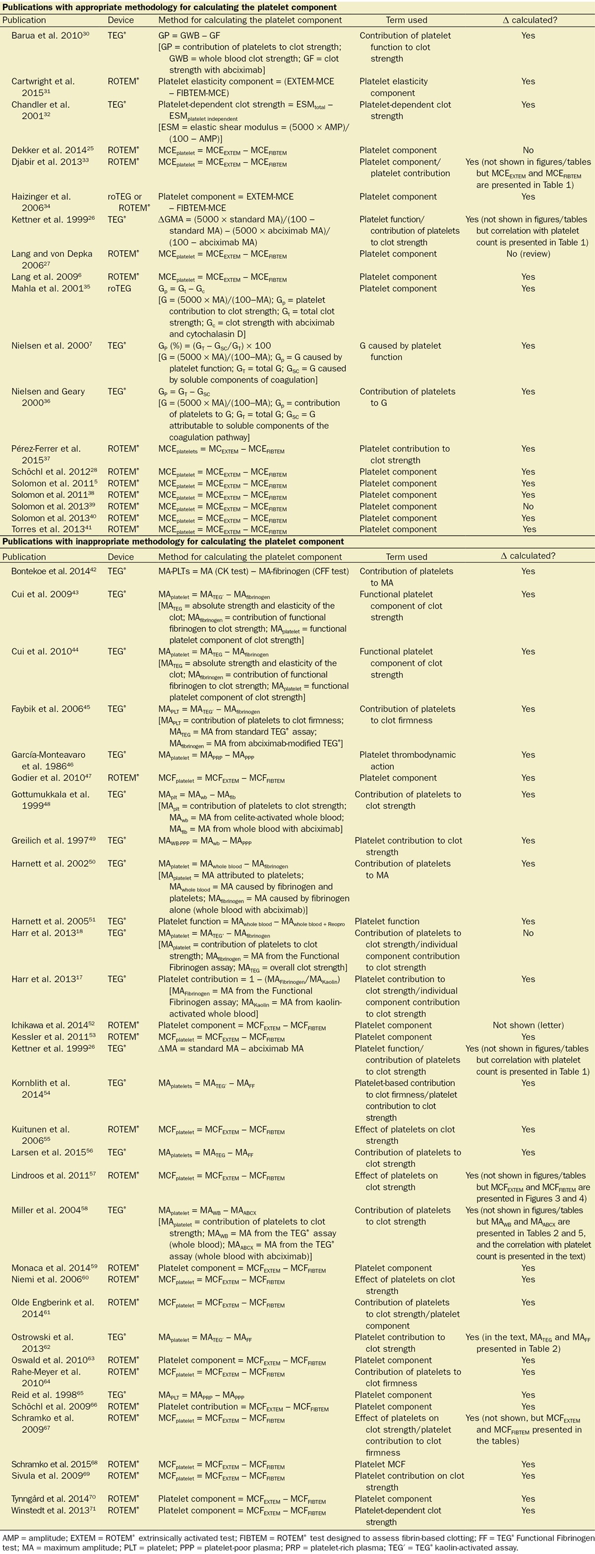
The platelet component is calculated from the elasticity results. First, CE (a dimensionless quantity) is obtained from clot amplitude (A) as shown in Table 1. MCE is calculated in the same way from maximum clot firmness (MCF) (Table 1). After such conversion, the platelet component can be calculated from EXTEM and FIBTEM results (Table 1).5,6,25 It is important that the calculation of platelet component be performed using elasticity (e.g., CE, MCE) as opposed to clot amplitude (e.g., A, MCF) because of the nonlinear relationship between clot amplitude and CE,6,7,21,26–28 as indicated in Figure 1 and Table 3. Unlike amplitude, CE may be considered a reflection of the force with which the blood clot resists rotation within the device. Where there is a nonlinear relationship between 2 variables X and Y, a specific increment of X (ΔX) will be associated with different increments of Y (ΔY), depending on the initial value of X. Therefore, an increment ΔX from baseline X′ cannot be considered as equivalent to the same increment ΔX from baseline X″. The European Society of Anaesthesiology guidelines for the management of perioperative bleeding highlight the fact that MCE and G have a curvilinear relationship with maximum amplitude (MA) and MCF.29 An illustration of the comparison between platelet component, correctly calculated from CE (in this case, EXTEM- and FIBTEM-MCE values) and incorrectly calculated from clot amplitude (MCF values), is presented in Table 3. This theoretical model shows that, across a range of platelet counts (from 10,000 to 100,000/μL), ΔMCF remains unchanged, whereas ΔMCE increases with platelet count. Therefore, it is clear that ΔMCF is not appropriate for calculating the platelet component.
Table 3.
Theoretical Data to Illustrate the Difference Between the Platelet Component (Based On the Difference in MCE Between EXTEM and FIBTEM) and the Difference in MCF Between EXTEM and FIBTEM

In the literature, there are publications where the contribution of platelets to clot strength has been calculated appropriately (i.e., using CE; Table 4). However, as also shown in Table 4, there are numerous examples where unsuitable methodology has been used, with calculations based on clot amplitude. Where the overall conclusions of a publication are based on possible incorrect calculation of platelet component (i.e., where the subtraction is performed using values for clot amplitude as opposed to CE), the findings should be interpreted with caution until the calculations have been repeated using correct methodology.
Table 4.
Methods Used in the Literature for Calculating the Platelet Component
TEG®
With the TEG® device, the standard kaolin-activated TEG® assay is most commonly used in relation to assessing the platelet contribution to clot strength. However, there is also a commercially available TEG® assay with platelet inhibition (Functional Fibrinogen assay), which is based on the same principle as FIBTEM. The platelet component of clot strength may be calculated by comparing elasticity results from the Functional Fibrinogen assay and from a standard assay without platelet inhibition. For example, the platelet component could be calculated as E (elasticity) obtained using the RapidTEG™ (Haemonetics Corp) assay minus E obtained using the Functional Fibrinogen assay, where E is a “normalized G parameter,”23 calculated in exactly the same way as Hartert’s shear modulus ε (Table 1). [Note that G is shear elastic modulus strength, with units dyne/cm2 (Table 1)].23 The parameter E may be considered as equivalent to the CE parameter of ROTEM®. The units of E are commonly referred to as dyne/cm2.23 However, CE is considered dimensionless and, because E is calculated in the same way as CE, we would argue that E should also be considered dimensionless. A is the equivalent of the ROTEM® parameter A (clot amplitude, in mm). For maximum values, EMX (E at maximum amplitude23; equivalent to MCE) would be used instead of E, in which case MA would be used instead of A. As an illustration of the need to use CE, Chandler14 stated that an increase in thrombelastograph clot amplitude from 50 to 67 mm (34% increase) corresponds with a 2-fold increase in CE. Thus, the principles discussed earlier in relation to ROTEM® apply to TEG® in the same way. Although the platelet component of clot strength can be derived from E or EMX, its calculation from G values may also be considered. The calculation of G by using the constant “5000,” and subsequent interpretation of G as an absolute value for shear elastic modulus may be flawed, however, for the following reasons: First, the strain imparted by the TEG® device is large enough to modify clot structure (i.e., the strain is too large for a linear viscoelastic response to be maintained).10,24 Second, the constant 5000 was derived from experiments reported by Hartert and Schaeder21 in 1962, and it is possible that differences in geometry or oscillation speed between today’s devices and that used by Hartert may mean that the value of 5000 should be redetermined experimentally using today’s devices. Until such experiments have been performed, using an elasticity parameter that does not rely on a constant (i.e., E) may, arguably, be considered preferable. Despite these considerations, as indicated earlier, G is mathematically a simple multiple of CE (e.g., G = 50 × E). This means that the mathematical approach for calculating the platelet component from G would be the same as that with E. However, we are not aware of published reports where the platelet component, based on TEG® data, has been correctly calculated and used to guide treatment for perioperative or trauma-related bleeding. As with ROTEM®, there are numerous examples in the literature where the platelet contribution to clot strength has been inappropriately calculated from TEG® values for clot amplitude instead of CE (Table 4).
The platelet mapping assay should enable more specific assessment of platelet function with the TEG® device. This assay involves up to 4 cuvettes.72,73 The first assesses citrated blood as in the standard method to determine the strength of the fully activated clot. The second cuvette is heparinized and contains reptilase plus factor XIIIa to measure strength of the full fibrin clot in the absence of platelet activation. The third and fourth cuvettes, also heparinized, measure the effects of adenosine diphosphate (ADP) and arachidonic acid stimulation on platelet aggregation. The platelet mapping assay was designed to provide an insight into the inhibitory effects of clopidogrel and aspirin. Conceivably, it could be used to guide platelet administration in a perioperative setting for patients who are bleeding without having taken platelet-inhibiting drugs. However, the results are analyzed by comparing MA values (e.g., percentage platelet inhibition in response to ADP = [(MAADP − MAFibrin)/(MAThrombin − MAFibrin) × 100]).73 As described earlier, it may be preferable to base such analyses on CE.
Platelet Component and Bleeding Management
The platelet component derived from ROTEM® and TEG® analysis provides a measurement of the contribution that platelets make to the strength of the whole blood clot. This is different from both platelet count and platelet function (measured by aggregometry). Nonetheless, there may be potential for using the platelet component to guide the transfusion of platelets in the treatment of coagulopathic bleeding.
At present, the platelet component of blood clot strength is not commonly used as a direct basis for treatment decisions. Instead, low whole blood clot strength (extrinsic activation), in the presence of adequate fibrin-based clot strength, is the typical criterion for administering platelets. The European Society of Anaesthesiology guidelines for the management of perioperative bleeding state that “adequate TEG® Functional Fibrinogen test/FIBTEM clot strength in the presence of decreased overall clot strength in bleeding patients may indicate platelet deficiency,” although specific thresholds for administering platelets are not provided.29 In a ROTEM®-based coagulation management algorithm for cardiovascular surgery patients, platelets are administered if EXTEM A10 is ≤40 mm and FIBTEM A10 is >10 mm.74 That is, these ROTEM® parameters suggest reduced overall clot strength in the setting of an adequate fibrinogen contribution to the clot. In a similar treatment algorithm for trauma-related bleeding, it is recommended that platelets are transfused if EXTEM CA10 <40 mm when FIBTEM CA10 >12 mm.75 Few bleeding management algorithms based on TEG® results have been published. In one example, platelet transfusion was based only on MA,76 a parameter with limited sensitivity to platelets. In the future, it is possible that platelet component, calculated from the difference in CE between the whole blood clot and the fibrin-based blood clot, could be integrated as one of the standard parameters of ROTEM® and that it might be validated against clinical parameters. The platelet component could then be used directly as a basis for quantitative treatment decisions.
Relationships Between Viscoelastic Coagulation Parameters and Platelet Count and Platelet Function
Correlations between EXTEM clot amplitude or CE and platelet count have been reported. In a prospective study involving patients undergoing cardiac surgery, EXTEM A5 significantly correlated with platelet count (Pearson correlation = 0.74; P < 0.001).61 Analyses on platelet-rich plasma samples from healthy volunteers demonstrated a positive correlation between changes in MCE and platelet count (r2 = 0.88; P < 0.001).6 In trauma patients, a statistically significant but weak correlation has been reported between ROTEM® platelet component (defined as MCEEXTEM − MCEFIBTEM) and platelet count (correlation coefficient = 0.44; P < 0.001).38 A more recent animal study also reported significant (P < 0.05) moderate correlation between these parameters.77 Variability in the correlation between ROTEM® platelet component and platelet count may be attributable to the platelet component being a measure of platelet function, which may be distinct from platelet count. Similar considerations apply to TEG®, where correlations between platelet count and TEG® MA have been documented.22,78,79 For example, a study on healthy volunteers and patients with peripheral arterial disease reported a strong correlation between log10 platelet count and TEG® MA in both groups (Pearson correlation = 0.97 and 0.89, respectively; P = 0.0001 in both groups).79 However, a previous study by Nielsen et al.7 in a rabbit whole blood model showed no significant correlation between the platelet contribution to G and the platelet count. We are not aware of clinical studies exploring the correlation between TEG® platelet component and platelet count. Overall, there is a need for additional investigation of the relationships between the platelet component (measured using either TEG® or ROTEM®) and the clinical status.
CONCLUSIONS
In this review, we provide evidence that the platelet component of clot strength should be calculated using CE as opposed to clot amplitude parameters from TEG® or ROTEM® analysis. This is because CE, unlike clot amplitude, reflects the force with which the blood clot resists rotation within the device, and the relationship between clot amplitude and CE is nonlinear. The platelet component has the potential to provide valuable insight into the clinical importance of a minor contribution of platelets to CE in emergency bleeding and might therefore help to guide treatment with platelet concentrate. However, this is conditional on the platelet component being calculated correctly. Certainly, clinical validation studies are needed to refine the interpretation of TEG® and ROTEM® results for the management of clinical bleeding.
DISCLOSURES
Name: Cristina Solomon, MD, MBA.
Contribution: This author helped design the study and wrote the manuscript.
Attestation: Cristina Solomon approved the final manuscript and attests to the integrity of the data presented. Cristina Solomon is the archival author.
Conflicts of Interest: Cristina Solomon is an employee of CSL Behring and previously received speaker honoraria and research support from Tem International and CSL Behring, and travel support from Haemoscope Ltd (former manufacturer of TEG®).
Name: Marco Ranucci, MD.
Contribution: This author helped prepare and critically revised the manuscript for important intellectual content.
Attestation: Marco Ranucci approved the final manuscript.
Conflicts of Interest: Marco Ranucci received speaker honoraria and research support from CSL Behring and Grifols, speaker honoraria from Medtronic, Haemoscope and Roche Diagnostics, research support from Tem International, and was on the Steering committee of a factor XIII study (until 2010) for Novo Nordisk.
Name: Gerald Hochleitner.
Contribution: This author helped in the design of the study and wrote the manuscript.
Attestation: Gerald Hochleitner approved the final manuscript.
Conflicts of Interest: Gerald Hochleitner is an employee of CSL Behring.
Name: Herbert Schöchl, MD.
Contribution: This author helped prepare and critically revised the manuscript for important intellectual content.
Attestation: Herbert Schöchl approved the final manuscript.
Conflicts of Interest: Herbert Schöchl has received research support and speaker fees from CSL Behring and from Tem International.
Name: Christoph J. Schlimp, MD.
Contribution: This author helped prepare and critically revised the manuscript for important intellectual content.
Attestation: Christoph J. Schlimp approved the final manuscript.
Conflicts of Interest: Christoph J. Schlimp has received research support and speaker fees from CSL Behring, and research support from Tem International.
This manuscript was handled by: Charles W. Hogue, Jr., MD.
ACKNOWLEDGMENTS
Editorial support was provided by Meridian HealthComms (Plumley, UK), funded by CSL Behring and Tem International.
Footnotes
Funding: This work was not funded by the National Institutes of Health (NIH), Howard Hughes Medical Institute (HHMI), Medical Research Council (MRC), and/or Wellcome Trust. Editorial assistance with manuscript preparation was provided by medical writers at Meridian HealthComms (Plumley, UK), funded by CSL Behring and Tem International.
Conflicts of Interest: See Disclosures at the end of the article.
Reprints will not be available from the authors.
REFERENCES
- 1.Bajd F, Vidmar J, Fabjan A, Blinc A, Kralj E, Bizjak N, Serša I. Impact of altered venous hemodynamic conditions on the formation of platelet layers in thromboemboli. Thromb Res. 2012;129:158–63. doi: 10.1016/j.thromres.2011.09.007. [DOI] [PubMed] [Google Scholar]
- 2.Khurana S, Mattson JC, Westley S, O’Neill WW, Timmis GC, Safian RD. Monitoring platelet glycoprotein IIb/IIIa-fibrin interaction with tissue factor-activated thromboelastography. J Lab Clin Med. 1997;130:401–11. doi: 10.1016/s0022-2143(97)90040-8. [DOI] [PubMed] [Google Scholar]
- 3.Lam WA, Chaudhuri O, Crow A, Webster KD, Li TD, Kita A, Huang J, Fletcher DA. Mechanics and contraction dynamics of single platelets and implications for clot stiffening. Nat Mater. 2011;10:61–6. doi: 10.1038/nmat2903. [DOI] [PMC free article] [PubMed] [Google Scholar]
- 4.Nielsen VG, Gurley WQ, Jr, Burch TM. The impact of factor XIII on coagulation kinetics and clot strength determined by thrombelastography. Anesth Analg. 2004;99:120–3. doi: 10.1213/01.ANE.0000123012.24871.62. [DOI] [PubMed] [Google Scholar]
- 5.Solomon C, Rahe-Meyer N, Sørensen B. Fibrin formation is more impaired than thrombin generation and platelets immediately following cardiac surgery. Thromb Res. 2011;128:277–82. doi: 10.1016/j.thromres.2011.02.022. [DOI] [PubMed] [Google Scholar]
- 6.Lang T, Johanning K, Metzler H, Piepenbrock S, Solomon C, Rahe-Meyer N, Tanaka KA. The effects of fibrinogen levels on thromboelastometric variables in the presence of thrombocytopenia. Anesth Analg. 2009;108:751–8. doi: 10.1213/ane.0b013e3181966675. [DOI] [PubMed] [Google Scholar]
- 7.Nielsen VG, Geary BT, Baird MS. Evaluation of the contribution of platelets to clot strength by thromboelastography in rabbits: the role of tissue factor and cytochalasin D. Anesth Analg. 2000;91:35–9. doi: 10.1097/00000539-200007000-00007. [DOI] [PubMed] [Google Scholar]
- 8.Riley JB, Stammers AH. A technique to give clinical relevance to parameters from the thromboelastograph. J Extra-Corpor Technol. 1992;23:112–24. [Google Scholar]
- 9.Ganter MT, Hofer CK. Coagulation monitoring: current techniques and clinical use of viscoelastic point-of-care coagulation devices. Anesth Analg. 2008;106:1366–75. doi: 10.1213/ane.0b013e318168b367. [DOI] [PubMed] [Google Scholar]
- 10.Burghardt WR, Goldstick TK, Leneschmidt J, Kempka K. Nonlinear viscoelasticity and the thrombelastograph: 1. Studies on bovine plasma clots. Biorheology. 1995;32:621–30. doi: 10.1016/0006-355X(95)00041-7. [DOI] [PubMed] [Google Scholar]
- 11.Scott-Blair GW. In: In: Flow Properties of Blood and Other Biological Systems. Copley AL, Stainsby G, editors. Oxford, UK: Pergamon; 1960. pp. 63–83. [Google Scholar]
- 12.Schmitt C, Hadj Henni A, Cloutier G. Characterization of blood clot viscoelasticity by dynamic ultrasound elastography and modeling of the rheological behavior. J Biomech. 2011;44:622–9. doi: 10.1016/j.jbiomech.2010.11.015. [DOI] [PubMed] [Google Scholar]
- 13.Miguel B, Gennisson J-L, Fink M, Tanter M. Presented at Institute of Electrical and Electronics Engineers (IEEE) International Conference, Prague; 2013. Jul, Cross validation of Supersonic Shear Wave Imaging (SSI) with classical rheometry during blood coagulation over a very large bandwidth. [Google Scholar]
- 14.Chandler WL. The thromboelastography and the thromboelastograph technique. Semin Thromb Hemost. 1995;21(suppl 4):1–6. [PubMed] [Google Scholar]
- 15.Ranucci M, Laddomada T, Ranucci M, Baryshnikova E. Blood viscosity during coagulation at different shear rates. Physiol Rep. 2014;2:pii:e12065. doi: 10.14814/phy2.12065. [DOI] [PMC free article] [PubMed] [Google Scholar]
- 16.Cines DB, Lebedeva T, Nagaswami C, Hayes V, Massefski W, Litvinov RI, Rauova L, Lowery TJ, Weisel JW. Clot contraction: compression of erythrocytes into tightly packed polyhedra and redistribution of platelets and fibrin. Blood. 2014;123:1596–603. doi: 10.1182/blood-2013-08-523860. [DOI] [PMC free article] [PubMed] [Google Scholar]
- 17.Harr JN, Moore EE, Chin TL, Ghasabyan A, Gonzalez E, Wohlauer MV, Banerjee A, Silliman CC, Sauaia A. Platelets are dominant contributors to hypercoagulability after injury. J Trauma Acute Care Surg. 2013;74:756–62. doi: 10.1097/TA.0b013e3182826d7e. [DOI] [PMC free article] [PubMed] [Google Scholar]
- 18.Harr JN, Moore EE, Ghasabyan A, Chin TL, Sauaia A, Banerjee A, Silliman CC. Functional fibrinogen assay indicates that fibrinogen is critical in correcting abnormal clot strength following trauma. Shock. 2013;39:45–9. doi: 10.1097/SHK.0b013e3182787122. [DOI] [PMC free article] [PubMed] [Google Scholar]
- 19.Hartert H. [Blutgerinnungstudien mit der Thrombelastographic einen neven Untersuchungsverfahren.]. Klin Wochenschr. 1948;26:577–83. doi: 10.1007/BF01697545. [DOI] [PubMed] [Google Scholar]
- 20.Hartert H. Thrombelastography: physical and physiological aspects. In: Copley AL, Stainsby G, editors. In: Flow Properties of Blood and Other Biological Systems. Oxford, UK: Pergamon; 1960. pp. 186–98. [Google Scholar]
- 21.Hartert H, Schaeder JA. The physical and biological constants of thrombelastography. Biorheology. 1962;1:31–9. [Google Scholar]
- 22.da Luz LT, Nascimento B, Rizoli S. Thrombelastography (TEG(R)): practical considerations on its clinical use in trauma resuscitation. Scand J Trauma Resusc Emerg Med. 2013;21:29. doi: 10.1186/1757-7241-21-29. [DOI] [PMC free article] [PubMed] [Google Scholar]
- 23.Haemoscope Corporation. TEG 5000 Thrombelastograph hemostasis system: user manual. Available at: http://diamedil.info/MediGal/Technical%20Support/PN06-510_TEG_UserManual_v4_3_2008-10.PDF. Accessed August 2014. [Google Scholar]
- 24.Evans PA, Hawkins K, Williams PR. Rheometry for blood coagulation studies. Rheology Rev. 2006:255–91. [Google Scholar]
- 25.Dekker SE, Viersen VA, Duvekot A, de Jong M, van den Brom CE, van de Ven PM, Schober P, Boer C. Lysis onset time as diagnostic rotational thromboelastometry parameter for fast detection of hyperfibrinolysis. Anesthesiology. 2014;121:89–97. doi: 10.1097/ALN.0000000000000229. [DOI] [PubMed] [Google Scholar]
- 26.Kettner SC, Panzer OP, Kozek SA, Seibt FA, Stoiser B, Kofler J, Locker GJ, Zimpfer M. Use of abciximab-modified thrombelastography in patients undergoing cardiac surgery. Anesth Analg. 1999;89:580–4. doi: 10.1097/00000539-199909000-00007. [DOI] [PubMed] [Google Scholar]
- 27.Lang T, von Depka M. Possibilities and limitations of thromboelastometry/thromboelastography. Hamostaseologie. 2006;26(suppl 1):S21–9. [PubMed] [Google Scholar]
- 28.Schöchl H, Solomon C, Laux V, Heitmeier S, Bahrami S, Redl H. Similarities in thromboelastometric (ROTEM®) findings between humans and baboons. Thromb Res. 2012;130:e107–12. doi: 10.1016/j.thromres.2012.03.006. [DOI] [PubMed] [Google Scholar]
- 29.Kozek-Langenecker SA, Afshari A, Albaladejo P, Santullano CA, De Robertis E, Filipescu DC, Fries D, Görlinger K, Haas T, Imberger G, Jacob M, Lancé M, Llau J, Mallett S, Meier J, Rahe-Meyer N, Samama CM, Smith A, Solomon C, Van der Linden P, Wikkelsø AJ, Wouters P, Wyffels P. Management of severe perioperative bleeding: guidelines from the European Society of Anaesthesiology. Eur J Anaesthesiol. 2013;30:270–382. doi: 10.1097/EJA.0b013e32835f4d5b. [DOI] [PubMed] [Google Scholar]
- 30.Barua RS, Sy F, Srikanth S, Huang G, Javed U, Buhari C, Margosan D, Ambrose JA. Effects of cigarette smoke exposure on clot dynamics and fibrin structure: an ex vivo investigation. Arterioscler Thromb Vasc Biol. 2010;30:75–9. doi: 10.1161/ATVBAHA.109.195024. [DOI] [PubMed] [Google Scholar]
- 31.Cartwright BL, Kam P, Yang K. Efficacy of fibrinogen concentrate compared with cryoprecipitate for reversal of the antiplatelet effect of clopidogrel in an in vitro model, as assessed by multiple electrode platelet aggregometry, thromboelastometry, and modified thromboelastography. J Cardiothorac Vasc Anesth. 2015;29:694–702. doi: 10.1053/j.jvca.2014.12.010. [DOI] [PubMed] [Google Scholar]
- 32.Chandler WL, Patel MA, Gravelle L, Soltow LO, Lewis K, Bishop PD, Spiess BD. Factor XIIIA and clot strength after cardiopulmonary bypass. Blood Coagul Fibrinolysis. 2001;12:101–8. doi: 10.1097/00001721-200103000-00003. [DOI] [PubMed] [Google Scholar]
- 33.Djabir Y, Letson HL, Dobson GP. Adenosine, lidocaine, and Mg2+ (ALM™) increases survival and corrects coagulopathy after eight-minute asphyxial cardiac arrest in the rat. Shock. 2013;40:222–32. doi: 10.1097/SHK.0b013e3182a03566. [DOI] [PubMed] [Google Scholar]
- 34.Haizinger B, Gombotz H, Rehak P, Geiselseder G, Mair R. Activated thrombelastogram in neonates and infants with complex congenital heart disease in comparison with healthy children. Br J Anaesth. 2006;97:545–52. doi: 10.1093/bja/ael206. [DOI] [PubMed] [Google Scholar]
- 35.Mahla E, Lang T, Vicenzi MN, Werkgartner G, Maier R, Probst C, Metzler H. Thromboelastography for monitoring prolonged hypercoagulability after major abdominal surgery. Anesth Analg. 2001;92:572–7. doi: 10.1097/00000539-200103000-00004. [DOI] [PubMed] [Google Scholar]
- 36.Nielsen VG, Geary BT. Thoracic aorta occlusion-reperfusion decreases hemostasis as assessed by thromboelastography in rabbits. Anesth Analg. 2000;91:517–21. doi: 10.1097/00000539-200009000-00003. [DOI] [PubMed] [Google Scholar]
- 37.Pérez-Ferrer A, Navarro-Suay R, Viejo-Llorente A, Alcaide-Martín MJ, de Vicente-Sánchez J, Butta N, de Prádena Y Lobón JM, Povo-Castilla J. In vitro thromboelastometric evaluation of the efficacy of frozen platelet transfusion. Thromb Res. 2015:pii. doi: 10.1016/j.thromres.2015.05.031. S0049-3848(15)30012-8. [DOI] [PubMed] [Google Scholar]
- 38.Solomon C, Traintinger S, Ziegler B, Hanke A, Rahe-Meyer N, Voelckel W, Schöchl H. Platelet function following trauma. A multiple electrode aggregometry study. Thromb Haemost. 2011;106:322–30. doi: 10.1160/TH11-03-0175. [DOI] [PubMed] [Google Scholar]
- 39.Solomon C, Baryshnikova E, Schlimp CJ, Schöchl H, Asmis LM, Ranucci M. FIBTEM PLUS provides an improved thromboelastometry test for measurement of fibrin-based clot quality in cardiac surgery patients. Anesth Analg. 2013;117:1054–62. doi: 10.1213/ANE.0b013e3182a1afac. [DOI] [PubMed] [Google Scholar]
- 40.Solomon C, Hagl C, Rahe-Meyer N. Time course of haemostatic effects of fibrinogen concentrate administration in aortic surgery. Br J Anaesth. 2013;110:947–56. doi: 10.1093/bja/aes576. [DOI] [PMC free article] [PubMed] [Google Scholar]
- 41.Torres LN, Sondeen JL, Ji L, Dubick MA, Torres Filho I. Evaluation of resuscitation fluids on endothelial glycocalyx, venular blood flow, and coagulation function after hemorrhagic shock in rats. J Trauma Acute Care Surg. 2013;75:759–66. doi: 10.1097/TA.0b013e3182a92514. [DOI] [PubMed] [Google Scholar]
- 42.Bontekoe IJ, van der Meer PF, de Korte D. Determination of thromboelastographic responsiveness in stored single-donor platelet concentrates. Transfusion. 2014;54:1610–8. doi: 10.1111/trf.12515. [DOI] [PubMed] [Google Scholar]
- 43.Cui Y, Hei F, Long C, Feng Z, Zhao J, Yan F, Wang Y, Liu J. Perioperative monitoring of thromboelastograph on hemostasis and therapy for cyanotic infants undergoing complex cardiac surgery. Artif Organs. 2009;33:909–14. doi: 10.1111/j.1525-1594.2009.00914.x. [DOI] [PubMed] [Google Scholar]
- 44.Cui Y, Hei F, Long C, Feng Z, Zhao J, Yan F, Wang Y, Liu J. Perioperative monitoring of thromboelastograph on blood protection and recovery for severely cyanotic patients undergoing complex cardiac surgery. Artif Organs. 2010;34:955–60. doi: 10.1111/j.1525-1594.2010.01148.x. [DOI] [PubMed] [Google Scholar]
- 45.Faybik P, Bacher A, Kozek-Langenecker SA, Steltzer H, Krenn CG, Unger S, Hetz H. Molecular adsorbent recirculating system and hemostasis in patients at high risk of bleeding: an observational study. Crit Care. 2006;10:R24. doi: 10.1186/cc3985. [DOI] [PMC free article] [PubMed] [Google Scholar]
- 46.García-Monteavaro ML, Rodríguez M, Díez ML, Navarro A. Thromboelastographic assays of the clotting process in situations of obesity and caloric restriction. Rev Esp Fisiol. 1986;42:57–62. [PubMed] [Google Scholar]
- 47.Godier A, Durand M, Smadja D, Jeandel T, Emmerich J, Samama CM. Maize- or potato-derived hydroxyethyl starches: is there any thromboelastometric difference? Acta Anaesthesiol Scand. 2010;54:1241–7. doi: 10.1111/j.1399-6576.2010.02306.x. [DOI] [PubMed] [Google Scholar]
- 48.Gottumukkala VN, Sharma SK, Philip J. Assessing platelet and fibrinogen contribution to clot strength using modified thromboelastography in pregnant women. Anesth Analg. 1999;89:1453–5. doi: 10.1097/00000539-199912000-00024. [DOI] [PubMed] [Google Scholar]
- 49.Greilich PE, Alving BM, O’Neill KL, Chang AS, Reid TJ. A modified thromboelastographic method for monitoring c7E3 Fab in heparinized patients. Anesth Analg. 1997;84:31–8. doi: 10.1097/00000539-199701000-00006. [DOI] [PubMed] [Google Scholar]
- 50.Harnett MJ, Bhavani-Shankar K, Datta S, Tsen LC. In vitro fertilization-induced alterations in coagulation and fibrinolysis as measured by thromboelastography. Anesth Analg. 2002;95:1063–6. doi: 10.1097/00000539-200210000-00050. [DOI] [PubMed] [Google Scholar]
- 51.Harnett MJ, Hepner DL, Datta S, Kodali BS. Effect of amniotic fluid on coagulation and platelet function in pregnancy: an evaluation using thromboelastography. Anaesthesia. 2005;60:1068–72. doi: 10.1111/j.1365-2044.2005.04373.x. [DOI] [PubMed] [Google Scholar]
- 52.Ichikawa J, Kodaka M, Kaneko G. Use of ROTEM® and MEA in a cardiac surgical patient with ITP. J Anesth. 2014;28:310. doi: 10.1007/s00540-013-1690-9. [DOI] [PubMed] [Google Scholar]
- 53.Kessler U, Grau T, Gronchi F, Berger S, Brandt S, Bracht H, Marcucci C, Zachariou Z, Jakob SM. Comparison of porcine and human coagulation by thrombelastometry. Thromb Res. 2011;128:477–82. doi: 10.1016/j.thromres.2011.03.013. [DOI] [PubMed] [Google Scholar]
- 54.Kornblith LZ, Kutcher ME, Redick BJ, Calfee CS, Vilardi RF, Cohen MJ. Fibrinogen and platelet contributions to clot formation: implications for trauma resuscitation and thromboprophylaxis. J Trauma Acute Care Surg. 2014;76:255–63. doi: 10.1097/TA.0000000000000108. [DOI] [PMC free article] [PubMed] [Google Scholar]
- 55.Kuitunen AH, Suojaranta-Ylinen RT, Kukkonen SI, Niemi TT. Tranexamic acid does not correct the haemostatic impairment caused by hydroxyethyl starch (200 kDa/0.5) after cardiac surgery. Blood Coagul Fibrinolysis. 2006;17:639–45. doi: 10.1097/01.mbc.0000252598.25024.68. [DOI] [PubMed] [Google Scholar]
- 56.Larsen AM, Leinoe EB, Johansson PI, Birgens H, Ostrowski SR. Haemostatic function and biomarkers of endothelial damage before and after RBC transfusion in patients with haematologic disease. Vox Sang. 2015;109:52–61. doi: 10.1111/vox.12249. [DOI] [PubMed] [Google Scholar]
- 57.Lindroos AC, Schramko AA, Niiya T, Suojaranta-Ylinen RT, Niemi TT. Effects of combined balanced colloid and crystalloid on rotational thromboelastometry in vitro. Perfusion. 2011;26:422–7. doi: 10.1177/0267659111409277. [DOI] [PubMed] [Google Scholar]
- 58.Miller BE, Tosone SR, Guzzetta NA, Miller JL, Brosius KK. Fibrinogen in children undergoing cardiac surgery: is it effective? Anesth Analg. 2004;99:1341–6. doi: 10.1213/01.ANE.0000134811.27812.F0. [DOI] [PubMed] [Google Scholar]
- 59.Monaca E, Strelow H, Jüttner T, Hoffmann T, Potempa T, Windolf J, Winterhalter M. Assessment of hemostaseologic alterations induced by hyperbaric oxygen therapy using point-of-care analyzers. Undersea Hyperb Med. 2014;41:17–26. [PubMed] [Google Scholar]
- 60.Niemi TT, Suojaranta-Ylinen RT, Kukkonen SI, Kuitunen AH. Gelatin and hydroxyethyl starch, but not albumin, impair hemostasis after cardiac surgery. Anesth Analg. 2006;102:998–1006. doi: 10.1213/01.ane.0000200285.20510.b6. [DOI] [PubMed] [Google Scholar]
- 61.Olde Engberink RH, Kuiper GJ, Wetzels RJ, Nelemans PJ, Lance MD, Beckers EA, Henskens YM. Rapid and correct prediction of thrombocytopenia and hypofibrinogenemia with rotational thromboelastometry in cardiac surgery. J Cardiothorac Vasc Anesth. 2014;28:210–6. doi: 10.1053/j.jvca.2013.12.004. [DOI] [PubMed] [Google Scholar]
- 62.Ostrowski SR, Berg RM, Windeløv NA, Meyer MA, Plovsing RR, Møller K, Johansson PI. Discrepant fibrinolytic response in plasma and whole blood during experimental endotoxemia in healthy volunteers. PLoS One. 2013;8:e59368. doi: 10.1371/journal.pone.0059368. [DOI] [PMC free article] [PubMed] [Google Scholar]
- 63.Oswald E, Streif W, Hermann M, Hengster P, Mittermayr M, Innerhofer P. Intraoperatively salvaged red blood cells contain nearly no functionally active platelets, but exhibit formation of microparticles: results of a pilot study in orthopedic patients. Transfusion. 2010;50:400–6. doi: 10.1111/j.1537-2995.2009.02393.x. [DOI] [PubMed] [Google Scholar]
- 64.Rahe-Meyer N, Solomon C, Tokuno ML, Winterhalter M, Shrestha M, Hahn A, Tanaka K. Comparative assessment of coagulation changes induced by two different types of heart-lung machine. Artif Organs. 2010;34:3–12. doi: 10.1111/j.1525-1594.2009.00792.x. [DOI] [PubMed] [Google Scholar]
- 65.Reid TJ, Snider R, Hartman K, Greilich PE, Carr ME, Alving BM. A method for the quantitative assessment of platelet-induced clot retraction and clot strength in fresh and stored platelets. Vox Sang. 1998;75:270–7. [PubMed] [Google Scholar]
- 66.Schöchl H, Frietsch T, Pavelka M, Jámbor C. Hyperfibrinolysis after major trauma: differential diagnosis of lysis patterns and prognostic value of thrombelastometry. J Trauma. 2009;67:125–31. doi: 10.1097/TA.0b013e31818b2483. [DOI] [PubMed] [Google Scholar]
- 67.Schramko AA, Suojaranta-Ylinen RT, Kuitunen AH, Kukkonen SI, Niemi TT. Rapidly degradable hydroxyethyl starch solutions impair blood coagulation after cardiac surgery: a prospective randomized trial. Anesth Analg. 2009;108:30–6. doi: 10.1213/ane.0b013e31818c1282. [DOI] [PubMed] [Google Scholar]
- 68.Schramko A, Suojaranta-Ylinen R, Niemi T, Pesonen E, Kuitunen A, Raivio P, Salmenperä M. The use of balanced HES 130/0.42 during complex cardiac surgery; effect on blood coagulation and fluid balance: a randomized controlled trial. Perfusion. 2015;30:224–32. doi: 10.1177/0267659114540022. [DOI] [PubMed] [Google Scholar]
- 69.Sivula M, Pettilä V, Niemi TT, Varpula M, Kuitunen AH. Thromboelastometry in patients with severe sepsis and disseminated intravascular coagulation. Blood Coagul Fibrinolysis. 2009;20:419–26. doi: 10.1097/MBC.0b013e32832a76e1. [DOI] [PubMed] [Google Scholar]
- 70.Tynngård N, Berlin G, Samuelsson A, Berg S. Low dose of hydroxyethyl starch impairs clot formation as assessed by viscoelastic devices. Scand J Clin Lab Invest. 2014;74:344–50. doi: 10.3109/00365513.2014.891259. [DOI] [PubMed] [Google Scholar]
- 71.Winstedt D, Hanna J, Schött U. Albumin-induced coagulopathy is less severe and more effectively reversed with fibrinogen concentrate than is synthetic colloid-induced coagulopathy. Scand J Clin Lab Invest. 2013;73:161–9. doi: 10.3109/00365513.2012.762114. [DOI] [PubMed] [Google Scholar]
- 72.Westbrook AJ, Olsen J, Bailey M, Bates J, Scully M, Salamonsen RF. Protocol based on thromboelastograph (TEG) out-performs physician preference using laboratory coagulation tests to guide blood replacement during and after cardiac surgery: a pilot study. Heart Lung Circ. 2009;18:277–88. doi: 10.1016/j.hlc.2008.08.016. [DOI] [PubMed] [Google Scholar]
- 73.Bochsen L, Wiinberg B, Kjelgaard-Hansen M, Steinbrüchel DA, Johansson PI. Evaluation of the TEG platelet mapping assay in blood donors. Thromb J. 2007;5:3. doi: 10.1186/1477-9560-5-3. [DOI] [PMC free article] [PubMed] [Google Scholar]
- 74.Görlinger K, Dirkmann D, Hanke AA, Kamler M, Kottenberg E, Thielmann M, Jakob H, Peters J. First-line therapy with coagulation factor concentrates combined with point-of-care coagulation testing is associated with decreased allogeneic blood transfusion in cardiovascular surgery: a retrospective, single-center cohort study. Anesthesiology. 2011;115:1179–91. doi: 10.1097/ALN.0b013e31823497dd. [DOI] [PubMed] [Google Scholar]
- 75.Schöchl H, Voelckel W, Grassetto A, Schlimp CJ. Practical application of point-of-care coagulation testing to guide treatment decisions in trauma. J Trauma Acute Care Surg. 2013;74:1587–98. doi: 10.1097/TA.0b013e31828c3171. [DOI] [PubMed] [Google Scholar]
- 76.Johansson PI, Stensballe J. Effect of haemostatic control resuscitation on mortality in massively bleeding patients: a before and after study. Vox Sang. 2009;96:111–8. doi: 10.1111/j.1423-0410.2008.01130.x. [DOI] [PMC free article] [PubMed] [Google Scholar]
- 77.Falco S, Zanatta R, Bruno B, Maurella C, Scalone A, Tarducci A, Borrelli A. Thromboelastometry used for evaluation of blood coagulability in dogs with kidney diseases. Acta Vet Brno. 2013;82:209–14. [Google Scholar]
- 78.Bolliger D, Seeberger MD, Tanaka KA. Principles and practice of thromboelastography in clinical coagulation management and transfusion practice. Transfus Med Rev. 2012;26:1–13. doi: 10.1016/j.tmrv.2011.07.005. [DOI] [PubMed] [Google Scholar]
- 79.Bowbrick VA, Mikhailidis DP, Stansby G. Influence of platelet count and activity on thromboelastography parameters. Platelets. 2003;14:219–24. doi: 10.1080/0953710031000118849. [DOI] [PubMed] [Google Scholar]


