Abstract
Background
The transversus abdominis (TrA), which is considered to be involved in controlling spinal stability, is covered by two muscles (i.e. the external and internal oblique muscles) as well as subcutaneous fat. Therefore, there were doubts about whether it was possible to perform highly reliable measurements of muscle elasticity.
Purpose
To investigate the reliability of ultrasound elastography for the quantification of elasticity of the TrA.
Material and Methods
A skilled and an unskilled operator of ultrasound elastography measured TrA elasticity in 10 healthy men (age, 24 ± 4 years; height, 172.0 ± 5.2 cm; weight, 72.3 ± 12.0 kg) in the supine position. The tests were repeated six times; of the six measured values, a group of four measurements showing the lowest coefficient of variation (CV) was adopted and the mean values were used for further analysis. This procedure was repeated twice on each participant on two different days.
Results
The intra-class correlation coefficients (ICCs) between days for skilled and unskilled operators were 0.86 (P < 0.00) and 0.59 (P = 0.02), respectively, and the CVs were 8.7% and 13.8%, respectively. The ICCs between operators and CVs were in the range of 0.56–0.57 (P = 0.02–0.03) and 13.5–15.5%, respectively. No systematic error was found for any comparison.
Conclusion
The reliabilities of the skilled and unskilled operators were high and moderate, respectively.
Keywords: Shear wave, shear modulus, inter-class correlation, coefficient of variation
Introduction
Ultrasound elastography is a non-invasive technique used to measure muscle elasticity, and it has recently been used in a variety of research and clinical settings. Muscle elasticity is an important index for both muscle condition (1) and contraction (2–4). The present modality has been used to measure the elasticity of muscles of the deep layer (e.g. the soleus muscle) as well as muscles of the surface layer (e.g. the gastrocnemius muscle) (5). However, we have little information about the reliability of such measurements for the former. The transversus abdominis (TrA) is located in the deepest layer of the abdominal musculature, and its contraction is considered to be involved in controlling spinal stability (6,7). Although many researchers, clinicians, and co-workers take notice of the function of TrA, it is not easy to quantify or inform patients about TrA activity or condition. TrA is covered by two muscles (i.e. the external and internal oblique muscles), as well as by thicker subcutaneous fat compared with the extremities. Therefore, there were doubts about whether it was possible to perform highly reliable measurements.
The reliability of measurements using ultrasound elastography is also affected by operator technique because the ultrasound probe is manually operated. Therefore, it is inadequate to quantify this reliability using just one operator and we should evaluate the influence of the operators’ empirical value. The purpose of the present study was to investigate the reliability of the quantification of TrA elasticity at rest condition by both skilled and unskilled operators.
Material and Methods
Participants
Ten healthy men (mean age, 24 ± 4 years; mean height, 172.0 ± 5.2 cm; mean weight, 72.3 ± 12.0 kg) participated in this study. All participants provided written consent to participate in the study after they were informed about the purpose, details of the experiment, potential risks, and their rights. The study was approved by the local Ethical Committee, consistent with the Declaration of Helsinki.
Procedure
Tests were conducted in a quiet room with an ambient temperature of approximately 23℃. Participants were assessed on two occasions at the same time of day, at a 1-day interval. During the interval, participants were not allowed to exercise. On each occasion, measurements were performed by both a skilled and an unskilled operator. The skilled operator has been using ultrasound elastography for 1 year and has published articles on this method. The unskilled operator has been accustomed to B-mode ultrasonography to some extent; however, the operator used elastography for the first time. Before the experiment, the unskilled operator was instructed by the skilled operator on how to use the device.
Each participant lay on a bed in a resting supine position. The target region (the right side of the umbilicus, which was 2 cm inside of the infra-axillary line) was marked with permanent marker, which was kept till the end of the experiment after the second occasion. The participant was asked to inspire naturally and then hold his breath at the end of expiration.
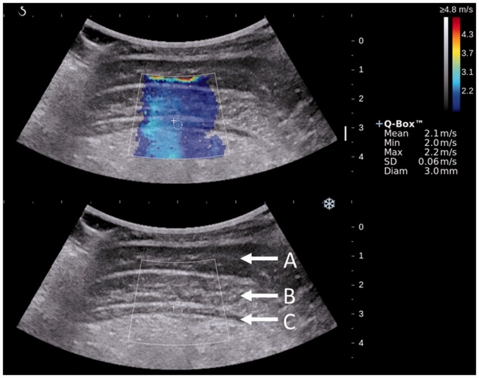
Muscle shear wave speed of TrA at the target region was measured using shear wave ultrasound elastography images obtained by an ultrasonic apparatus (Aixplorer, SuperSonic Imagine, Aix-en-Provence, France). An electronic convex probe (SuperCurved 6-1, SuperSonic Imagine, Aix-en-Provence, France) at 11 Hz was placed on the target region transversely to the long axis direction of the body (i.e. along the line of muscle fibres of TrA). Because we intended to measure the muscle located in the deepest layer of the trunk abdominal musculature, we used a convex probe, which can search deeper layers compared with the linear probe. Shear wave ultrasound elastography generated color-coded images with a scale from blue (slow shear wave speed; soft) to red (fast shear wave speed; hard) depending on the shear wave speed (Fig. 1). From images stored in the ultrasonic apparatus, a single image that captured a stable color distribution at a specific time was selected to measure the muscle shear wave speed. In the image, an approximately 20 × 20-mm box region of interest was set on TrA. In addition, a 3-mm circle was set near the center of TrA and the shear wave speed within the circle was calculated automatically. The circle size was fit to the minimal TrA thickness of all the patients. The measurements were performed six times. Of the six measured values, a group of four measurements showing the lowest coefficient of variation (CV) among 12 possible groups was adopted and the mean values were used for further analysis. These procedures were repeated twice by skilled and unskilled operators in a random order. Neither operator knew the values that the other had measured.
Fig. 1.
Typical example of a colour-coded presentation of transversus abdominis elasticity (shear wave speed) superimposed on the B-mode ultrasound image: (A) external abdominal oblique, (B) internal abdominal oblique, and (C) transversus abdominis.
Statistical analysis
The descriptive data are presented as mean ± standard deviation. The Student’s paired t-test was used to test the difference in the muscle shear wave speed between the first and second occasions and skilled and unskilled operators. The intra-class correlation coefficients (ICCs) were calculated to examine the reliability. The relative reliability between the two occasions was calculated using a one-way analysis of variance (ANOVA) model with consistency (ICC [1, 1]). The relative reliability between the two operators was calculated using a two-way random-effects ANOVA model with absolute agreement (ICC [3, 1]). ICCs that were obtained ranged from 0 (no correlation) to 1 (perfect correlation). The strength of the correlation was interpreted as follows: an ICC of ≤ 0.20 was “slight”, of 0.21–0.40 was “fair”, of 0.41–0.60 was “moderate”, of 0.61–0.80 was “substantial”, and of ≥0.81 was “almost perfect” (8). The CV for every comparison was also determined. Bland-Altman plots were constructed to determine absolute reliability. A P value of <0.05 was considered statistically significant.
Results
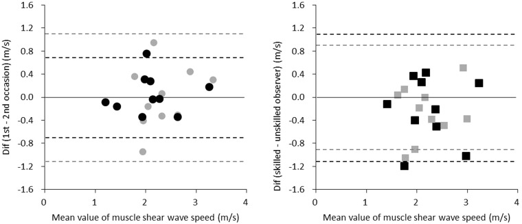
The muscle shear wave speed of TrA measured by the skilled operator was 2.1 ± 0.6 m/s on the first occasion and 2.1 ± 0.6 m/s on the second occasion, and there was no significant difference between the two occasions. The ICC and CV between the first and second occasions were 0.86 (P < 0.00, 95% confidence interval [CI] = 0.57–0.96) and 8.7%, respectively, and no systematic errors were found (Fig. 2). The muscle shear wave speed of TrA measured by the unskilled operator was 2.3 ± 0.6 m/s on the first occasion and 2.3 ± 0.5 m/s on the second occasion, and there was no significant difference between the two occasions. The ICC and CV between the first and second occasions were 0.59 (P = 0.02, 95% CI = 0.14–0.88) and 13.8%, respectively, and no systematic errors were found (Fig. 2). The ICC between the two operators was 0.57 (P = 0.03, 95% CI = 0.11–0.87) on the first day and 0.56 (P = 0.02, 95% CI = 0.01–0.87) on the second day. The CV between the two operators was 15.5% on the first day and 13.5% on the second day. No systematic errors were observed for these comparisons (Fig. 2).
Fig. 2.
Bland-Altman plots. The left and right panels represent the inter-day reliability (black circle: skilled operator, gray circle: unskilled operator) and inter-operator reliability (black square: first occasion, gray square: second occasion), respectively. Dotted lines represent 1.96 standard deviations.
Discussion
To the best of our knowledge, the present study is the first to quantify TrA elasticity using ultrasound elastography. For the skilled operator of ultrasound elastography, the ICC between the two occasions was 0.86 (i.e. almost perfect), and a high reliability was confirmed. This result indicates that ultrasound elastography is a highly reliable modality even for deep-layer muscles, if operated by a skilled operator. Our results showed that even the unskilled operator could measure muscle elasticity with moderate reliability (ICC = 0.59) implying that ultrasound elastography is a straightforward method to measure the elasticity of muscles located in the deepest layer of the trunk musculature. The slightly high CV (skilled operator: 8.7%, unskilled operator: 13.8%) for the high or moderate ICC was likely due to the small values of the muscle shear speed (2.1–2.3 m/s on average); it has previously been suggested that a CV of <15% was acceptably low for a biological measurement (9). This recommendation combined with the high ICC values that we calculated does not suggest that the reliability obtained in the present study was low because the CV values were slightly high.
Although systematic error was not found in the inter-operator comparison, the ICC remained moderate. This result could be due to the unskilled operator’s crude technique. We did not make a comparison between two skilled operators; thus, we cannot draw conclusions about the inter-operator reliability. We can say with certainty that it is possible to perform the necessary measurements with high reliability if one skilled operator performs the measurements throughout the procedure.
The present study had some limitations. The participants of this study were young men who were not obese. Therefore, it is unclear whether the present study findings are applicable to other populations. Although muscle atrophy is less in deep abdominal muscles compared with superficial ones (10), muscle elasticity changes with age (11); thus, we cannot apply the conclusion of the present study to the elderly population. Similarly, it is unclear at the moment whether the present findings can be applied to athletes with hypertrophied muscles. In addition, the number of participants in the present study was relatively low. The present findings will be more robust by increasing the number of participants. The range of the ultrasound is limited. Thus, it would be difficult to measure the TrA muscle elasticity in obese individuals who have a greater abdominal subcutaneous fat area (12). Because TrA locates in the deepest layer of the trunk abdominal musculature, we used a convex probe, which can search deeper layers, instead of a linear probe, which cannot search deeper layers. According to the manufacturer, although there is no musculoskeletal preset on the convex probe, there is no significant error in the measurement of soft structures until approximately 200 kPa of elasticity. However, we avoided using elasticity in our measurements and used velocity instead to promote the accuracy of the results.
In conclusion, the reliabilities of the skilled and unskilled operators were high and moderate, respectively, and the inter-operator reliability remained moderate. Although further studies that examine the validity of estimating TrA force and/or condition using ultrasound elastography are needed, our results can increase the feasibility of ultrasound elastography in future studies on trunk deep musculature.
Declaration of conflicting interests
The authors declared no potential conflicts of interest with respect to the research, authorship, and/or publication of this article.
Funding
This study was supported by a Grant-in-Aid for Young Scientists (B) (No. 25750323).
References
- 1.Akagi R, Takahashi H. Effect of a 5-week static stretching program on hardness of the gastrocnemius muscle. Scand J Med Sci Sports 2014; 24: 950–957. [DOI] [PubMed] [Google Scholar]
- 2.Bouillard K, Nordez A, Hug F, et al. Estimation of individual muscle force using elastography. PLoS One 2011; 6: e29261–e29261. [DOI] [PMC free article] [PubMed] [Google Scholar]
- 3.Yoshitake Y, Takai Y, Kanehisa H, et al. Muscle shear modulus measured with ultrasound shear-wave elastography across a wide range of contraction intensity. Muscle Nerve 2014; 50: 103–113. [DOI] [PubMed] [Google Scholar]
- 4.Bouillard K, Hug F, Guével A, et al. Shear elastic modulus can be used to estimate an index of individual muscle force during a submaximal isometric fatiguing contraction. J Appl Physiol 2012; 113: 1353–1361. [DOI] [PubMed] [Google Scholar]
- 5.Shinohara M, Sabra K, Gennisson JL, et al. Real-time visualization of muscle stiffness distribution with ultrasound shear wave imaging during muscle contraction. Muscle Nerve 2010; 42: 438–441. [DOI] [PubMed] [Google Scholar]
- 6.Hodges PW, Richardson CA. Contraction of the abdominal muscles associated with movement of the lower limb. Phys Ther 1997; 77: 132–142. [DOI] [PubMed] [Google Scholar]
- 7.Hodges PW, Richardson CA. Delayed postural contraction of transversus abdominis in low back pain associated with movement of the lower limb. J Spinal Disord 1998; 11: 46–56. [PubMed] [Google Scholar]
- 8.Landis JR, Koch GG. The measurement of observer agreement for categorical data. Biometrics 1997; 33: 159–174. [PubMed] [Google Scholar]
- 9.Martinson H, Stokes MJ. Measurement of anterior tibial muscle size using real-time ultrasound imaging. Eur J Appl Physiol Occup Physiol 1991; 63: 250–254. [DOI] [PubMed] [Google Scholar]
- 10.Ota M, Ikezoe T, Kaneoka K, et al. Age-related changes in the thickness of the deep and superficial abdominal muscles in women. Arch Gerontol Geriatr 2012; 55: e26–30. [DOI] [PubMed] [Google Scholar]
- 11.Wang CZ, Li TJ, Zheng YP. Shear modulus estimation on vastus intermedius of elderly and young females over the entire range of isometric contraction. PLoS One 2014; 9: e101769–e101769. [DOI] [PMC free article] [PubMed] [Google Scholar]
- 12.Kanehisa H, Miyatani M, Azuma K, et al. Influences of age and sex on abdominal muscle and subcutaneous fat thickness. Eur J Appl Physiol 2004; 91: 534–537. [DOI] [PubMed] [Google Scholar]


