Abstract
Objectives
The composition of dental plaque has been well defined, whereas currently there is limited understanding of the composition of denture plaque and how it directly influences denture related stomatitis (DS). The aims of this study were to compare the microbiomes of denture wearers, and to understand the implications of these towards inter-kingdom and host-pathogen interactions within the oral cavity.
Methods
Swab samples were obtained from 123 participants wearing either a complete or partial denture; the bacterial composition of each sample was determined using bar-coded illumina MiSeq sequencing of the bacterial hypervariable V4 region of 16S rDNA. Sequencing data processing was undertaken using QIIME, clustered in Operational Taxonomic Units (OTUs) and assigned to taxonomy. The dentures were sonicated to remove the microbial flora residing on the prosthesis, sonicate was then cultured using diagnostic colorex Candida media. Samples of unstimulated saliva were obtained and antimicrobial peptides (AMP) levels were measured by ELISA.
Results
We have shown that dental and denture plaques are significantly distinct both in composition and diversity and that the oral microbiome composition of a denture wearer is variable and is influenced by the location within the mouth. Dentures and mucosa were predominantly made up of Bacilli and Actinobacteria. Moreover, the presence of natural teeth has a significant impact on the overall microbial composition, when compared to the fully edentulous. Furthermore, increasing levels of Candida spp. positively correlate with Lactobacillus spp. AMPs were quantified, though showed no specific correlations.
Conclusions
This is the first study to provide a detailed understanding of the oral microbiome of denture wearers and has provided evidence that DS development is more complex than simply a candidal infection. Both fungal and bacterial kingdoms clearly play a role in defining the progression of DS, though we were unable to show a defined role for AMPs.
Introduction
Healthcare improvements in the last century have led to an increasingly elderly population. Worldwide, 810 million people are aged 60 years or over, which is predicted to increase to at least two billion by 2050 (22% of the entire global population) [1]. In the EU alone the proportion of the population who are 65 years and older is predicted to reach 53% by the year 2025 [2]. This demographic change will result in significant challenges for oral healthcare delivery, to an increasingly aged population with declining oral health. As the population ages then oral diseases become more relevant with respect to their local and systemic impact, which can have profound implications for healthcare provision [3, 4].
The oral cavity is a complex environment that is continually exposed to numerous opportunistic microbial pathogens. These are kept in check by a robust arsenal of immune factors that maintain a healthy oral environment and prevent the development of disease. This arena has gradually become a key area of biomedical research, which has led to a greater understanding of the causes, pathogenesis and host response against oral disease, with the majority of research focussing on diseases affecting dentate individuals, such as gingivitis, periodontitis and caries. Conversely, there is very limited research regarding denture related disease. Despite major improvements in oral health worldwide, recent estimates report that the rate of edentulouness still varies from 7 to 69% of the worlds adult population [5, 6], and in the US and UK populations around one fifth wear some form of removable denture [7, 8]. This continued high prevalence should convince researchers that, there is a requirement to develop an understanding of the implications of dentures on oral and systemic health.
The primary condition denture wearers suffer from is denture stomatitis (DS). This refers to inflammation of the oral mucosa and pathological changes associated with the wearing of dentures [9]. DS can be classified according to the severity of inflammation using a scale first described by Newton [10, 11]. The aetiology of DS is related to a variety of factors including poorly fitting dentures causing trauma and biological factors such as poor salivary flow, smoking or antibiotic treatment as well as microbial infection [12]. However, Candida albicans is generally attributed as being the main causative agent in DS affecting approximately 30–70% of denture wearers [9]. Candida spp. colonise the denture surface, forming co-aggregates with bacteria to build complex microbial communities known as biofilms. The majority of literature in this area focuses solely on Candida as the primary cause of infection, however, there is increasing evidence to suggest that this is very much a polymicrobial disease in which bacterial and fungal interactions play a role in disease pathogenesis [13, 14]. Several studies have isolated bacteria directly from the surface of dentures using standard microbial culturing techniques, primarily streptococci and staphylococci species [15–18]. However, these culture based methods are largely inadequate and unlikely to give a comprehensive representation of the polymicrobial population, which can contain up to 1011 microbes per milligram of denture plaque [19]. The advent of high throughput sequencing however has revolutionised our understanding of microbial ecosystems, and thus using this superior method we can for the first time gain an insight into the oral microbiome of denture wearers.
In addition to an incomplete understanding of the composition of denture plaque, we also have limited knowledge and understanding of the local host immune response. It has been established that the immune response is gradually impaired with increasing age [20, 21], but with the addition of loss of natural teeth an even greater rapid decline in host protective responses in the oral cavity is reported [22]. Antimicrobial peptides (AMP) including cathelicidin LL-37, histatins and defensins, which exhibit antimicrobial and immunoregulatory properties and protect mucosal surfaces against pathogens, have been found to be present in saliva. However, no studies have yet investigated salivary AMPs in denture wearers and the potential role they may play in the protection against denture plaque. Thus, a more in depth investigation is required into the host-microbiome relationship in the denture wearing individuals, which may help towards understanding the pathogenesis of DS.
The aim of this study was therefore to carry out a detailed site specific analysis of the bacterial composition of the oral microbiome of denture wearers using high throughput 16S rRNA gene sequencing technology. Furthermore, to assess changes in the diversity and composition of the microbiome against candidal load to aid understanding of the potential bacterial-fungal interactions that may be important in DS. In addition, the relationship between host antimicrobial peptides and denture plaque composition was also investigated. This is the first study to provide such a detailed microbial analyses of denture biofilms.
Materials and Methods
Study participants
123 denture wearing patients attending the University of Glasgow Dental School and Hospital were enrolled in the study. Convenience sampling was used based on the patient availability on recruitment days. Patients were recruited by a designated research nurse or a PhD student. Written informed consent was obtained from all participants. Ethical approval for the study was granted by the West of Scotland Research Ethics Service (12/WS/0121). All patients wore full or partial removable dentures. A team of qualified dental clinician were responsible for the collection of samples and the recording of clinical features. Data on patient age, gender, smoking status, routine oral hygiene regimens and any history of recent antimicrobial medication were recorded on a patient questionnaire. There was no age related exclusion criteria for this study. Newton’s classification method for DS was used to score the appearance of the patient’s palatal mucosa [11]. The following scores were applied; 0 = healthy mucosa, 1 = pin-point hyperaemic lesions (localized erythema), 2 = diffuse erythema (generalized simple inflammation), and 3 = hyperplastic granular surface (inflammatory papillary hyperplasia). For standardisation, all clinicians received training to calibrate scoring the extent of erythema. Patients were excluded from the study if they were pregnant, had previous radiotherapy for the treatment of head and neck malignancy, had been receiving antimicrobial/antifungal treatment, using prescription mouthwashes or had received immunosuppressant therapy within six months previous to sampling.
Clinical sample collection
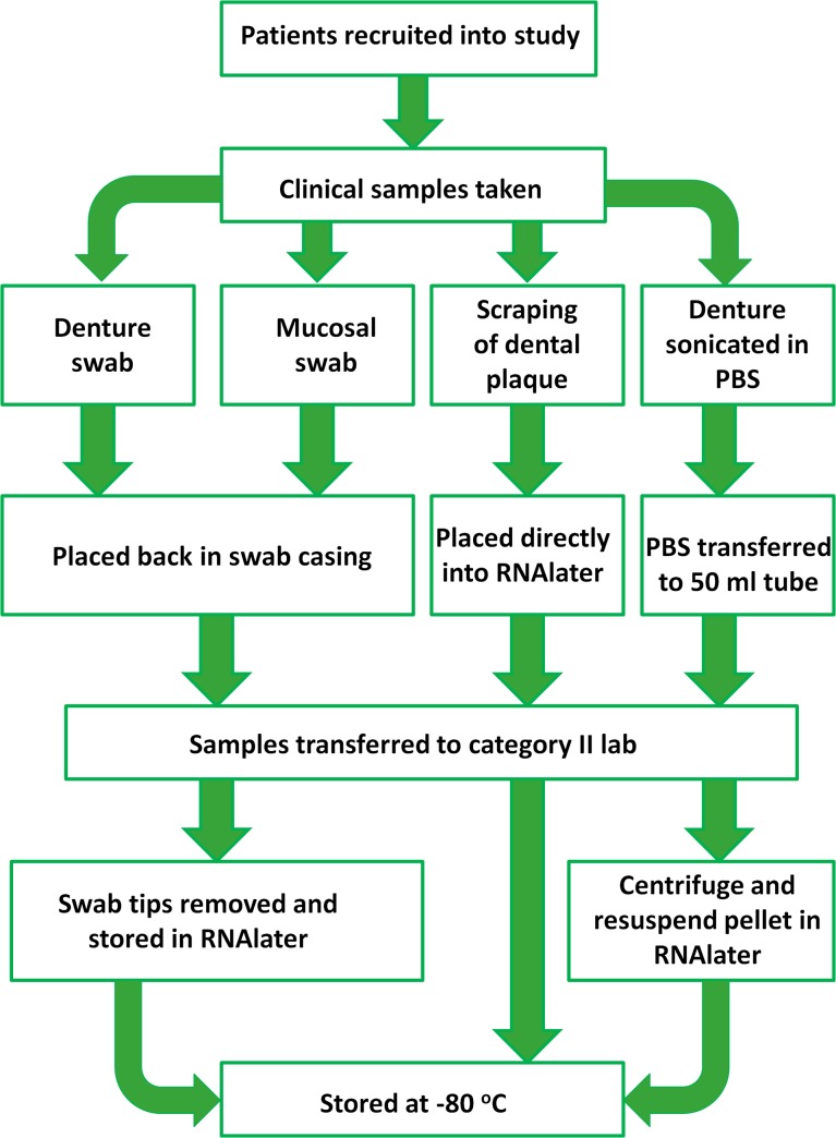
Clinical samples were collected as shown in Fig 1. Ethylene oxide sterilised swabs (Fisher Scientific, Loughborough, UK) were used to collect samples from the denture surface in contact with the palatal mucosa and the palatal mucosal surface covered by the denture. If any natural were teeth present the clinician took a supragingival plaque sample from a single tooth using a sterile dental probe, which was immediately placed into a 2 ml collection tube (Fisher Scientific) containing RNAlater® (QIAgen, Manchester, UK). Dentures were removed from the patients’ mouth and placed in sterile bags (Fisher Scientific) filled with 50 ml PBS (Sigma-Aldrich, Dorset, UK), then placed in a sonic bath (Ultrawave, Cardiff, UK) for 5 min to remove adherent denture plaque. The denture sonicate was then transferred to a 50 ml tube. Denture sonicate was centrifuged for 10 min at 3700 x g, and the plaque pellet re-suspended in 2 ml of RNAlater (QIAgen), as previously described [23]. Swab tips were removed and stored in RNAlater. Denture plaque, dental plaque and swab samples were all stored at -80°C. In total samples from 131 patients were collected, which included 131 denture swabs, 131 mucosal swabs and 79 dental plaque samples. During DNA extraction process not all samples had sufficient DNA to use for sequencing, therefore DNA from 108 denture samples, 87 mucosal samples and 63 dental samples remained for sequencing, collectively all these samples originated from 123 patients. Samples were further separated into distinct groups for direct comparison and analysis: health and DS groups, dentate and edentate groups and complete denture and partial denture groups.
Fig 1. Flow chart demonstrating the process of clinical sample collection and processing in the study.
Collection of saliva
Samples of whole unstimulated saliva were obtained by expectorating into a SaivaBio collection tube (Salimetrics, Suffolk, UK). Patients were given a maximum of 5 min to provide a sample. Saliva was clarified by centrifugation at 10,000 g for 10 min and then stored at -80°C.
Candida isolation
One ml of denture sonicate was used to prepare ten-fold serial dilutions ranging from 100–10−3. The dilutions were then used to perform colony forming unit (CFU) counts using Colorex Candida plates on which C. albicans colonies appeared green, C. glabrata were pink and C. tropicalis were blue. (E&O Laboratories, Bonnybridge, UK). One hundred μl of each serial dilution was spread across each plate, and then incubated at 30°C for 48 hrs. CFUs forming on plates were then counted and the average number of Candida cells colonising each denture was calculated.
DNA isolation
Dental plaque samples were centrifuged for 15 min @ 13000 x g the supernatant was removed and the sample resuspended in 150 μl TE buffer. Swab samples were sonicated for 30 sec, and then the sonicated fluid was transferred into a deep well plate and centrifuged for 15 min at 13000 x g. The precipitate was then resuspended in 150 μl TE buffer. All samples were then transferred to a plate with each well containing 0.25 ml of lysis buffer (AGOWA mag Mini DNA Isolation Kit, AGOWA, Berlin, Germany), 0.3 g zirconium beads (diameter, 0.1 mm; Biospec Products, Bartlesville, OK, USA) and 0.2 ml phenol. The samples were homogenized with a Mini-beadbeater (Biospec Products) for 2 min. DNA was extracted with the AGOWA mag Mini DNA Isolation Kit.
Quantitative PCR
Real time qPCR was performed to determine the concentration of bacterial DNA per sample as the original DNA extracted may contain DNA from other microbes such as fungi. Primers and a probe for the 16S rRNA gene were used, (F: TCCTACGGGAGGCAGCAGT, R: GGACTACCAGGGTATCTAATCCTGTT Probe: 6FAM-CGTATTACCGCGGCTGCTGGCAC-BBQ). As an internal control for PCR inhibition a qPCR of PhHV (Phocid herpesvirus type 1 gB gene) was also performed as described by Watzinger et al (2004) [24]. The total reaction volume was 20 μl, including 3 μl of DNA. Reactions contained a 26PCR Probe Master Mix (Roche), for 16S rRNA, 7.5 pmol primers and 3.8 pmol probe. For PhHV 1.8 pmol primers and 0.4 pmol probe of each primer was used. qPCR was carried out using the Light cycler LC480-II (Roche Diagnostics, Switzerland) under the following conditions: an activation step of 10 min at 95°C, followed by 50 cycles consisting of a denaturation step at 95°C for 30 sec, an annealing step at 60°C for 30 sec, and an extension step at 72°C for 30 sec. Bacterial 16S rDNA concentrations (CFU/ml) were determined from standard curves of E. coli K12 cultures.
PCR amplification and Illumina sequencing
Amplicon libraries of the V4 hypervariable region of the 16S rRNA gene were generated for each of the individual samples. PCR was performed using the forward primer 515F (GTGCCAGCMGCCGCGGTAA) and the reverse primer 806R (GGACTACHVGGGTWTCTAAT) [25]. The primers included Illumina adapters and a unique 8-nt sample index sequence key [26]. The amplification mix contained 2 units of Phusion HotStart II High fidelity polymerase (Thermoscientific), 1 unit Buffer Phusion HS II [5x], including 1.5 mM MgCl2 (Thermoscientific), 0.2 mM dNTP (Thermoscientific, Germany) and 1 μM of each primer. To each reaction 1 ng of DNA template was added. After denaturation (98°C; 30 sec), 35 cycles of denaturation (98°C; 10 sec), annealing (55°C; 30 sec), and extension (72°C; 30 sec) were performed. Individual amplicon libraries were analyzed for DNA content with the fluorescent Quant-iT™ PicoGreen® dsDNA Assay Kit (Invitrogen). The libraries were pooled in equimolar amounts. The amplicons were purified by means of the IllustraTM GFXTM PCR DNA and Gel Band Purification Kit (GE Healthcare, Eindhoven, the Netherlands). The quality and the size of the amplicons were analyzed on the Agilent 2100 (Santa Clara, CA, USA). The amplicon was sequenced in paired end mode on a MiSeq sequencing system (Illumina, Eindhoven, the Netherlands) with the v2 kit (Illumina) [26, 27].
Sequencing data analysis
Reads were first quality filtered using Trimmomatic v0.32, [28]. Next, the reads were merged using fastq-join implemented in QIIME v.1.8.0 [29]. Sequences were clustered into operational taxonomic units (OTUs) using USEARCH v7.01090 [30], after quality filtered with usearch (maxee 0.5).The representative sequence of each cluster was assigned a taxonomy using the RDP classifier [31]. The Ribosomal Database Project: improved alignments and new tools for rRNA analysis. Nucl Acids Res 37: D141–145. doi: 10.1093/nar/gkn879) (QIIME v.1.8.0) (Greengenes v13.8 97_otus set) with a minimum confidence of 0.8.
ELISAs
A range of antimicrobial peptides which are commonly associated with the oral cavity were selected for assessment in our patient’s saliva [32–34]. The following AMPs were assessed by ELISA, LL-37, Calprotectin, Lactoferrin, HNP1-3 (Hycult biotech, The Netherlands), Histatin 5 (Stratech scientific, Suffolk, UK) and Beta defensin 1 (BD1) (Peprotech, London, UK) as per manufacturer’s instructions. Clarified saliva samples were diluted 1:5 in assay buffer (PBS, 0.5% BSA, 0.1% Tween20). Results were calculated using a 4-parameter curve fit, quantifying colorimetric changes at 630 nm (BMG-Labtech, Ortenberg, Germany).
Statistical analyses
The data set was randomly sub-sampled to 770 reads per sample (minimum number of reads per sample was 776) to include the maximum number of samples for analysis. OTU datasets were reduced by log2 transformation so as to carry out principal component analysis (PCA) and diversity statistics (Shannon diversity index and Dominance index); the analysis was carried out using PAST software [35].
A one-way ANOVA test was applied to compare diversity statistics at oral microbiome sites using GraphPad Prism software (version 4; La Jolla, CA, USA). PCA was used to reduce the dimensionality of the OTU dataset. 258 OTU’s were entered into the PCA. A scree plot was used to determine how many components emerged. Factor loadings above 0.15 on a component were considered to have a strong association with that component and were deemed to be the most informative in describing the microbiome components. To determine if distinct clusters formed for each group on the PCA plots, new variables were created for each principle component by using the factor loadings as regression coefficients, producing a score for each sample. These scores were then used as outcome variables to compare between groups (using t-tests where appropriate- dentate/edentate and complete/partial dentures groups).
The contribution of each bacterial class was calculated in terms of proportion to the overall sample, percentages were log transformed and a t-test was used to compare health and DS groups. Diversity statistics were compared via a t-test with GraphPad Prism v5. Spearman’s rank correlation was used to assess correlations between the abundance of individual bacterial classes or genera with the proportion of Candida found on dentures (CFU counts), using SPSS version 20.
Salivary concentrations of AMPs were compared between healthy and DS groups, dentate/edentate groups and complete/partial groups (S1 Table). Data were log transformed and analysed using a t-test with prism. Furthermore Spearman’s rank correlation was used to assess correlations between the abundance of individual bacterial classes or genera with the concentrations of salivary AMPs found on dentures.
To visualize the relationships and associations of the microbiomes with environmental variables canonical correspondence analysis (CCA) was applied. This form of analysis, carried out using PAST, allows the visualisation of OTU distribution and sample group distribution in relation to a number of environmental variables. Environmental variables included were Candida CFU counts and salivary concentration of a number of AMPs. The significance of each of the CCA axes was calculated by permuting the data 999 times.
Candida CFU counts were compared between healthy and DS groups, dentate/edentate groups and complete/partial groups. Data were log transformed and analysed using a t-test with GraphPad Prism v5.
Study design
The study was designed as a pilot study and was initially only powered to detect a biologically meaningful association between diseased and healthy mouths and microbiome composition, and therefore was not originally powered to detect differences between additional variables including, denture type, dentate status, Candida levels and salivary AMP levels. Thus, non-significant results between these variables are not necessarily absence of effect, but a result of not achieving the full sample size required. S2 Table provides a list of the statistics carried for all the analysis.
Dentate and edentate groups were compared separately from complete and partial denture groups, as having a complete denture only applied to the upper denture from which the sample was taken. The patient however may have had natural teeth remaining on the mandible and therefore could not be classed as edentulous. The inter-correlation between complete and edentulous or partial and dentate groups are shown in Table 1. Thus given the variation in group numbers, analysis was carried out on both datasets.
Table 1. Patient demographics.
| Gender n (%) | Mean age | Type of Denture n (%) | Dentate status | Disease status n (%) | Mean age of denture | |
|---|---|---|---|---|---|---|
| (Male) | (years) | (Complete upper) | (Edentulous) | (Healthy) | (years) | |
| (Female) | (Partial upper) | (Dentate) | (Disease) | |||
| Patients n = 123 | 43 (35) | 70.7 ± 11.5 (min: 33, max: 95) | 82 (67) | 56 (46) | 78 (63) | 4.6 ± 5.3 (min:0.2, max: 40) |
| 80 (65) | 41 (33) | 67 (54) | 45 (37) |
Results
Patient demographics
Of the 123 patients that participated in this study, 43 were male and 80 were female (Table 1). The mean (SD) patient age was 70.7 (11.5) years (min: 33, max: 95) with an average (SD) denture age of 4.6 (5.3) years (min: 0.2 max: 40). Clinical diagnoses indicated that 63% of participants had a healthy oral mucosa and the remaining 37% had an inflamed mucosa of varying severity, and were suffering from DS. The majority of patients (67%) wore complete upper dentures, while the remainder (33%) had a partial upper denture with ≥1 natural teeth remaining.
Illumina sequencing output
Across all of the samples 632 OTUs were identified, with an average of 94 OTUs per sample (SD 39; min 25, max 254), 536 of which contained a minimum of 5 reads. The data was sub-sampled to 770 reads per sample in order to avoid bias of variable sample size. After sub-sampling 502 OTUs remained with an average of 46 OTUs per sample (SD 18; min 11, max 121). The samples were categorised into five main phyla which represented 99.6% of the reads: Firmicutes (40.6%), Actinobacteria (23%), Bacteriodetes (22.2%), Proteobacteria (9.8%) and Fusobacteria (4%).
Oral microbiome by sample site
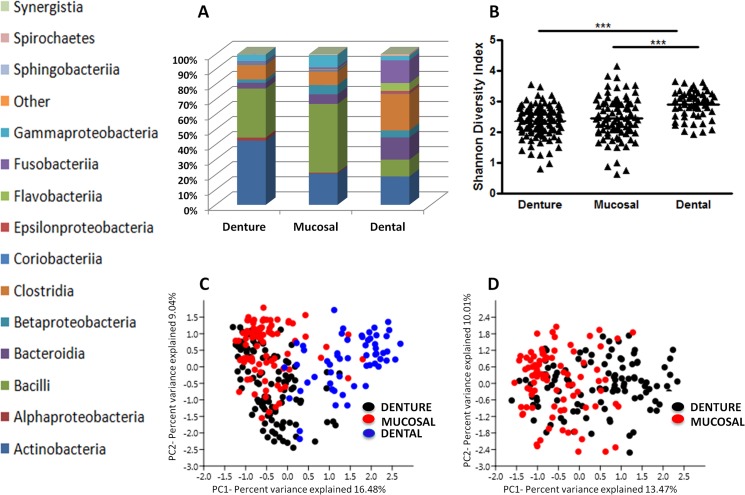
Samples were categorised into groups according to sample site: denture (n = 108), mucosal (n = 87) and dental (n = 63), with 337, 414 and 306 OTUs identified, respectively. On average, denture samples had 42 OTUs (SD 14; min 13, max 87), mucosal 49 OTUs (SD 20; min 11, max 121) and dental 51 OTUs (SD 37; min 16, max 306). Interestingly, denture plaque samples were found to have significantly less OTUs compared to dental, (p<0.01), and mucosal, (p<0.05), plaque. Fig 2A shows the relative abundance of bacterial taxa at the class level present in each sample type, and demonstrates considerable variation in taxonomic profiles between sites. Actinobacteria and Bacilli were the two predominant classes found at denture and mucosal sites, comprising 75.2% and 66.4%, respectively, of the overall composition when combined. However, Actinobacteria and Bacilli only contributed only 30% to dental plaque. Diversity statistics applied across samples revealed that dental plaque is more diverse (Fig 2B) than both denture plaque (Mean: 2.35 v 2.89, p<0.001), and mucosal surfaces (Mean: 2.44 v 2.89, p<0.001), with less dominant taxa according to the dominance index (p<0.001) (S1 Fig).
Fig 2. Microbial composition varies between sample sites in the oral cavity of denture wearers.
One hundred and eight denture swabs, 87 mucosal swabs and 63 dental plaque samples underwent pyrosequencing of the 16S hypervariable V4 region to determine the bacterial composition of denture, palatal mucosal and dental plaque. A) Relative abundance of bacterial taxa at the class level per sample site. B) The taxonomic diversity of each group was analysed and compared via a Shannon Diversity Index. These diversity statistics show that both denture and mucosal plaque are significantly less diverse when compared to dental plaque. ***p<0.001. Principal component analysis (PCA) was applied to all sample types to reduce the multidimensionality of the data and to determine variances between C) Denture, Mucosal and Dental plaque. D) Denture and Mucosal plaque.
PCA was applied to reduce the multidimensionality of the dataset. Two primary principal components emerged from this analysis. On average, dental samples scored higher on PC1 than denture or mucosal samples (p = 0.0001) (Fig 2C), indicating higher frequencies of Fusobacterium, Corynebacterium, Selenomonas, Campylobacter and Prevotella, and lower of Streptococcus and Rothia. Furthermore, denture and mucosal samples were directly compared where denture samples had a higher average score on PC1 than mucosal samples (p = 0.0006) (Fig 2D). This indicated higher frequencies of Lactobacillus, Actinomyces, Atopobium and Scardovia, and lower frequencies of Streptococcus, Rothia and Haemophilus.
Dentate versus edentate
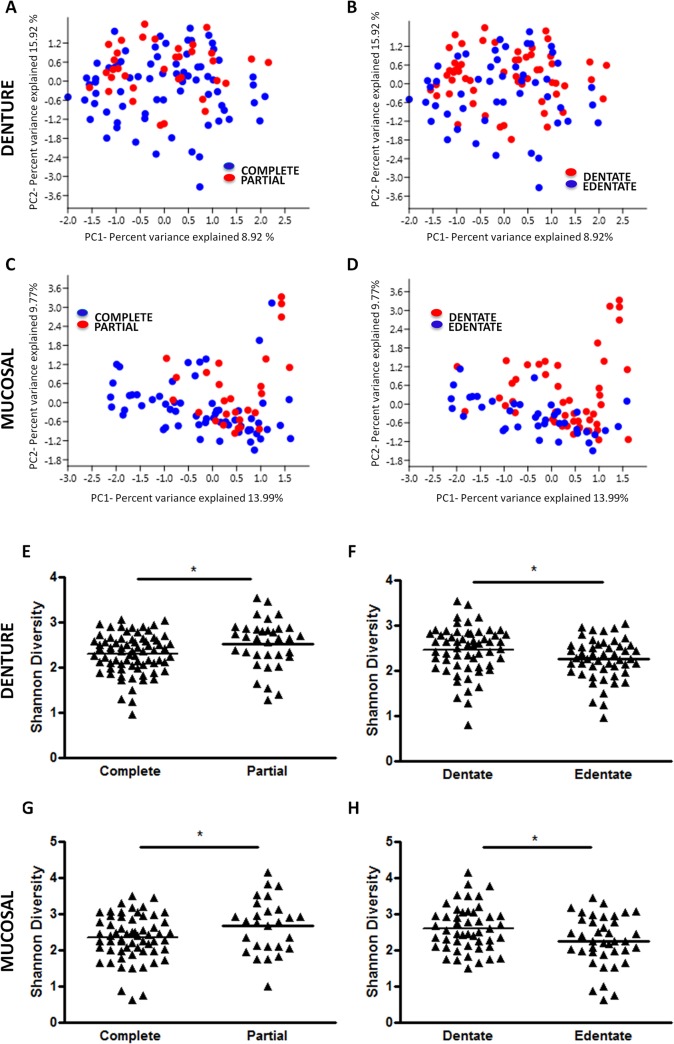
Samples were separated into groups depending on denture type (complete/partial) and on the presence or absence of teeth (dentate or edentate), and the microbiomes were compared at the denture and mucosal sites. PCA was applied to the data, and as previous, two principal components emerged from this analysis. When comparing denture type on the denture samples (Fig 3A), partial dentures scored higher on PC2 than complete denture samples (p = 0.0073), revealing higher frequencies of Actinomyces, Haemophilus, Corynebacterium and Veillonella and lower frequencies of Lactobacillus and Streptococcus. Denture samples were then split into dentate and edentate groups (Fig 3B). Dentate samples scored higher along PC2 than edentate (p = 0.0194), and as with the partial denture group, they had higher frequencies of Actinomyces, Haemophilus, Corynebacterium and Veillonella and lower frequencies of Lactobacillus and Streptococcus. At the mucosal microbiome partial dentures scored higher on PC1 than complete denture samples (Fig 3C) (p = 0.0001), and showed higher frequencies of Actinomyces, Prevotella, Haemophilus and Neisseria and lower frequencies of Streptococcus, Lactobacillus and Jathinobacterium. No differences in scores were found between dentate and edentate samples (Fig 3D).
Fig 3. The presence of natural teeth and type of denture have significant impact of microbiome diversity.
Samples were separated into groups based on denture type, (complete or partial) and dentition status (dentate or edentate) and principal component analysis was applied to the data for the denture (A and B) and mucosal microbiome (C and D) respectively. The taxonomic diversity of each group was analysed and compared via a Shannon Diversity Index for the denture (E and F) and mucosal microbiome (G and H) *p<0.05.
The diversity of the same sample groups were compared and revealed that partial denture samples were significantly more diverse than those from complete dentures at both the denture, (Mean: 2.29 v 2.52, p<0.05) and mucosal surfaces, (Mean: 2.34 v 2.67, p<0.05], (Fig 3E and 3G). Furthermore, the dentate patients had a significantly more diverse microbiome than the edentate, (Fig 3F and 3H); these results were found at both the denture, (Mean: 2.45 v 2.25, p<0.05), and mucosal surfaces, (Mean: 2.6 v 2.25, p<0.05).
Healthy versus diseased microbiome
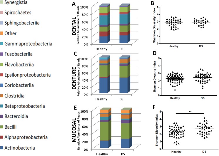
Based on Newton’s inflammation scores, patients were separated into health and DS groups, and their microbiomes analysed. When comparing the relative abundance (%) of bacterial classes between health and DS groups, there were no differences found in dental plaque (Fig 4A). Denture plaque showed significantly higher proportion of Bacteroidia (Mean: 0.14 v 0.41, p<0.05), in DS patients (Fig 4C), and when investigated at the genus level, further increases in the DS group could be attributed to Prevotella spp. (Mean: 0.09 v 0.37, p<0.05) and Veillonella spp (Mean: 0.55 v 0.76, p<0.05). The mucosal microbiome showed an altered composition between health and disease, with classes Actinobacteria (Mean: 1.08 v 1.28, p<0.05) and Bacteroidia (Mean: 0.43 v 0.74, p<0.05), increasing in the latter (Fig 4E). Furthermore, Shannon diversity index statistics indicated that mucosal samples from DS patients were significantly more diverse than their healthy counterparts (Mean: 2.27 v 2.69, p<0.01) (Fig 4F), while no differences were found in dental, and denture plaque (Fig 4B and 4D). Further analysis of diversity was carried out by sub-grouping into mild inflammation (Newton’s grade 1) and severe inflammation (Newton’s grade 2&3), however, no differences that were statistically significant were observed.
Fig 4. Changes in mucosal microbiome are associated with disease status.
The microbiome composition was assessed for differences between health and disease in A) dental plaque C) denture plaque E) mucosal plaque at the class level. Differences in taxonomic diversity between health and DS were assessed. No significant differences were found in diversity in B) dental or D) denture plaque. Mucosal plaque saw significant changes in diversity as disease progressed F). *p<0.05, **p<0.01.
Influence of Candida on the microbiome
It was found that 72% of patient’s dentures were colonised by Candida. The prevalence of Candida species isolated from dentures of healthy and DS patients is shown in Table 2. The overall prevalence of Candida was higher on dentures from DS sufferers (78%) when compared with their healthy counterparts (64%). At the species level C. albicans was more predominant on DS individuals’ dentures, whereas for C. glabrata there were no differences in prevalence between health and disease. Furthermore, the number of patients in which C. albicans and C. glabrata were co-isolated was also more common in DS sufferers (36%).
Table 2. Prevalence of Candida species isolated from dentures of healthy and diseased patients.
| Candida+ | C. albicans+ | C. glabrata+ | Mixed C. albicans / C. glabrata+ | |
|---|---|---|---|---|
| Healthy n(%) n = 78 | 50 (64) | 32 (41) | 32 (41) | 14 (18) |
| DS n(%) n = 45 | 35 (78) | 34 (76) | 18(40) | 16 (36) |
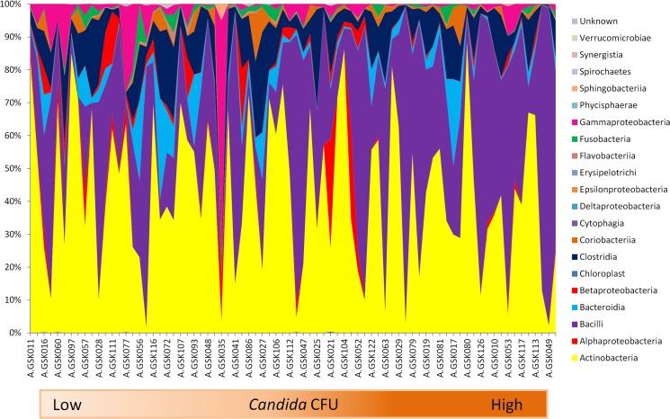
The influence of increasing Candida CFU on the bacterial microbiome of each individual was assessed at the denture and mucosal surfaces. A positive correlation was found between Candida CFU and Bacilli class on dentures (p = 0.01; Spearman’s rho 0.387), but negatively with Fusobacteria (p = 0.01; Spearman’s rho -0.470) (Fig 5). This was reflected at the genus level with Lactobacillus (p = 0.0001; Spearman’s rho 0.502) and Fusobacteria (p = 0.025; Spearman’s rho -0.417). No significant correlations were found between bacterial classes and Candida on the mucosal surface when assessing the complete cohort.
Fig 5. The influence of increasing Candida spp. CFU on the microbiome of the denture.
At the denture site the relative abundance of bacterial taxa within each sample was measured against increasing Candida CFUs at the class level.
In addition to the influence of denture type and edentulism on microbiome composition and diversity, other factors were also important, including Candida CFU count and salivary AMP concentration. The average CFU Candida count found on dentures was significantly higher in individuals with a complete denture (Mean: 5.77 v 5.02, p>0.01) (S2A Fig) and in edentate individuals (Mean: 5.86 v 5.25, p<0.05) (S2B Fig), when compared to partial dentures and dentate individual respectively.
Antimicrobial peptide detection in saliva of healthy and diseased patients
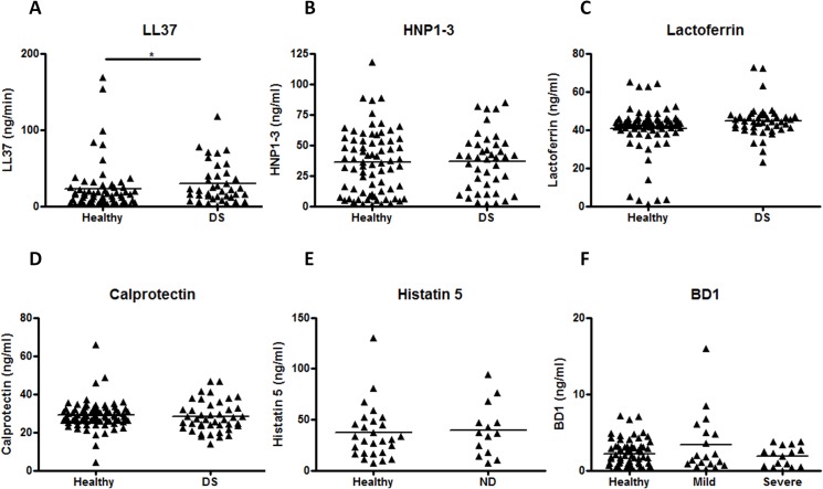
The levels of each AMP were compared between healthy and DS groups, and only LL-37 showed significantly elevated levels in DS patients (Mean: 1.04 v 1.28, p<0.05) (Fig 6A). HNP 1–3, lactoferrin, calprotectin, Histatin 5 and BD1, showed no statistically significant differences between groups (Fig 6B–6F). Furthermore, certain salivary AMPs, namely LL37, HNP 1–3 and Histatin 5 all showed significantly higher concentrations in patients with partial dentures (S3A–S3C Fig), and also in dentate individuals (S3G–S3I Fig). Calprotectin, Lactoferrin and BD1 all showed no differences between complete/partial (S3D–S3E Fig) or dentate/edentate groups (S3J–S3L Fig).
Fig 6. Concentrations of salivary AMPs increases with severity of inflammation.
The salivary concentrations of A) LL37, B) HNP1-3, C) Lactoferrin, D) Calprotectin, E) Histatin 5 and F) BD1 were compared between healthy and diseased individuals with mild (Newton’s grade 1) or severe inflammation (Newton’s grade2&3) *p<0.05.
Influence of salivary antimicrobial peptides on the microbiome
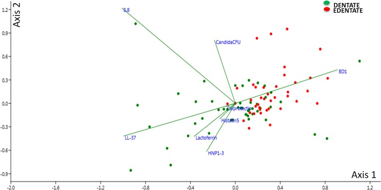
Finally, we investigated the influence of the AMPs on the denture and mucosal microbiome. The relative abundance of bacterial classes and genera for the complete patient cohort was compared against increasing levels of each AMP. However, no significant AMP/ bacterial correlations were found at the denture or mucosal surface. The AMP saliva concentration was also compared against increasing Candida CFU, but again no significant correlations were identified. Nonetheless, CCA analysis was performed and found that at the mucosal microbiome distinct clusters formed and showed that the dentate group related stronger to the AMPs (Fig 7).
Fig 7. Cannonical Correspondence Analysis of the denture microbiome.
CCA biplot allowing visualisation of dentate and edentate samples, represented by dots, in relation to environmental factors, including Candida CFU and salivary AMPs, which are represented by vectors. The variability in the samples was explained on axis 1 with 26.5% and on axis 2 with 19.3%.
Discussion
With the continued advancements in healthcare, the average lifespan is ever increasing, meaning that there is a larger elderly population than ever before [36]. Improving oral health has become a primary focus in medical research, and thus significant amount of time and money has been spent on investigating the pathogenesis and prevention of oral disease [6, 37]. However, this research has focused primarily on dental-related disease, whereas denture-related disease, a problem affecting a large proportion of the ever-increasing elderly population [3, 38], has been relatively understudied. Therefore, further investigation is required to elucidate its pathogenesis.
The majority of studies investigating DS focus primarily on the role of Candida spp. [9, 39], however given that up to 1011 microbes are capable of colonising the denture surface [19], then it is likely that bacteria also play a role in disease. Other studies have investigated the bacterial composition of denture plaque, however, these studies have been limited by the method of investigation, such as culturing and DNA-DNA hybridisation, which limit the number of bacterial species that can be identified [18, 40–43]. For example, Campos et al (2008) used PCR and identified 82 bacterial species, this however only made up approximately 50% of the bacterial flora detected in the samples, as the remainder were not-yet cultivated phylotypes and thus remained unidentified [41]. The study described herein has for the first time used detailed high-throughput 16S rRNA gene analysis of the oral microbiomes of a range of denture wearers, which gives a more comprehensive and global representation of the bacterial microbiome.
We first assessed the bacterial microbiome of three independent sites from the oral cavity of denture wearers, the denture, the palatal mucosa and dental plaque. Comparing the relative abundance of bacterial classes at the different sites showed the more diverse nature of the dental plaque in comparison to the denture and mucosal sites, both of which were predominantly composed of two classes, Actinobacteria and Bacilli. This result is unsurprising as both of these are associated with being amongst the primary colonisers within the oral cavity, particularly Actinomyces and Streptococcus species [44–46]. These results are supported by a recent study comparing biofilms forming on natural teeth against those forming on denture teeth, which demonstrated that biofilms forming on natural teeth develop quicker and have a more abundant proportion of species present [43]. PCA indicated that denture, mucosal and dental groups formed distinct clusters, thus indicating not only are they distinct in terms of diversity, but also compositionally separate. The different anatomical locations of these microbiomes likely explains these differences as the adherent surfaces and surrounding environment of dental plaque may be more suitable for the growth of a more diverse range of microorganisms [47]. For example, saliva is an important source of nutrients for microbes in the oral cavity [48], however, the presence of a denture acts as a barrier, thus with reduced nutrients available fewer organisms can form a sustainable niche.
This study has shown that the presence of natural dentition has a profound impact on the composition and diversity of the oral microbiome of a denture wearer. This suggests that this increased microbial diversity is not restricted to the anatomical sample site of the tooth, but also appears to affect the entire oral cavity. Patients included in this study that still had natural teeth remaining, ranged from 1 to 28 teeth, yet despite this wide range, PCA analysis indicated that they formed distinct groups from one another, indicating that even the presence of only one tooth is sufficient to have a profound impact of the microbiome composition. The lack of obvious groups between dentate and edentate samples at the mucosa could perhaps be explained by the nature of the surface. As we have already shown, the microbiome composition within the oral cavity is very much dependant on the location, therefore as the mucosal surface is biotic, microbes in contact are likely to be exposed to pathogen recognition receptors (PRRs) or host defence peptides from the epithelial cells [49–51]. As many of the dentate associated bacteria are not traditional commensals, they may have limited ability to colonise such a niche as well as the potential to initiate an immune response [52, 53]. Dentures, on the other hand are an abiotic surface, therefore biofilms from dentate individuals forming on the surface are likely to have less exposure to host defences and thus bacteria may grow unimpeded and have sufficient time to develop into a compositionally distinct plaque microcosm.
One of the primary aims of this study was to identify any compositional changes in the microbiome between health and disease. Denture plaque revealed a number of notable changes between health and disease, with significantly higher proportions of Prevotella and Veionella species found in denture stomatitis sufferers. This suggests that DS microbiomes have a composition more comparable to that of a dental plaque. Furthermore, at the mucosa, bacterial classes Actinobacteria and Bacteroidia also increased significantly in DS patients, further strengthening this hypothesis. A study supporting these findings by Campos et al (2008) investigated the microbial biofilm communities of denture stomatitis sufferers [41]. They found 32 bacterial phylotypes that were unique to denture stomatitis biofilms, a large proportion of which fell under the genera Atopobium (16%) and Prevotella (11%), both of which fall into the classes Actinobacteria and Bacteroidia, respectively. Moreover the mucosal microbiome of DS individuals is significantly more diverse than their healthy counterparts, suggesting that the compositional changes responsible for disease progression are occurring at the mucosa. However, whether these changes are cause or effect of disease is an area that merits further investigation.
We have established that the presence of natural teeth alters the composition and diversity of the microbiome, and our results suggest that the DS microbiome contains increased proportions of microbes more commonly associated with a dentate status. Therefore, we looked at the ratio of dentate to edentate within the health and DS groups and we found that both groups were equally represented within the health category (50%); however the dentate group made up a considerably larger proportion of the DS category, (61%) than the edentate (39%). Therefore, this may explain why we are seeing higher levels of these, normally dentate-associated microbes, within the microbiomes of DS individuals. Within the DS group it is likely that dentate and edentate individuals form two distinct biofilms, both with pathogenic and invasive potential, however, given that the majority of those with severe inflammation had teeth (67%), the presence of natural teeth may exacerbate denture stomatitis infection, creating a more pathogenic biofilm than those found in the edentulous.
Denture related disease is almost always attributed to infection with C. albicans, however, given the vast range of bacterial species identified on dentures and the surrounding mucosa in this study; it is unlikely that the infection can be attributed solely to Candida spp. Our study identified a 72% prevalence of Candida species on dentures, of which C. albicans was the most predominant, and was the only species in which we saw a significantly higher CFU count in DS individuals (S4A Fig). However, C. glabrata was also isolated in a high number of patients (40%), which is in line with studies showing the increasing emergence of this species, as it is currently responsible for approximately 13% of invasive candidosis cases [54–56]. In addition, the frequency of C. glabrata oral carriage rate has been shown to increase with age. Of particular interest was the number of patients in which both C. albicans and C. glabrata were co-isolated, this was high particularly amongst DS sufferers (35%). Recent studies have suggested that when co-colonised, C. albicans and C. glabrata form a more pathogenic and invasive biofilm than either species alone, thus may contribute to more severe cases of DS [57]. However, despite showing that DS is more common in dentate individuals, we found significantly higher Candida CFU counts on edentate patients and complete dentures when compared to dentate and partial dentures, respectively (S3 Fig). Nonetheless, this is most likely due to the less diverse microbiome of the edentulous, as with fewer microorganisms, this opens up a niche for Candida spp. to colonise.
Correlation analysis was carried out to investigate the affect of increasing Candida load (CFU counts) on the bacterial microbiome both at the class and genus level. At the denture and mucosal microbiome, there was a positive correlation with the class Bacilli and a negative correlation with Fusobacteria at the denture, these findings are in line with Kraneveld et al (2012), where they identified the similar correlations when analysing the effect of increasing Candida load on the salivary microbiome of the elderly [58]. At the genus level these correlations could be attributed to Lactobacillus species. This finding was surprising as the majority of literature regarding these species, indicates that they have an antagonistic relationship [59]. The mechanisms by which Lactobacillus inhibits growth of Candida spp are not fully understood. Investigations have suggested production of hydrogen peroxide by lactobacilli leads to anti-candidal activity. Furthermore lactobacilli can modulate the host response, up-regulating cytokines when co-cultured with C. albicans, which could be associated with the clearance of candidal infection. Nevertheless, denture-wearing patients have been shown to have higher levels of Lactobacillus species in their saliva, but they are more commonly isolated in the saliva of DS sufferers (87%) in comparison to healthy controls (65%) [60]. Yet, in spite of the vast evidence of an antagonistic interaction, certain species of oral Lactobacillus (namely L. casei) have demonstrated a stimulatory effect on C. albicans hyphal growth [59]. Furthermore, it has been shown that Candida hyphae can co-aggregate to lactobacilli and sustain their levels in patients with advanced oral diseases [60]. Nonetheless, most of these studies have focused on Lactobacillus spp. inhibitory effects on C. albicans, but their inhibition of other Candida species such as C. glabrata has proven less effective, as Jiang et al (2014) demonstrated that only 1 of six probiotic Lactobacillus species used in the study had an inhibitory effect on C. glabrata growth [61].
As well as the microbial interactions in the oral cavity of denture wearers, we were also interested in gaining a wider understanding of the host-microbial interactions. From the range of AMPs measured, only LL37 showed any significant increases between health and inflammation. This could be explained by the fact that the majority of those with inflammation (66%) were in the dentate category, whereas the healthy group were predominantly edentate (95%), and as studies have shown the concentration of salivary AMPs declines with the loss of natural teeth [22]. However, gaining a true understanding of salivary AMP concentration is more difficult with an elderly population, as the vast majority take one or more types of oral medication, many of which have been shown to cause xerostomia and thus there is the potential that they may affect salivary AMP concentrations [62]. Yet, to our knowledge the direct implication of oral medication on AMP production has not yet been elucidated.
In order to identify any potential relationships or associations between AMPs and the microbiome, correlation analysis was carried out. Each AMP was correlated with the denture and mucosal microbiome at both the class and genus level. However, no significant correlations were identified. Nonetheless, this is not to say that the AMPs are not interacting with specific microbes, it is more likely due to their broad spectrum activity [63]. Furthermore, Bals et al (2000) stated that establishing the individual contribution of an AMP to host defence is extremely difficult due to the complexity of the environment and host-microbial interactions [64], as we have clearly demonstrated from the data described herein.
One of the main limitations of this study is that the majority of OTUs detected have only been identified up to genus level. Therefore, this will make it more difficult to decide on the correct species to include in devising an appropriate biofilm model of DS. However, given the vast number of studies regarding the composition of the oral microbiome, there is already an excellent understanding of the bacterial species normally found in the oral cavity based on the genuses we have identified [44, 47]. Nonetheless, we still have to proceed with caution when selecting species as different species within the same genus could have drastically different physiological properties and distinct pathogenic potential.
In conclusion, given the complexity of the oral environment, comprehending the intricate interactions of this ecosystem is a difficult task, and the addition of a denture further adds to the complexity. However, this study has taken a significant step forward towards understanding this environment. With a detailed knowledge of the microbial composition, research in this area can progress further, with a more in depth focus on how these organisms interact with one another as well as the host, allowing us to develop meaningful biofilm models of denture plaque. From this study we can conclude that the bacterial microbiome composition of denture wearers is not consistent throughout the mouth and varies depending on sample site. Moreover, the diversity of dental plaque is unsurprisingly more diverse than the denture and mucosal microbiome, and with this we have shown that the presence of natural teeth has a significant impact on the overall microbial composition. Furthermore, higher levels of Candida spp. appear to induce a compositional switch to a more aciduric environment. Thus, we have deduced that there are a vast number of environmental variables with the potential to alter this environment, which ultimately means that the oral microbiome of each denture wearer is unique to the individual. Nonetheless, with the detailed knowledge we have gained from this study we can achieve a better understanding of the pathogenesis of denture related disease and which will aid towards the development of potential therapeutics.
Author information: Glasgow Dental School, School of Medicine, College of Medical, Veterinary and Life Sciences, University of Glasgow.
Supporting Information
The taxonomic dominance of bacterial groups was analysed within each sample and compared across sample sites via a Dominance Index. Statistics indicate dental samples are more diverse than both denture and mucosal, samples. ***p<0.001.
(TIF)
The average CFU Candida counts found on dentures were compared between complete and partial A) and dentate and edentate B) patients. *p<0.05, **p<0.01.
(TIF)
The average AMP concentrations found in saliva was compared between complete and partial A), B) C), D), E) and F) and dentate and edentate G), H), I), J), K) and L) for LL37, HNP 1–3, Histatin 5, Calprotectin, Lactoferrin and BD1 respectively patients. **p<0.01, **p<0.001.
(TIF)
The average CFU counts of A) C. albicans, and B) C. glabrata found on dentures were compared between healthy and diseased patients. **p<0.01.
(TIF)
(DOCX)
(DOCX)
Acknowledgments
Victoria Hannah, Jamie Dickie, John Gibson, Chris McCurry (University of Glasgow), Laura Murdoch, and Christine McAllister (NHS Greater Glasgow and Clyde) for their assistance in the collection of clinical samples. We also thank Aurea Simon-Soro (University of Valencia) for her helpful comments on study design and David F Lappin (University of Glasgow) for statistical support.
Data Availability
All relevant data are within the paper and its Supporting Information files.
Funding Statement
This study was funded by the BBSRC CASE studentship (BB/K501013/1) awarded to Prof Gordon Ramage and Dr Douglas Robertson. GlaxoSmithKline (GSK) also supported this project. GSK were involved in the initial discussions at the conception of the study. GlaxoSmithKline provided support in the form of salaries for authors DB and ML, but did not have any additional role in the study design, data collection and analysis, decision to publish, or preparation of the manuscript. The specific roles of these authors are articulated in the "author contributions" section.
References
- 1. Guzmán JM, Pawliczko A, Beales S, Till C, Voelcker I. Ageing in the twenty-first century: a celebration and a challenge 2012. United Nations Population Fund (UNFPA), New York, and HelpAge International, London. [Google Scholar]
- 2. Muenz R. Aging and demographic change in european societies: main trends and alternative policy options Societal Protection—The World Bank, 2007. [Google Scholar]
- 3. Griffin SO, Jones JA, Brunson D, Griffin PM, Bailey WD. Burden of oral disease among older adults and implications for public health priorities. American journal of public health. 2012;102(3):411–8. 10.2105/AJPH.2011.300362 [DOI] [PMC free article] [PubMed] [Google Scholar]
- 4. Meurman JH, Hamalainen P. Oral health and morbidity—implications of oral infections on the elderly. Gerodontology. 2006;23(1):3–16. Epub 2006/01/26. 10.1111/j.1741-2358.2006.00102.x . [DOI] [PubMed] [Google Scholar]
- 5. Felton D, Cooper L, Duqum I, Minsley G, Guckes A, Haug S, et al. Evidence-based guidelines for the care and maintenance of complete dentures: a publication of the American College of Prosthodontists. Journal of prosthodontics: official journal of the American College of Prosthodontists. 2011;20 Suppl 1:S1–S12. 10.1111/j.1532-849X.2010.00683.x . [DOI] [PubMed] [Google Scholar]
- 6. Petersen PE. The World Oral Health Report 2003: continuous improvement of oral health in the 21st century—the approach of the WHO Global Oral Health Programme. Community dentistry and oral epidemiology. 2003;31 Suppl 1:3–23. . [DOI] [PubMed] [Google Scholar]
- 7. Coulthwaite L, Verran J. Potential pathogenic aspects of denture plaque. British journal of biomedical science. 2007;64(4):180–9. . [DOI] [PubMed] [Google Scholar]
- 8. Shulman JD, Beach MM, Rivera-Hidalgo F. The prevalence of oral mucosal lesions in U.S. adults: data from the Third National Health and Nutrition Examination Survey, 1988–1994. Journal of the American Dental Association. 2004;135(9):1279–86. . [DOI] [PubMed] [Google Scholar]
- 9. Gendreau L, Loewy ZG. Epidemiology and etiology of denture stomatitis. Journal of prosthodontics: official journal of the American College of Prosthodontists. 2011;20(4):251–60. Epub 2011/04/06. 10.1111/j.1532-849X.2011.00698.x . [DOI] [PubMed] [Google Scholar]
- 10. Jeganathan S, Lin CC. Denture stomatitis—a review of the aetiology, diagnosis and management. Australian dental journal. 1992;37(2):107–14. Epub 1992/04/01. . [DOI] [PubMed] [Google Scholar]
- 11. Newton A. Denture sore mouth. A possible etiology. Br dent J. 1962;(112):357–60. [Google Scholar]
- 12. Salerno C, Pascale M, Contaldo M, Esposito V, Busciolano M, Milillo L, et al. Candida-associated denture stomatitis. Medicina Oral Patología Oral y Cirugia Bucal. 2011:e139–e43. 10.4317/medoral.16.e139 [DOI] [PubMed] [Google Scholar]
- 13. Morales DK, Hogan DA. Candida albicans interactions with bacteria in the context of human health and disease. PLoS pathogens. 2010;6(4):e1000886 Epub 2010/05/06. 10.1371/journal.ppat.1000886 [DOI] [PMC free article] [PubMed] [Google Scholar]
- 14. Shirtliff ME, Peters BM, Jabra-Rizk MA. Cross-kingdom interactions: Candida albicans and bacteria. FEMS microbiology letters. 2009;299(1):1–8. Epub 2009/06/26. 10.1111/j.1574-6968.2009.01668.x . [DOI] [PMC free article] [PubMed] [Google Scholar]
- 15. Sumi Y, Miura H, Sunakawa M, Michiwaki Y, Sakagami N. Colonization of denture plaque by respiratory pathogens in dependent elderly. Gerodontology. 2002;19(1):25–9. Epub 2002/08/08. . [DOI] [PubMed] [Google Scholar]
- 16. Ealla KK, Ghanta SB, Motupalli NK, Bembalgi M, Madineni PK, Raju PK. Comparative analysis of colony counts of different species of oral streptococci in saliva of dentulous, edentulous and in those wearing partial and complete dentures. The journal of contemporary dental practice. 2013;14(4):601–4. Epub 2013/12/07. . [DOI] [PubMed] [Google Scholar]
- 17. Daniluk T, Fiedoruk K, Sciepuk M, Zaremba ML, Rozkiewicz D, Cylwik-Rokicka D, et al. Aerobic bacteria in the oral cavity of patients with removable dentures. Advances in medical sciences. 2006;51 Suppl 1:86–90. Epub 2007/04/27. . [PubMed] [Google Scholar]
- 18. Sumi Y, Kagami H, Ohtsuka Y, Kakinoki Y, Haruguchi Y, Miyamoto H. High correlation between the bacterial species in denture plaque and pharyngeal microflora. Gerodontology. 2003;20(2):84–7. Epub 2003/12/31. . [DOI] [PubMed] [Google Scholar]
- 19. Nikawa H, Hamada T, Yamamoto T. Denture plaque—past and recent concerns. J Dent. 1998;26(4):299–304. 10.1016/S0300-5712(97)00026-2 . [DOI] [PubMed] [Google Scholar]
- 20. Muller L, Pawelec G. Aging and immunity—impact of behavioral intervention. Brain, behavior, and immunity. 2014;39:8–22. 10.1016/j.bbi.2013.11.015 . [DOI] [PubMed] [Google Scholar]
- 21. Castelo-Branco C, Soveral I. The immune system and aging: a review. Gynecological endocrinology: the official journal of the International Society of Gynecological Endocrinology. 2014;30(1):16–22. 10.3109/09513590.2013.852531 . [DOI] [PubMed] [Google Scholar]
- 22. Davidopoulou S, Diza E, Sakellari D, Menexes G, Kalfas S. Salivary concentration of free LL-37 in edentulism, chronic periodontitis and healthy periodontium. Archives of oral biology. 2013;58(8):930–4. Epub 2013/06/20. 10.1016/j.archoralbio.2013.01.003 . [DOI] [PubMed] [Google Scholar]
- 23. Malcolm J, Sherriff A, Lappin DF, Ramage G, Conway DI, Macpherson LM, et al. Salivary antimicrobial proteins associate with age-related changes in streptococcal composition in dental plaque. Molecular oral microbiology. 2014;29(6):284–93. 10.1111/omi.12058 . [DOI] [PubMed] [Google Scholar]
- 24. Watzinger F, Suda M, Preuner S, Baumgartinger R, Ebner K, Baskova L, et al. Real-time quantitative PCR assays for detection and monitoring of pathogenic human viruses in immunosuppressed pediatric patients. Journal of clinical microbiology. 2004;42(11):5189–98. 10.1128/JCM.42.11.5189-5198.2004 [DOI] [PMC free article] [PubMed] [Google Scholar]
- 25. Caporaso JG, Lauber CL, Walters WA, Berg-Lyons D, Lozupone CA, Turnbaugh PJ, et al. Global patterns of 16S rRNA diversity at a depth of millions of sequences per sample. Proceedings of the National Academy of Sciences of the United States of America. 2011;108 Suppl 1:4516–22. Epub 2010/06/11. 10.1073/pnas.1000080107 ; PubMed Central PMCID: PMCPmc3063599. [DOI] [PMC free article] [PubMed] [Google Scholar]
- 26. Kozich JJ, Westcott SL, Baxter NT, Highlander SK, Schloss PD. Development of a dual-index sequencing strategy and curation pipeline for analyzing amplicon sequence data on the MiSeq Illumina sequencing platform. Applied and environmental microbiology. 2013;79(17):5112–20. 10.1128/AEM.01043-13 [DOI] [PMC free article] [PubMed] [Google Scholar]
- 27. Caporaso JG, Lauber CL, Walters WA, Berg-Lyons D, Huntley J, Fierer N, et al. Ultra-high-throughput microbial community analysis on the Illumina HiSeq and MiSeq platforms. The ISME journal. 2012;6(8):1621–4. 10.1038/ismej.2012.8 [DOI] [PMC free article] [PubMed] [Google Scholar]
- 28. Bolger AM, Lohse M, Usadel B. Trimmomatic: a flexible trimmer for Illumina sequence data. Bioinformatics (Oxford, England). 2014;30(15):2114–20. Epub 2014/04/04. 10.1093/bioinformatics/btu170 ; PubMed Central PMCID: PMCPmc4103590. [DOI] [PMC free article] [PubMed] [Google Scholar]
- 29. Caporaso JG, Kuczynski J, Stombaugh J, Bittinger K, Bushman FD, Costello EK, et al. QIIME allows analysis of high-throughput community sequencing data. Nature methods. 2010;7(5):335–6. 10.1038/nmeth.f.303 [DOI] [PMC free article] [PubMed] [Google Scholar]
- 30. Edgar RC. UPARSE: highly accurate OTU sequences from microbial amplicon reads. Nature methods. 2013;10(10):996–8. 10.1038/nmeth.2604 . [DOI] [PubMed] [Google Scholar]
- 31. Cole JR, Wang Q, Cardenas E, Fish J, Chai B, Farris RJ, et al. The Ribosomal Database Project: improved alignments and new tools for rRNA analysis. Nucleic acids research. 2009;37(Database issue):D141–5. Epub 2008/11/14. 10.1093/nar/gkn879 ; PubMed Central PMCID: PMCPmc2686447. [DOI] [PMC free article] [PubMed] [Google Scholar]
- 32. Hans M, Madaan Hans V. Epithelial antimicrobial peptides: guardian of the oral cavity. International journal of peptides. 2014;2014:370297 Epub 2014/12/02. 10.1155/2014/370297 ; PubMed Central PMCID: PMCPmc4243596. [DOI] [PMC free article] [PubMed] [Google Scholar]
- 33. Dale BA, Fredericks LP. Antimicrobial peptides in the oral environment: expression and function in health and disease. Current issues in molecular biology. 2005;7(2):119–33. Epub 2005/08/02. ; PubMed Central PMCID: PMCPmc1479858. [DOI] [PMC free article] [PubMed] [Google Scholar]
- 34. Peters BM, Zhu J, Fidel PL Jr., Scheper MA, Hackett W, El Shaye S, et al. Protection of the oral mucosa by salivary histatin-5 against Candida albicans in an ex vivo murine model of oral infection. FEMS yeast research. 2010;10(5):597–604. Epub 2010/05/25. 10.1111/j.1567-1364.2010.00632.x ; PubMed Central PMCID: PMCPmc2921938. [DOI] [PMC free article] [PubMed] [Google Scholar]
- 35. Hammer O, Harper D, Ryan PD. PAST: Paleontological statistics software package for education and data analysis.2001; (4):[1–9 pp.]. [Google Scholar]
- 36. Trends in aging—United States and worldwide. MMWR Morbidity and mortality weekly report. 2003;52(6):101–4, 6. Epub 2003/03/21. . [PubMed] [Google Scholar]
- 37. Petersen PE, Bourgeois D, Ogawa H, Estupinan-Day S, Ndiaye C. The global burden of oral diseases and risks to oral health. Bulletin of the World Health Organization. 2005;83(9):661–9. doi: /S0042-96862005000900011. [PMC free article] [PubMed] [Google Scholar]
- 38. Divaris K, Ntounis A, Marinis A, Polyzois G, Polychronopoulou A. Loss of natural dentition: multi-level effects among a geriatric population. Gerodontology. 2012;29(2):e192–9. 10.1111/j.1741-2358.2010.00440.x . [DOI] [PubMed] [Google Scholar]
- 39. Pereira-Cenci T, Del Bel Cury AA, Crielaard W, Ten Cate JM. Development of Candida-associated denture stomatitis: new insights. Journal of applied oral science: revista FOB. 2008;16(2):86–94. . [DOI] [PMC free article] [PubMed] [Google Scholar]
- 40. Lamfon H, Al-Karaawi Z, McCullough M, Porter SR, Pratten J. Composition of in vitro denture plaque biofilms and susceptibility to antifungals. FEMS microbiology letters. 2005;242(2):345–51. Epub 2004/12/29. 10.1016/j.femsle.2004.11.032 . [DOI] [PubMed] [Google Scholar]
- 41. Campos MS, Marchini L, Bernardes LA, Paulino LC, Nobrega FG. Biofilm microbial communities of denture stomatitis. Oral microbiology and immunology. 2008;23(5):419–24. Epub 2008/09/17. 10.1111/j.1399-302X.2008.00445.x . [DOI] [PubMed] [Google Scholar]
- 42. Sachdeo A, Haffajee AD, Socransky SS. Biofilms in the edentulous oral cavity. Journal of prosthodontics: official journal of the American College of Prosthodontists. 2008;17(5):348–56. Epub 2008/03/22. 10.1111/j.1532-849X.2008.00301.x . [DOI] [PubMed] [Google Scholar]
- 43. Teles FR, Teles RP, Sachdeo A, Uzel NG, Song XQ, Torresyap G, et al. Comparison of microbial changes in early redeveloping biofilms on natural teeth and dentures. Journal of periodontology. 2012;83(9):1139–48. Epub 2012/03/27. 10.1902/jop.2012.110506 [DOI] [PMC free article] [PubMed] [Google Scholar]
- 44. Wade WG. The oral microbiome in health and disease. Pharmacological research: the official journal of the Italian Pharmacological Society. 2013;69(1):137–43. 10.1016/j.phrs.2012.11.006 . [DOI] [PubMed] [Google Scholar]
- 45. Aas JA, Paster BJ, Stokes LN, Olsen I, Dewhirst FE. Defining the normal bacterial flora of the oral cavity. Journal of clinical microbiology. 2005;43(11):5721–32. 10.1128/JCM.43.11.5721-5732.2005 [DOI] [PMC free article] [PubMed] [Google Scholar]
- 46. Diaz PI, Chalmers NI, Rickard AH, Kong C, Milburn CL, Palmer RJ Jr., et al. Molecular characterization of subject-specific oral microflora during initial colonization of enamel. Applied and environmental microbiology. 2006;72(4):2837–48. 10.1128/AEM.72.4.2837-2848.2006 [DOI] [PMC free article] [PubMed] [Google Scholar]
- 47. Zaura E, Keijser BJ, Huse SM, Crielaard W. Defining the healthy "core microbiome" of oral microbial communities. BMC microbiology. 2009;9:259 10.1186/1471-2180-9-259 [DOI] [PMC free article] [PubMed] [Google Scholar]
- 48. Humphrey SP, Williamson RT. A review of saliva: normal composition, flow, and function. The Journal of prosthetic dentistry. 2001;85(2):162–9. 10.1067/mpr.2001.113778 . [DOI] [PubMed] [Google Scholar]
- 49. Diamond G, Beckloff N, Ryan LK. Host Defense Peptides in the Oral Cavity and the Lung: Similarities and Differences. Journal of dental research. 2008;87(10):915–27. . [DOI] [PMC free article] [PubMed] [Google Scholar]
- 50. Bingle CD, Gorr SU. Host defense in oral and airway epithelia: chromosome 20 contributes a new protein family. The international journal of biochemistry & cell biology. 2004;36(11):2144–52. Epub 2004/08/18. 10.1016/j.biocel.2004.05.002 . [DOI] [PubMed] [Google Scholar]
- 51. Dickinson BC, Moffatt CE, Hagerty D, Whitmore SE, Brown TA, Graves DT, et al. Interaction of oral bacteria with gingival epithelial cell multilayers. Molecular oral microbiology. 2011;26(3):210–20. Epub 2011/05/07. 10.1111/j.2041-1014.2011.00609.x ; PubMed Central PMCID: PMCPmc3248246. [DOI] [PMC free article] [PubMed] [Google Scholar]
- 52. Signat B, Roques C, Poulet P, Duffaut D. Fusobacterium nucleatum in periodontal health and disease. Current issues in molecular biology. 2011;13(2):25–36. Epub 2011/01/12. . [PubMed] [Google Scholar]
- 53. Kinane DF, Galicia JC, Gorr SU, Stathopoulou PG, Benakanakere M. P. gingivalis interactions with epithelial cells. Frontiers in bioscience: a journal and virtual library. 2008;13:966–84. Epub 2007/11/06. . [DOI] [PubMed] [Google Scholar]
- 54. Pfaller MA, Castanheira M, Lockhart SR, Ahlquist AM, Messer SA, Jones RN. Frequency of decreased susceptibility and resistance to echinocandins among fluconazole-resistant bloodstream isolates of Candida glabrata. Journal of clinical microbiology. 2012;50(4):1199–203. 10.1128/JCM.06112-11 [DOI] [PMC free article] [PubMed] [Google Scholar]
- 55. Lewis JS 2nd, Wiederhold NP, Wickes BL, Patterson TF, Jorgensen JH. Rapid emergence of echinocandin resistance in Candida glabrata resulting in clinical and microbiologic failure. Antimicrobial agents and chemotherapy. 2013;57(9):4559–61. 10.1128/AAC.01144-13 [DOI] [PMC free article] [PubMed] [Google Scholar]
- 56. Klingspor L, Tortorano AM, Peman J, Willinger B, Hamal P, Sendid B, et al. Invasive Candida infections in surgical patients in intensive care units: a prospective, multicentre survey initiated by the European Confederation of Medical Mycology (ECMM) (2006–2008). Clinical microbiology and infection: the official publication of the European Society of Clinical Microbiology and Infectious Diseases. 2015;21(1):87 e1- e10. 10.1016/j.cmi.2014.08.011 . [DOI] [PubMed] [Google Scholar]
- 57. Alves CT, Wei XQ, Silva S, Azeredo J, Henriques M, Williams DW. Candida albicans promotes invasion and colonisation of Candida glabrata in a reconstituted human vaginal epithelium. The Journal of infection. 2014;69(4):396–407. 10.1016/j.jinf.2014.06.002 . [DOI] [PubMed] [Google Scholar]
- 58. Kraneveld EA, Buijs MJ, Bonder MJ, Visser M, Keijser BJ, Crielaard W, et al. The relation between oral Candida load and bacterial microbiome profiles in Dutch older adults. PloS one. 2012;7(8):e42770 Epub 2012/08/18. 10.1371/journal.pone.0042770 [DOI] [PMC free article] [PubMed] [Google Scholar]
- 59. Orsi CF, Sabia C, Ardizzoni A, Colombari B, Neglia RG, Peppoloni S, et al. Inhibitory effects of different lactobacilli on Candida albicans hyphal formation and biofilm development. Journal of biological regulators and homeostatic agents. 2014;28(4):743–52. . [PubMed] [Google Scholar]
- 60. Bilhan H, Sulun T, Erkose G, Kurt H, Erturan Z, Kutay O, et al. The role of Candida albicans hyphae and Lactobacillus in denture-related stomatitis. Clinical oral investigations. 2009;13(4):363–8. 10.1007/s00784-008-0240-6 . [DOI] [PubMed] [Google Scholar]
- 61. Jiang Q, Stamatova I, Kari K, Meurman JH. Inhibitory activity in vitro of probiotic lactobacilli against oral Candida under different fermentation conditions. Beneficial microbes. 2014:1–8. 10.3920/BM2014.0054 . [DOI] [PubMed] [Google Scholar]
- 62. Visvanathan V, Nix P. Managing the patient presenting with xerostomia: a review. International journal of clinical practice. 2010;64(3):404–7. Epub 2009/10/13. 10.1111/j.1742-1241.2009.02132.x . [DOI] [PubMed] [Google Scholar]
- 63. Radek K, Gallo R. Antimicrobial peptides: natural effectors of the innate immune system. Seminars in immunopathology. 2007;29(1):27–43. . [DOI] [PubMed] [Google Scholar]
- 64. Bals R. Epithelial antimicrobial peptides in host defense against infection. Respiratory research. 2000;1(3):141–50. 10.1186/rr25 [DOI] [PMC free article] [PubMed] [Google Scholar]
Associated Data
This section collects any data citations, data availability statements, or supplementary materials included in this article.
Supplementary Materials
The taxonomic dominance of bacterial groups was analysed within each sample and compared across sample sites via a Dominance Index. Statistics indicate dental samples are more diverse than both denture and mucosal, samples. ***p<0.001.
(TIF)
The average CFU Candida counts found on dentures were compared between complete and partial A) and dentate and edentate B) patients. *p<0.05, **p<0.01.
(TIF)
The average AMP concentrations found in saliva was compared between complete and partial A), B) C), D), E) and F) and dentate and edentate G), H), I), J), K) and L) for LL37, HNP 1–3, Histatin 5, Calprotectin, Lactoferrin and BD1 respectively patients. **p<0.01, **p<0.001.
(TIF)
The average CFU counts of A) C. albicans, and B) C. glabrata found on dentures were compared between healthy and diseased patients. **p<0.01.
(TIF)
(DOCX)
(DOCX)
Data Availability Statement
All relevant data are within the paper and its Supporting Information files.


