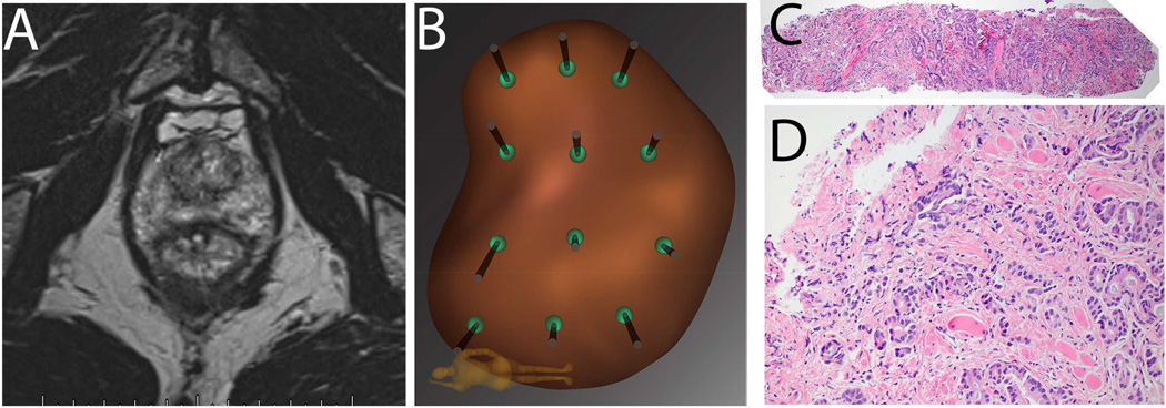
Figure 2. Example of Falsely Negative MRI in Patient P.M.
Patient is a 68 year old Caucasian male (PSA 3.8 ng/ml), who on a previous conventional biopsy was found to have a microfocus of Gleason 3+3=6 prostate cancer. He was considered for active surveillance, and mpMRI of prostate was obtained (A): prostate volume was found to be 35cc; no region of interest (ROI) was identified, even retrospectively. Mapping biopsy was performed by following the 12-point template of the Artemis device (B). A tissue core from the left lateral apex revealed 6 mm of Gleason 3+5=8 prostate cancer (C, 4×; D, 20×). Radical prostatectomy was performed, revealing a tumor on the left side of the prostate with diameters of 15mm × 12mm × 9 mm.
In our experience, the incidence of falsely negative MRI (i.e., Gleason Score ≥7 with no MRI evidence of tumor) is 15% when using biopsy evidence29 and approaches 30% when using whole mount prostatectomy evidence30. When biopsy is clinically indicated, a negative MRI should not preclude mapping biopsy.

