Abstract
Bursaphelenchus xylophilus is the pathogen of pine wilt disease. Bursaphelenchus mucronatus is similar to B. xylophilus in morphology. Both species share a common niche, but they are quite different in pathogenicity. Presently, the role of bacteria in pine wilt disease development has been widely speculated. The diversity of bacteria associated with B. xylophilus and B. mucronatus with different virulence remains unclear. In this study, virulence of four B. xylophilus and four B. mucronatus strains were evaluated by inoculating Pinus thunbergii. High-throughput sequencing targeted 16S rDNA of different virulence nematode strains was carried out. The associated bacterial community structures of the eight strains were analyzed. The results showed that 634,051 high-quality sequences were obtained from the eight nematode strains. The number of OTUs of bacteria associated with B. mucronatus was generally greater than those of B. xylophilus. The richness of the community of bacteria associated with high virulent B. xylophilus ZL1 and AmA3 was higher than moderately virulent B. xylophilus AA3, HE2, and all B. mucronatus strains. While the diversity of bacteria associated with B. mucronatus was higher than B. xylophilus. Stenotrophomonas, Pseudomonadaceae_Unclassified or Rhizobiaceae_Unclassified were predominant in the nematode strains with different virulence. Oxalobacteraceae and Achromobacter were found more abundant in the low virulent B. xylophilus and non-virulent B. mucronatus strains.
Introduction
Pine wilt disease (PWD) is a destructive disease caused by the pine wood nematode (PWN), Bursaphelenchus xylophilus. It is native to North America and has been spread to Japan, China, South Korea and Europe, subsequently causing huge economic losses and decimating ecological systems in these countries. Bursaphelenchus mucronatus is similar to B. xylophilus in morphology and distributed in Eastern Asia, Americas and Europe [1].
The onset of pine wilt disease is rapid and susceptible pines can be killed within a year [2]. As for the symptoms and infection rate after infection of B. xylophilus, there is a difference in virulence among different B. xylophilus strains. The strains are divided into highly virulent, moderately virulent and low virulent strains [3]. B. mucronatus has been found in dead pines in the areas without pine wilt nematode infection in China. The pathogenicity of B. mucronatus has attracted increasing attention [4–6]. Some studies suggested that B. mucronatus is not pathogenic [7–9], but more researchers opined that B. mucronatus had low virulence [10, 11], potential virulence [12] or a certain degree of virulence [13–15]. The pathogenicity of the nematodes is different according to the differences of the population sources, strains and developing stages of the nematodes, hosts, and temperature [16–18].
The pathogenesis of pine wilt disease is still unclear. Occurrence of the disease involves nematodes, insect vectors, bacteria, hosts and a number of other factors. Recent studies on the relationship between B. xylophilus and bacteria found that bacteria may play a role in pine wilt disease [11, 19–22]. Researches on species of bacteria associated with B. xylophilus have shown that B. xylophilus from different countries and regions carried different bacterial diversities [23, 24, 25]. The role of bacterial community in PWD has been studied [25–30]. New findings from B. xylophilus (transcriptomics and secretomics) also show the existence of bacterial putative effectors that may contribute for nematode pathogenicity in the tree [31–33]. However, little research has been done on the relationship between bacteria and B. mucronatus [34, 35]. At present, differences of bacteria associated with B. xylophilus and B. mucronatus and their relationships with the virulence of nematodes have not been reported.
Yuan et al. found endo-bacteria in B. xylophilus and B. mucronatus [36]. Wu et al. isolated 15 species of culturable endo-bacteria from B. xylophilus in China and found that Stenotrophomonas maltophilia and Achromobacter xylosoxidans subsp. xylosoxidans may relate to the differences in virulent of the nematode strains [26]. Traditional researches on bacterial diversity use culture-depended methods, but only less than 1% microorganisms in nature can be isolated [37]. Culture methods fail to determine overall situation of bacteria associated with B. xylophilus and are inadequate in analyzing relationship between virulence of B. xylophilus and bacteria. Molecular analysis of the microbial community associated with the nematodes isolated from Pinus pinaster was performed by Proença [38]. Tian identified 64 species of bacteria from two 16S rDNA clone libraries constructed from a Chinese and a Japanese B. mucronatus populations [35]. Culture-independent methods showed their advantage in analyzing the bacteria associated with nematodes. Therefore, identifications of the isolated strains obtained only by biochemical methods maybe incomplete [39].
This study used high-throughput sequencing targeted 16S rDNA to analyze diversity of bacteria associated with B. xylophilus and B. mucronatus in combination with virulence of different nematode strains on pine seedlings to better understand the diversity of bacteria from B. xylophilus and B. mucronatus with different virulence.
Materials and Methods
Bursaphelenchus xylophilus and B. mucronatus strains
Four B. xylophilus strains and four B. mucronatus strains with different virulence were selected. All of the nematode strains were deposited in Jiangsu Key Laboratory for Prevention and Management of Invasive Species, China. The origins and hosts of eight strains were listed in Table 1.
Table 1. Bursaphelenchus xylophilus and B. mucronatus strains for virulence test.
| Nematodes | Nematode strains | Pine hosts | Year of collection | Sampling region |
|---|---|---|---|---|
| B. xylophilus | ZL1 | Pinus massoniana | 2004 | Linhai, Zhejiang |
| AmA3 | P. massoniana | 2004 | Ma’anshan, Anhui | |
| HE2 | P. thunbergii | 2004 | Enshi, Hubei | |
| AA3 | P. taiwanensis | 2004 | Anqing, Anhui | |
| B. mucronatus | CFS1 | P. elliottii | 2005 | Fushun, Sichuan |
| SD1 | P. massoniana | 2005 | Dazhu, Sichuan | |
| JNL10 | P. taeda | 2004 | Nanjing, Jiangsu | |
| GHB3 | P. massoniana | 2004 | Huizhou, Guangdong |
Virulence test of the nematode strains
Bursaphelenchus xylophilus and B. mucronatus strains were cultured on a fungus Botrytis cinerea at 25°C for 7 days. The propagated nematodes were collected with a Baermann funnel and were rinsed three times with sterile deionized water. The nematode suspension was prepared with sterile deionized water.
Four-year-old Pinus thunbergii seedlings with a similar growing condition were used. P. thunbergii stems were cut 15 cm above the soil level with a sterilized scalpel. A piece of sterile absorbent cotton was placed on each wound and 200 μL nematodes suspension (about 5,000 individuals) was pipetted into wounds. Then the wounds were covered by parafilm. Control was P. thunbergii seedlings inoculated with 200 μL sterile deionized water. Each treatment had 5 replicates. The disease development of P. thunbergii seedlings was observed at an interval of two days. The infection rate is the prevalence of the disease. The disease severity of P. thunbergii seedlings was divided into five levels: 0, the seedlings healthy with green needles and growing well; I, a few needles turning brown; II, half of the needles turning brown and the terminal shoots of seedlings bending; III, most of the needles turning brown and dead and the terminal shoots of seedlings drooping; IV, all of the needles turning brown and the whole seedling wilt. The infection rates and the disease severity index were calculated according to Fang [40].
Surface sterilization of nematodes
A Baermann funnel was used to extract the nematodes. The nematodes were washed three times with sterile water, followed by sterilization of shaking the nematodes in 1% mercury bichloride for 30 min. After that, the nematodes were washed additional three times with sterile water, followed by shaking in a mixture of 1% streptomycin sulphate and 1% gentamicin for 30 min. Then, the nematodes were washed three times with sterile water. The nematode suspension was centrifuged and the supernatant was discarded. The precipitate containing nematode was resuspended with 1 mL sterile water. One hundred μL of the nematode suspension (about 2,500 individuals) was poured onto NA Petri dishes [26]. The plates were incubated at 28°C for 48 h to check presence of any bacterial colonies.
DNA extraction and PCR amplification of bacteria associated with nematodes
Based on our preliminary research (unpublished data), 50,000 individual nematodes would be sufficient for DNA extraction and suitable for subsequent high throughput sequencing (e.g: concentration >50ng/μL, OD260/280 ranging from 1.8 to 2.0, and total amount >5000ng). The surfaces of nematodes were checked to be aseptic and 50,000 individual nematodes were ground in liquid nitrogen with a sterile mortar and a pestle. The ground nematodes were transferred to a 1.5 mL centrifuge tube and the total genomic DNA (PWN+PWN-associated bacteria) was extracted by E.Z.N.A. Mullusc DNA Kit (Norcross, OMEGA, USA).
To study the diversity and composition of bacteria associated with nematodes, a pair of PCR primers 515f/806r targeting bacterial 16S rRNA V4 region was applied in PCR amplification [41]. To distinguish the different samples, a Barcoded-tag with six nucleotide bases was randomly added to the upstream of the universal primer. The primers which added with Barcoded-tag sequences were Barcoded-tag fusion primers (BFP). After quantification and quality control, PCR products were gradually diluted and quantified. 16S rDNA PCR products were sequenced using 300bp paired-end model with the MiSeq system (Illumina, USA). The EzTaxon-e database (http://eztaxon-e.ezbiocloud.net/) was used for bacterial taxonomic identification.
Bioinformatic analysis
The lengths of short reads were extended by finding the overlap between paired-end reads by the FLASH software [41]. Low quality data were filtered out using QIIME software [42]. The reads were sorted according to barcode sequences and the sample sources. The number of reads of each sample was counted. Sequences were clustered to operational taxonomic unit (OTU) at 97% sequence similarity by using UPARSE software [43].
To calculate the Alpha diversity, species richness (Chao), species coverage (Coverage, C) and species diversity (Simpson's diversity index, 1-D / Shannon-Wiener Index, H) were calculated. Python script alpha_rarefaction.py from QIIME—1.7.0 was used for diversity index analysis with default parameter setting. The richness index Chao, was used to estimate the number of OTUs in the bacterial communities. Simpson and Shannon indexes were used to estimate the diversity of the OTUs. Community structure analyses were based on the phylum and genus taxonomy levels.
Nucleotide Sequence Accession Numbers
Nucleotide Sequences were deposited at NCBI SRA database under the accession numbers SRX876433 and SRX876463-SRX876469.
Results
Pathogenicity of B. xylophilus and B. mucronatus on P. thunbergii
P. thunbergii seedlings of all treatments had a similar growing condition. After inoculated with nematodes, the P. thunbergii seedlings showed differences in symptoms. After 20 DAI (days after inoculation), infection rates of P. thunbergii inoculated with B. xylophilus strains ZL1, AmA3, AA3 and HE2 were 100%, 80%, 80% and 60%, respectively. The infection rates of P. thunbergii inoculated with B. mucronatus strains CFS1 and SD1 were 40% and 20%, respectively. Thus, six nematode strains were able to infect and induce wilting symptoms. No symptom was found when inoculated P. thunbergii seedlings with B. mucronatus strains GHB3 and JNL10. On the 30 DAI, P. thunbergii seedlings inoculated with the four B. xylophilus strains and B. mucronatus strains CFS1 and SD1 all showed symptoms. While the disease severity index of P. thunbergii seedlings were different. All of the P. thunbergii seedlings which inoculated with four B. xylophilus strains were wilted in 40 DAI, and all of the disease severity index were 100 (Table 2). The wilting process of P. thunbergii seedlings after inoculated with nematodes was different. Wilt symptoms were observed when inoculated with B. xylophilus strains ZL1 and AmA3, and the first P. thunbergii seedling was dead in 24 days. The P. thunbergii seedlings started to die in the 31 and 33 days after inoculated with B. xylophilus strains AA3 and HE2, respectively. It indicated that ZL1 and AmA3 caused death to P. thunbergii seedlings at a faster rate than AA3 and HE2. All of the P. thunbergii seedlings were infected when inoculated with B. mucronatus strains CFS1 and SD1, but no seedling was dead. Therefore, the pathogenicity of CFS1 and SD1 was weaker than those of other four B. xylophilus strains. A mild wilt symptom was observed after inoculated with B. mucronatus strain JNL10, and the infection index was 5. No wilt symptom was observed after inoculated with B. mucronatus strain GHB3 (Table 2). Thus, JNL10 showed rather weak pathogenicity, while GHB3 was not pathogenic to P. thunbergii. In summary, B. xylophilus strains ZL1 and AmA3 were highly virulent (HV) and strains AA3 and HE2 were moderately virulent (MV) to P. thunbergii. B. mucronatus strains CFS1 and SD1 were moderately virulent (MV), and strain JNL10 had low virulence (LV) to P. thunbergii, and the GHB3 strain was not virulent (NV).
Table 2. The symptoms of P. thunbergii after inoculated with B. xylophilus and B. mucronatus.
| Infection rates/ % | DSI | |||||||
|---|---|---|---|---|---|---|---|---|
| Nematode strains | 20th day | 30th day | 40th day | 20th day | 30th day | 40th day | Days of symptoms appeared (d) | Days of P. thunbergii wilted (d) |
| ZL1 | 100 | 100 | 100 | 30 | 85 | 100 | 11 | 24 |
| AMA3 | 80 | 100 | 100 | 35 | 70 | 100 | 11 | 24 |
| AA3 | 80 | 100 | 100 | 25 | 45 | 100 | 13 | 31 |
| HE2 | 60 | 100 | 100 | 20 | 45 | 100 | 17 | 33 |
| CFS1 | 40 | 100 | 100 | 20 | 45 | 75 | 19 | - |
| SD1 | 20 | 100 | 100 | 10 | 35 | 60 | 19 | - |
| JNL10 | 0 | 0 | 20 | 0 | 0 | 5 | 31 | - |
| GHB3 | 0 | 0 | 0 | 0 | 0 | 0 | - | - |
| CK | 0 | 0 | 0 | 0 | 0 | 0 | - | - |
Species abundance analysis of bacteria associated with B. xylophilus and B. mucronatus
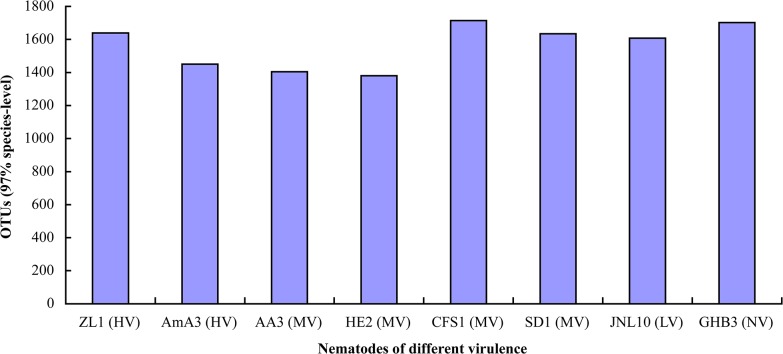
Sequences of the bacteria associated with B. xylophilus and B. mucronatus with different virulence were filtered using QIIME software. The statistical results showed that 634,051 high-quality sequences were obtained from the eight nematode strains. The average sequencing results of the eight samples were 79,256. The average length of the assembled reads was 252 bp. The OTUs for ZL1, AmA3, AA3 and HE2 were 1639, 1450, 1405 and 1380, respectively. The OTUs for CFS1, SD1, JNL10 and GHB3 were 1714, 1635, 1608 and 1702, respectively. The HE2 strain contained the smallest number of OTUs, while CFS1 contained the most abundant OTUs. The number of OTUs of bacteria associated with B. mucronatus was generally greater than those of B. xylophilus (Fig 1).
Fig 1. OTU numbers of bacteria associated with B. xylophilus and B. mucronatus.
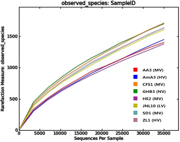
The rarefaction curves of all nematode-bacteria communities were flat and stable but did not reach maximum (Fig 2). It indicated that the sampling method was reasonable, reliable and able to represent the actual bacteria communities but a very small number of bacteria still remain undetected.
Fig 2. Rarefaction analysis of bacteria associated with B. xylophilus and B. mucronatus.
All of the coverage estimations were 100 of the bacteria associated with B. xylophilus and B. mucronatus with different virulence. The results indicated a high coverage of the sequencing libraries, and showed the diversity of the associated bacteria. Chao indexes showed that the richness of the community of bacteria associated with highly virulent B. xylophilus ZL1 and AmA3 was higher than moderately virulent B. xylophilus AA3, HE2, and all B. mucronatus strains. The Simpson and Shannon indexes of bacteria associated with B. mucronatus were both higher than those with B. xylophilus, indicating that diversity of bacteria associated with B. mucronatus was higher than those with B. xylophilus (Table 3).
Table 3. The diversity index of bacteria associated with B. xylophilus and B. mucronatus.
| Nematodes | Nematode strains | Coverage (C) / % | Chao (97%) | Simpson (1-D) (97%) | Shannon (H) (97%) |
|---|---|---|---|---|---|
| B. xylophilus | ZL1 (HV) | 100 | 7289 | 0.769 | 4.126 |
| AmA3 (HV) | 100 | 7276 | 0.871 | 4.995 | |
| AA3 (MV) | 100 | 4098 | 0.848 | 5.121 | |
| HE2 (MV) | 100 | 4663 | 0.829 | 4.552 | |
| B. mucronatus | CFS1 (MV) | 100 | 6319 | 0.919 | 6.140 |
| SD1 (MV) | 100 | 5967 | 0.898 | 5.602 | |
| JNL10 (LV) | 100 | 6743 | 0.896 | 5.588 | |
| GHB3 (NV) | 100 | 4786 | 0.933 | 6.132 |
Community structure analysis of bacterial associated with B. xylophilus and B. mucronatus at phylum level
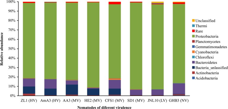
The OTUs of bacteria associated with the nematodes were classified and organized. At phylum level, bacteria associated with the eight strains of nematodes were classified to 9–12 taxonomic phyla. The bacteria associated with different nematode strains were mainly Proteobacteria, Bacteroidetes, Acidobacteria, Actinobacteria, Chloroflexi, Cyanobacteria, Firmicutes, Gemmatimonadetes, Planctomycetes and Verrucomicrobia. Proteobacteria was the most predominant phylum in the eight libraries, followed by Bacteroidetes. Some OTUs were not identified as phylum in the sequencing results. For the bacteria associated with B. xylophilus, the proportion of unidentified OTUs was more than 7%. Unidentified OTUs of bacteria was 6.6% in B. mucronatus strain CFS1, and the abundance was less than 0.4% in B. mucronatus strain SD1, JNL10 and GHB3 (Fig 3).
Fig 3. The bacterial composition of B. xylophilus and B. mucronatus at phylum level.
Community structures of bacteria associated with B. xylophilus and B. mucronatus at genus level
There are at least 24 groups of bacteria associated with B. xylophilus and B. mucronatus at genus level by 16S rDNA high-throughput sequencing.
Predominant bacteria associated with B. xylophilus
Bacteria associated with B. xylophilus were sorted by their abundance. The abundance higher than 10% were the predominant bacteria associated with the nematode strains. As shown in Table 4, the predominant bacteria associated with the highly virulent B. xylophilus ZL1 were Pseudomonadaceae_Unclassified, Pseudomonas and Stenotrophomonas. The predominant bacteria associated with the highly virulent B. xylophilus AmA3 were Stenotrophomonas and Rhizobiaceae_Unclassified. The predominant bacteria associated with moderately virulent B. xylophilus AA3 were Stenotrophomonas and Achromobacter. The predominant bacteria associated with moderately virulent B. xylophilus strain HE2 were Rhizobiaceae_Unclassified, Achromobacter and Luteibacter.
Table 4. The sorting of PWN-associated bacteria according to the relative abundance.
| The abundance of nematode associated bacteria | |||||
|---|---|---|---|---|---|
| Nematode strains | 1 | 2 | 3 | 4 | 5 |
| ZL1 (HV) | Pseudomonadaceae_Unclassified (35.9%) | Pseudomonas (19.7%) | Stenotrophomonas (12.4%) | Achromobacter (6.6%) | Rhizobiaceae_Unclassifie (6.1%) |
| AmA3 (HV) | Stenotrophomonas (35.9%) | Rhizobiaceae_Unclassifie (24.4%) | Chitinophaga (8.2%) | Oxalobacteraceae_Unclassified (4.5%) | Ochrobactrum (2.5%) |
| AA3 (MV) | Stenotrophomonas (48.7%) | Achromobacter (9.2%) | Enterobacteriaceae_Unclassified (4.2%) | Oxalobacteraceae_Unclassified (2.9%) | Escherichia (2.9%) |
| HE2 (MV) | Rhizobiaceae_Unclassified (40.2%) | Achromobacter (16.6%) | Luteibacter (16.5%) | Stenotrophomonas (3.7%) | Pseudomonadaceae_Unclassified (2.7%) |
| CFS1 (MV) | Pseudomonadaceae_Unclassified (31.2%) | Rhizobiaceae_Unclassified(10.1%) | Stenotrophomonas (9.5%) | Oxalobacteraceae_Unclassified (7.4%) | Sphingobacteriaceae_Unclassified (6.3%) |
| SD1 (MV) | Stenotrophomonas (27.7%) | Pseudomonadaceae_Unclassified (24.2%) | Oxalobacteraceae_Unclassified (14.7%) | Sphingobacteriaceae_Unclassified (5.1%) | Enterobacteriaceae_Unclassified (4.5%) |
| JNL10 (LV) | Oxalobacteraceae_Unclassified (23.3%) | Achromobacter (19.5%) | Pseudomonadaceae_Unclassified (10.9%) | Rhizobiaceae_Unclassifie (9.7%) | Pseudomonas (6.2%) |
| GHB3 (NV) | Stenotrophomonas (21.5%) | Oxalobacteraceae_Unclassified (12.1%) | Rhizobiaceae_Unclassified(10.1%) | Sphingobacteriaceae_Unclassified (9.4%) | Enterobacteriaceae_Unclassified (8.2%) |
Predominant bacteria associated with B. mucronatus
As shown in Table 4, the predominant bacteria associated with moderately virulent B. mucronatus CFS1 were Pseudomonadaceae_Unclassified and Rhizobiaceae_Unclassified. The predominant bacteria associated with moderately virulent B. mucronatus SD1 were Stenotrophomonas and Pseudomonadaceae_Unclassified. The predominant bacteria associated with the lowly virulent B. mucronatus JNL10 were Oxalobacteraceae_Unclassified and Achromobacter. Meanwhile, the predominant bacteria associated with the non-virulent B. mucronatus GHB3 were Stenotrophomonas and Oxalobacteraceae_Unclassified.
Community structures of bacterial associated with B. xylophilus and B. mucronatus with different virulence
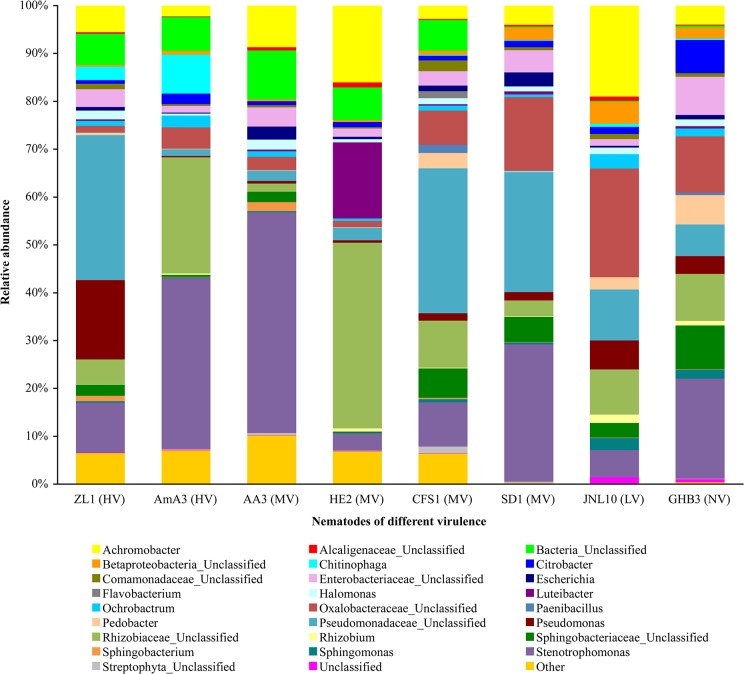
As well as the species and abundance of predominant bacteria associated with the nematodes which had different virulence were different, and the bacteria associated with each nematode were different. Stenotrophomonas, Rhizobiaceae_Unclassified, Achromobacter, Enterobacteriaceae_Unclassified and Pseudomonadaceae_Unclassified occupied large proportions of the bacteria associated with the nematodes (Fig 4). However, the proportions were different. The abundance of other associated bacteria, for example, Streptophyta_Unclassified, Sphingomonas, Sphingobacterium, Rhizobium, Paenibacillus, Flavobacterium, Escherichia and Comamonadaceae_Unclassified, were similar in B. xylophilus and B. mucronatus with different virulence.
Fig 4. The bacterial composition of B. xylophilus and B. mucronatus at genus level.
The abundance of Betaproteobacteria_Unclassified, Sphingobacteriaceae_Unclassified, Oxalobacteraceae_Unclassified and Pedobacter in B. mucronatus was significantly higher than those in B. xylophilus. The abundance of Bacteria_Unclassified associated with B. xylophilus and virulent B. mucronatus strain CFS1 were significantly higher than those in B. mucronatus strain SD1, JNL10 and GHB3. The proportions of Pseudomonadaceae_Unclassified in highly virulent B. xylophilus strain ZL1 and moderately virulent B. mucronatus strain CFS1 and SD1 were significantly higher than those in other nematodes. The proportions of Chitinophaga in highly virulent B. xylophilus strain ZL1 and AmA3 were 3.4% and 8.2%, respectively. However, there were no Chitinophaga in moderately virulent B. xylophilus strain AA3 and strain HE2. Only a few Chitinophaga existed in the bacteria associated with B. mucronatus, and the proportions were less than 0.7%. The abundance of Sphingomonas in lowly virulent B. mucronatus strain JNL10 and non-virulent B. mucronatus strain GHB3 were 2.6% and 1.9%, respectively, and they were significantly higher than those in other nematodes. Achromobacter occupied a certain proportion in all of the nematode-associated bacteria, and the proportions were relatively high in moderately virulent B. xylophilus strain AA3 and non-virulent B. mucronatus strains JNL10 and GHB3 (Fig 4).
Discussion
Advantages of 16S rDNA high-throughput sequencing technology
In this study, the diversities of bacteria associated with B. xylophilus and B. mucronatus with different virulence were analyzed by 16S rDNA high-throughput sequencing. Comparing to traditional culture method, it enabled us to analyze unculturable bacteria. Twenty four groups of nematode-associated bacteria with different virulence, such as Stenotrophomonas, Pseudomonadaceae_Unclassified and Rhizobiaceae_Unclassified, had relatively high abundance. This indicated that the species of bacteria associated with B. xylophilus and B. mucronatus were very abundant. Wu et al. isolated 15 endo-bacteria from B. xylophilus including Stenotrophomonas and Achromobacter [26]. Stenotrophomonas maltophilia and Myroides were isolated from B. mucronatus [44]. Through the construction of clone libraries and 454 sequencing, 21 genera of bacteria were associated with B. xylophilus were found [44]. Bacterial strains were grouped into 38 RAPD-types on basis of visual similarities, these bacterial strains belonged to 16 genera [39]. While only a small number of culturable bacteria could be obtained from the culture-depended methods [26, 34]. Therefore, compared to these techniques, the bacteria associated with B. xylophilus and B. mucronatus obtained by 16S rDNA high-throughput sequencing were more abundant. Bacteria communities isolated and identified by different methodologies from the nematodes occurring in USA, Japan, China, Korea and Portugal were considerably diverse and ubiquitous [28]. In comparison with the afore-mentioned the same predominant species in B. xylophilus and B. mucronatus, the new phyla found was Chloroflexi, Cyanobacteria, Gemmatimonadetes, Planctomycetes/Verrucomicrobia. Although we could not exclude the possibility that a trace number of ecto-bacteria would remain in the sample of bacterial community for high throughput sequencing, we deduce that the major component analyzed should be endo-bacteria, because most of ecto-bacteria would be removed in the course of multiple rinsing.
The virulence of B. xylophilus and B. mucronatus remained stable in the long-term subculture
Large differences of virulence were found between B. xylophilus and B. mucronatus strains [16–18]. The pathogenicity of nematodes in this study showed that all P. thunbergii seedlings inoculated with the four B. xylophilus strains were dead. The disease development and dying process of B. xylophilus strains ZL1 and AmA3 took shorter time than the strains AA3 and HE2. The result was consistent with that by Liu (2007), who determined that the B. xylophilus ZL1 and AmA3 were high virulent strains, and AA3 and HE2 were moderately virulent strains [45]. Measurement of the pathogenicity of B. mucronatus showed that the strains CFS1 and SD1 were pathogenic on P. thunbergii seedlings, and all of the seedlings were infected after inoculation by the two strains. However, the B. mucronatus’ infection of P. thunbergii seedlings was lower than those inoculated with B. xylophilus, and the wilt symptoms took longer time to develop. Thirty one days after P. thunbergii seedlings inoculated with B. mucronatus JNL10, the wilt symptoms were observed for the first time. The disease severity index was very low. These results indicated that the pathogenicity of B. mucronatus JNL10 was weak, and the GHB3 strain was not pathogenic, which were consistent with previous reports [26]. Our findings indicated that the virulence of B. xylophilus and B. mucronatus remained stable in the long-term subculture.
The diversity of bacteria from B. xylophilus and B. mucronatus with different virulence
Many studies suggested that the pathogenicity of B. xylophilus was related with the associated bacteria [27–28, 31]. In our study, some OTUs were annotated as Bacteria_Unclassified in the sequencing results, indicating that some species of bacteria associated with B. xylophilus and B. mucronatus could not be identified. The abundances of Bacteria_Unclassified in the four B. xylophilus strains and virulent B. mucronatus strain CFS1 were significantly higher than those in B. mucronatus strains SD1, JNL10 and GHB3. The richness indexes of highly virulent B. xylophilus strains ZL1 and AmA3 were significantly higher than those of other nematode strains. This indicated that the higher virulence a nematode had, the more richness. It suggested that the adaptability of high virulence B. xylophilus to ecological environment may be better. The study also found high virulence B. xylophilus ZL1 and AA3 had different associated-bacterial compositions. Although the two strains were belonged to high virulent nematode and isolated from the same pine species. However, the sampling places were different, one from Zhejiang and another from Anhui. It suggested that the bacteria were associated with the sampling geographic difference, soils, pine species and vectors. Thus, the differences found in these two high virulent strains would be reasonable.
Tian [44] made a comparison of community differences of the associated bacteria between M- and R-type B. xylophilus. Certain bacteria, such as Stenotrophomonas and Pseudomonas existed among the bacteria associated with R-type B. xylophilus and were absent among the ones with the M-type. In our study Stenotrophomonas or Pseudomonadaceae_Unclassified or Rhizobiaceae_Unclassified were found in the bacteria associated with virulent B. xylophilus and B. mucronatus. The afore-mentioned bacteria generally exist in B. xylophilus and B. mucronatus with high levels of relative abundances. The results indicated that the three kinds of bacteria mentioned above were predominant in the nematode-associated bacteria. Meanwhile, the proportions of Chitinophaga in highly virulent B. xylophilus strain ZL1 and AmA3 were significantly higher than other strains. Oxalobacteraceae was more abundant in the bacterial communities associated with lowly virulent B. mucronatus strain JNL10 and non-virulent B. mucronatus strain GHB3. The amount of Achromobacter was relatively high in moderately virulent B. xylophilus strain AA3, HE2 and non-virulent B. mucronatus strains JNL10 and GHB3. This suggested that the above PWN-associated bacteria may be related to the virulence of the nematodes. Our study also found the differences of abundance of other eight genera of bacteria associated with B. xylophilus and B. mucronatus with different virulence, such as Sphingomonas, were not significant. This suggests that these bacteria existed generally in the nematode strains and may have no direct relationship with the virulence of the nematodes. The evidence of these bacteria involved in the pathogenic processes of B. xylophilus need to be further studied.
Acknowledgments
We are grateful to Dr. De-Wei Li, The Connecticut Agricultural Experiment Station, USA, for reviewing the manuscript.
Data Availability
Relevant data are available at the NCBI SRA database under the accession numbers SRX876433 and SRX876463-SRX876469.
Funding Statement
This work was supported by the National Natural Science Foundation of China (NO.31270683), Southern China Collaborative Innovation Center of Sustainable Forestry, and the Priority Academic Program Development of Jiangsu Higher Education Institutions (PAPD). The funders had no role in study design, data collection and analysis, decision to publish, or preparation of the manuscript.
References
- 1.PPQ, New Pest Advisory Group (2001) NPGA data: Bursaphelenchus mucronatus a pine wood nematode, potential introduction. Electronic publication by USDA, APHIS code: Nem ParBmD01. pdf.
- 2. Mamiya Y (1983) Pathology of the pine wilt disease caused by Bursaphelenchus xylophilus . Annual Review of Phytopathology 21: 201–220. 10.1146/annurev.py.21.090183.001221 [DOI] [PubMed] [Google Scholar]
- 3.Li H (2008) Identification and pathogenicity of Bursaphelenchus species (Nematoda: Parasitaphelenchidae). Thesis, Ghent University.
- 4. Zhang ZY, Lin MS, Yu BY (2004) Pathogenicity of Bursaphelenchus mucronatus on the seedlings of Pinus thunbergii . Journal of Nanjing Agricultural University 27: 46–50. [Google Scholar]
- 5. Zhang ZY, Lin MS, Yu BY (2001) Evaluation of the pathogenicity of Bursaphelenchus mucronatus on black pine. Journal of Shenyang Agricultural University 32: 28–28. [Google Scholar]
- 6. Zhao YX, Xu ZH, Wang F, Yu SF, Fang WC, Li GX. (2003) Study on the dead pine of Pinus yunnanensisin occurrence area and non-occurrence area of Bursaphelenchus mucronatus . Journal of Southwest Forestry College 23: 62–66. [Google Scholar]
- 7. Mamiya Y, Enda N (1979) Bursaphelenchus mucronatus n. sp.(Nematoda: Aphelenchoididae) from pine wood and its biology and pathogenicity to pine trees. Nematologica 25: 353–361. [Google Scholar]
- 8. McNamara DG, Støen M (1988) A survey for Bursaphelenchus spp. in pine forests in Norway. EPPO Bulletin 18: 353–363. [Google Scholar]
- 9. Tomminen J (1993) Pathogenicity studies with Bursaphelenchus mucronatus in Scots pine in Finland. European Journal of Forest Pathology 23: 236–243. [Google Scholar]
- 10. Futai K (1980) Developmental rate and population growth of Bursaphelenchus lignicolus (Nematoda: Aphelenchoididae) and B. mucronatus . Applied Entomology and Zoology 15: 115–122. [Google Scholar]
- 11. Cheng HR, Lin MS, Li WQ, Fang ZD (1983) Pine wilt disease occured in Pinus thunbergii in Nanjing. Forest Pest and Disease 4: 1–5. [Google Scholar]
- 12. Braasch H (1997) Host and pathogenicity tests with pine wood nematode (Bursaphelenchus xylophilus) from North America under Central European weather conditions. Nachrichtenblatt des Deutschen Pflanzenschutzdienstes 49: 209–214. [Google Scholar]
- 13. Bakke A, Anderson RV, Kvamme T (1991) Pathogenicity of the nematodes Bursaphelenchus xylophilus and B. mucronatus to Pinus sylvestris seedlings: a greenhouse test. Scandinavian Journal of Forest Research 6: 407–412. [Google Scholar]
- 14. Zhang ZY, Zhang KY, Lin MS, Luo HW, Xu FY (2002) Pathogenicity determination of Bursaphelenchus xylophilus isolates to Pinus thunbergii . Journal of Nanjing Agricultural University 25: 43–46. [Google Scholar]
- 15. Kanzaki N, Futai K (2006) Is a weak pathogen to the Japanese red pine? Nematology 8: 485–489. [Google Scholar]
- 16. Wu G, Tang G, Luo X (2006) Comparative analysis of pathogenicity of pine wood nematode. Acta Agriculture Shanghai 22: 61–64. [Google Scholar]
- 17. Shen PY, Jiao GY, Li HQ (1995) Comparative analysis of pathogenicity of pine wood nematode from Japan and China. Forest Pest and Disease 4: 1–2. [Google Scholar]
- 18. Kiyohara T, Bolla RI (1990) Pathogenic variability among populations of the pinewood nematode, Bursaphelenchus xylophilus . Forest Science 36: 1061–1076. [Google Scholar]
- 19. Kawazu K, Kaneko N (1997) Asepsis of the pine wood nematode isolate OKD-3 causes it to lose its pathogenicity. Japanese Journal of Nematology 27. [Google Scholar]
- 20. Kawazu K, Kaneko N, Kanzaki H (1999) What factors govern the pathogenicity of the pine wood nematode, Bursaphelenchus xylophilus? Shokado Shoten. pp. 42–46. [Google Scholar]
- 21. Zhao BG, Liu Y, Lin F (2005) Mutual influences between Bursaphelenchus xylophilus and bacteria carries. Journal of Nanjing Forestry University 29: 1–4. [Google Scholar]
- 22. Tan JJ, Xiang HQ, Feng ZX (2008) A Preliminary study on isolation, identification and pathogenicity of the bacterium accompanying Bursaphelenchus xylophilus . China Forestry Science and Technology 22: 23–26. [Google Scholar]
- 23. Ju YW, Xie LF, Yang XY, Zhao BG (2008) Varieties of bacteria carried by pine wood nematode from different sources. Journal of Northeast Forestry University 36: 84–85. [Google Scholar]
- 24. Vicente CS, Nascimento F, Espada M, Mota M, Oliveira S (2011) Bacteria associated with the pinewood nematode Bursaphelenchus xylophilus collected in Portugal. Antonie van Leeuwenhoek 100: 477–481. 10.1007/s10482-011-9602-1 [DOI] [PubMed] [Google Scholar]
- 25. Proença DN, Francisco R, Paiva G, Santos SS, Abrantes IMO, Morais PV. (2011) Bacteria and Archaea: complexity of endophytic microbial community in pine trees in areas subject to pine wilt disease Braga, Portugal: Microbiotec’11. [Google Scholar]
- 26. Wu XQ, Yuan WM, Tian XJ, Fan B, Fang X, Ye JR, et al. (2013) Specific and functional diversity of endophytic bacteria from pine wood nematode Bursaphelenchus xylophilus with different virulence. International Journal of Biological Sciences 9: 34–44. 10.7150/ijbs.5071 [DOI] [PMC free article] [PubMed] [Google Scholar]
- 27. Vicente CS, Nascimento F, Espada M, Barbosa P, Mota M, Glick BR, et al. (2012) Characterization of bacteria associated with pinewood nematode Bursaphelenchus xylophilus . PloS ONE 7: e46661 10.1371/journal.pone.0046661 [DOI] [PMC free article] [PubMed] [Google Scholar]
- 28. Nascimento FX, Hasegawa K, Mota M, Vicente CS. (2014) Bacterial role in pine wilt disease development review and future perspectives. Environmental Microbiology Reports 10: 1111/1758-2229.12202. [DOI] [PubMed] [Google Scholar]
- 29. Vicente CSL, Ikuyo Y, Mota M, Hasegawa K (2013) Pine wood nematode-associated bacteria contribute to oxidative stress resistance of Bursaphelenchus xylophilus . BMC Microbiol 13: 299 10.1186/1471-2180-13-299 [DOI] [PMC free article] [PubMed] [Google Scholar]
- 30. Paiva G, Proença DN, Francisco R, Verissimo P, Santos SS, Fonseca L, et al. (2013) Nematicidal bacteria associated to pinewood nematode produce extracellular proteases. PLoS ONE 8: e79705 10.1371/journal.pone.0079705 [DOI] [PMC free article] [PubMed] [Google Scholar]
- 31. Kikuchi T, Cotton JA, Dalzell JJ, Hasegawa K, Kanzaki N, McVeigh P, et al. (2011) Genomic insights into the origin of parasitism in the emerging plant pathogen Bursaphelenchus xylophilus . PLoS Pathog 7(9): e1002219 10.1371/journal.ppat.1002219 [DOI] [PMC free article] [PubMed] [Google Scholar]
- 32. Cheng XY, Tian XL, Wang YS, Lin RM, Mao ZC, Chen N, et al. (2013) Metagenomic analysis of the pinewood nematode microbiome reveals a symbiotic relationship critical for xenobiotics degradation. Science Report 3: 1869. [DOI] [PMC free article] [PubMed] [Google Scholar]
- 33. Shinya R, Morisaka H, Kikuchi T, Takeuchi Y, Ueda M, Futai K. (2013) Secretome analysis of the pine wood nematode Bursaphelenchus xylophilus reveals the tangled roots of parasitism and its potential for molecular mimicry. PLoS ONE 8(6): e67377 [DOI] [PMC free article] [PubMed] [Google Scholar]
- 34.Chi SY (2003) Studies on the pathogenicity of the bacteria carried by pine wood nematode and the relationship between the bacteria and the nematode. Thesis, Nanjing Foresty University.
- 35. Tian XL, Cheng XY, Mao ZC, Chen GH, Yang JR, Xie BY. (2011) Composition of bacterial communities associated with a plant–parasitic nematode Bursaphelenchus mucronatus . Current Microbiology 62: 117–125. 10.1007/s00284-010-9681-7 [DOI] [PubMed] [Google Scholar]
- 36. Yuan WM, Wu XQ, Ye JR, Tian XJ (2011) Observation by transmission electron microscope and identification of endophytic bacteria isolated from Bursaphelenchus xylophilus and B. Mucronatus . Acta Microbiologica Sinica 51 (8): 1071–1077. [PubMed] [Google Scholar]
- 37. Yue XJ, Yu LY, Zhang YQ (2004) Progress in research of microorganisms of natural environments in the viable but non-culturable state. Microbiology China 31(2): 108–111. [Google Scholar]
- 38. Proença DN, Francisco R, Santos CV, Lopes A, Fonseca L, Abrantes IMO, et al. (2010) Diversity of bacteria associated with Bursaphelenchus xylophilus and other nematodes isolated from Pinus pinaster trees with pine wilt disease. PLoS One 5: e15191 10.1371/journal.pone.0015191 [DOI] [PMC free article] [PubMed] [Google Scholar]
- 39. Proença DN, Fonseca L, Powers TO, Abrantes IM, Morais PV. (2014) Diversity of bacteria carried by pinewood nematode in USA and phylogenetic comparison with isolates from other countries. PLoS One 9(8): e105190 10.1371/journal.pone.0105190 [DOI] [PMC free article] [PubMed] [Google Scholar]
- 40. Fang ZD (1998) Methods in research of plant disease Beijing, China: Chinese Agricultural Publishing House. 11pp. [Google Scholar]
- 41. Magoč T, Salzberg SL (2011) FLASH: Fast length adjustment of short reads to improve genome assemblies. Bioinformatics 27: 2957–2963. 10.1093/bioinformatics/btr507 [DOI] [PMC free article] [PubMed] [Google Scholar]
- 42. Caporaso JG, Kuczynski J, Stombaugh J, Bittinger K, Bushman FD, Costello EK, et al. (2010) QIIME allows analysis of high-throughput community sequencing data. Nature Methods 7: 335–336. 10.1038/nmeth.f.303 [DOI] [PMC free article] [PubMed] [Google Scholar]
- 43. Edgar RC (2013) UPARSE: Highly accurate OTU sequences from microbial amplicon reads,Nature Methods 10: 996–998. 10.1038/nmeth.2604 [DOI] [PubMed] [Google Scholar]
- 44.Tian XL (2010) Diversity and Ecological role of bacteria associated with Bursaphelenchus xylophilus and B Mucronatus. Thesis, Northwest A&F University.
- 45.Liu X (2007) Studies on morphology and pathogenic mutation of Chinese group of pine wood nematode. Thesis, Nanjing Forestry University.
Associated Data
This section collects any data citations, data availability statements, or supplementary materials included in this article.
Data Availability Statement
Relevant data are available at the NCBI SRA database under the accession numbers SRX876433 and SRX876463-SRX876469.


