Abstract
The aim of this study is to assess the pneumatization of the paranasal sinuses (PNS) and other parts of temporal bone such as mastoid air cells and to investigate if there was any association between the aeration of these structures among the three major ethnic groups in Malaysia (Malay, Chinese, Indian) as this would be representative of Asia. A retrospective review of 150 computed tomography (CT) scans of PNS and temporal bones was done and analysed. The pneumatization of each area was obtained and compared using statistical analysis. Patients with a history of previous medical or surgical problems in the intended areas were excluded from the study. The pneumatization of the mastoid air cells and other temporal bone parts were noted to be symmetrical in more than 75 %. There was a positive correlation between the pneumatization of mastoid air cells and that of the sphenoid sinus. The prevalence of Agger nasi, Haller’s and Onodi cells was observed to be significantly higher in the Chinese group. Preoperative assessment of the temporal bone and PNS with CT scan may be helpful in the evaluation of their anatomical landmark and decrease the possibility of surgical complications related to 3D structures.
Keywords: Pneumatization, Mastoid, Ethmoid, Sphenoid, Sinuses
Introduction
The paranasal sinuses (PNS) and other parts of temporal bone such as the mastoid air cells system are the best characterized structures for aeration in humans. These structures develop by gradual pneumatization of solid tissue, which is achieved by positive pressure on the naso-pharynx through the Eustachian tube opening [1].
Several factors including environmental conditions, genetic diseases, and past infections may affect the process of pneumatization which is the main distinctive feature of these complexes.
Temporal bone pneumatization has been divided into 5 compartments: Middle ear, Mastoid, Peri-labyrinthine, Petrous apex and Accessory regions which include squamous, zygomatic, occipital and styloid cells. Pneumatization cease around puberty, which is an adult stage, with the development of the last air cells in the petrous apex [2].
The endoscopic sinus surgery (ESS) techniques deal with the sinuses and structures around the orbit, pituitary and clivus. The sphenoid sinus is a critical sinus with its anatomic variations and relationships with the adjacent structures, Further the sphenoid sinus has been classified as Sellar, Pre-sellar and Conchal type. It’s importance is stressed in pituitary surgery, as even minimal damage to adjacent structures can lead to complications. Agger nasi, Haller’s cells and Onodi cells are some variants described.
The mastoid pneumatization and its measurement has also been studied earlier [3, 4]. In a multiracial country like Malaysia it is an advantage to do racial comparison studies with regards to the pneumatization of these structures as these should apply to the most common Asian races in general.
The aims of the present study were
To compare the pneumatization of the temporal bone, ethmoid and sphenoid sinuses based on analysing the conventional two-dimensional CT scan images with Osirix software station.
The prevalence of the following common anatomical variants including; Agger nasi cells, Haller’s cells, concha bullosa, Onodi cells, pneumatization of the crista galli, and to evaluate if there are any correlations in the degree of pneumatization of these variants.
Materials and Methods
Data Collection
This study was based on a retrospective review of the PNS and temporal bone CT scans which was obtained from the CT scan image archives of a tertiary medical care centre in Malaysia in the last 5 years (July 2006–June 2011). A total of 150 CT scans were selected with 50 subjects in each major ethnic group (Malay, Chinese and Indian) among Malaysian population, 25 each of male and female. Based on the inclusion and the exclusion criteria.
Image Acquisition and Analysis
Each patient’s skull was scanned and performed on the 16-slice multi-detector CT scan (Siemens Somatom Sensation 16; Germany and General Electric, USA). The exposure settings were 80–120 kV and 100–250 mA, which were optimised for each patient. Continuous non-overlapping sections with acquisition parameters of 1.0-mm or less slice thickness were used with axial slice field of view, 150 mm; high resolution. These CT scan data was then downloaded into the multiplanar computer workstation which was analysed using OsiriX software (Version 3.8.1; 32 bit; OsiriX Foundation, Geneva) All of the subjects’ data were then viewed and reformatted into coronal and sagittal sections.
Inclusion Criteria
Patients 18-year old and above without history of head trauma, severe sinusitis, middle ear disease, or previous sinus or mastoid surgery.
Exclusion Criteria
Patients with a history of chronic rhinosinusitis and/or nasal polyps, middle ear surgery or sinonasal surgery and those with a CT scan findings compatible with chronic otitis media and/or mastoiditis.
Definitions of Variants that can be Observed
Pneumatization of Mastoid Air Cells System
In adult life, the normal mastoid may be fully pneumatized, diploic (partial pneumatized), or sclerotic (non pneumatized) [5] (Fig. 1a–c) as described.
Fig. 1.
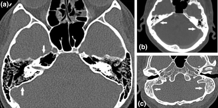
Axial view of the temporal bone showing: a well pneumatised mastoid regions, b poorly pneumatised mastoid air cell, c none pneumatised mastoids (sclerotic type)
Perilabyrinthine and Petrous Apex
The perilabyrinthine region is subdivided into: (1) the supralabyrinthine area and (2) The infralabyrinthine area which lie superior and inferior to the labyrinth, respectively. The petrous apex region of the temporal bone is divided into: (1) the peritubal area which surrounds the osseous portion of the Eustachian tube and lies anterolateral to the carotid canal, and (2) the apical area which lies anteromedial to the carotid canal. Air cells may be found extending from the mastoid, middle ear, and following tracts above and below the cochlea and into the petrous apex [5] (Fig. 1a).
Pneumatization of Sphenoid Sinus
The degree of pneumatization of the sphenoid sinuses may vary considerably ranging from absent to extensive [6]. Sphenoid sinuses are classified into pre-sellar, post-sellar (Fig. 2a, b) and conchal [3]. The post-sellar or sellar types are further subdivided into complete and incomplete types depending on the pneumatization reaching the clivus. The conchal type of sphenoid is known as non-pneumatized or rudimentary, the sinus does not reach into the body of the sphenoid bone.
Fig. 2.
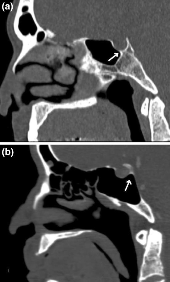
a Presellar type of sphenoid sinus, b postsellar sphenoid sinus in which the pneumatization extends back beneath the pituitary fossa
Agger Nasi Cells
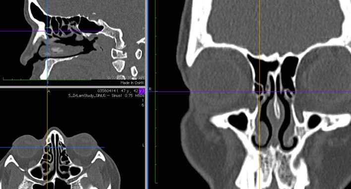
Agger nasi cells are the most anterior ethmoid air chamber below the frontal sinus, situated in the lateral nasal wall, anterior and superior to the insertion of the middle concha. It is intimately related to the frontonasal recess, reaching the lacrimal fossa inferolaterally, and is anterolaterally arched by the nasal bones [7] (Fig. 3).
Fig. 3.
Multiplanar view of the CT scan demonstrating Agger nasi cell on the right side
Haller (Infraorbital) Cells
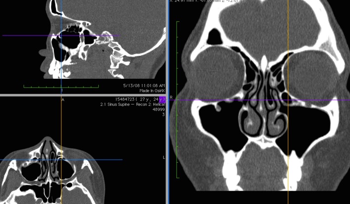
These are air cells from the anterior or posterior ethmoid sinuses that may extend and located in the roof of the maxillary sinus and inferior portion of the lamina papyracea including air cells located within the ethmoidal infundibulum below the ethmoid bulla [7] (Fig. 4).
Fig. 4.
Multiplanar view of the CT scan demonstrating Haller cell on the left side
Concha Bullosa
Concha bullosa is a cystic distension of the middle nasal concha when it is aerated in both horizontal and vertical parts. Pneumatization of the middle turbinate may originate from the frontal recess, the anterior ethmoids or directly from the middle meatus [8] (Fig. 5).
Fig. 5.
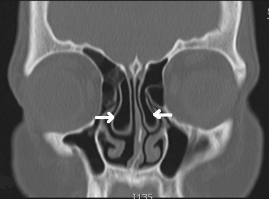
Coronal CT scan image showing bilaterally pneumatized middle turbinate (concha bullosa)
Onodi (Sphenoethmoid) Cell
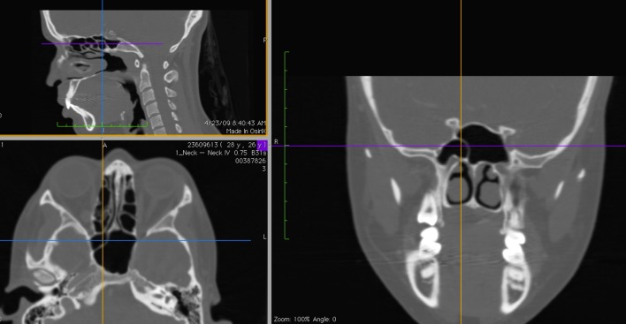
Is defined as the posterior most ethmoid cell that pneumatizes into the region normally occupied by the sphenoid sinus and positioned posterolateral to it. As a result of its location the optic nerve, and less commonly, the internal carotid artery (ICA), are very closely related with a very thin bone (0.03 mm in thickness) separating them [9] (Fig. 6).
Fig. 6.
Multiplanar view of CT scan demonstrating Onodi cell (right side)
Pneumatization of the Crista Galli
The crista galli is a median ridge of bone that projects from the cribriform plate of the ethmoid bone. Pneumatization of the crista usually occurs from the frontal sinus [10], however it might be expected that aeration of the crista would come from ethmoid cells too (Fig. 7).
Fig. 7.
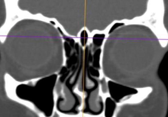
Coronal CT scan image showing pneumatized crista galli
Results
150 CT scans were selected by using stratified randomization so as to attain 50 subjects in each racial group, namely the Malay, Chinese and Indian.
There were equal numbers of men and women (75) in each group. The subjects’ ages ranged from 18 to 89 years old with a mean age of 47.28 years old with a standard deviation of 16.33.
Pneumatization of Mastoid Air Cells System
Mastoid air cells were found to be fully pneumatized in 136 (90.7 %) patients of whom 45 (30 %), 44 (29.3 %) and 47 (31.3 %) were Malay, Chinese and Indian respectively. Diploic or poorly pneumatized mastoid were recorded in 13 (8.7 %) patients while sclerotic type was limited to only 1 (0.7 %) Indian patient. No statistical differences were observed in mastoid pneumatization among the three races in the Malaysian population (P value 0.392).
Pneumatization of the Perilabyrinthine Cells and Petrous Apex
Perilabyrinthine and petrous apex regions were studied as one area for each. Pneumatization of the perilabyrinthine cells was noted in 59 (39.9 %) patients. 47 (31.3 %) patients found to have pneumatised petrous apex. However, there were no statistical significant differences neither between right and left sides nor among the three races. (P = 0.072) for perilabyrinthine cells or (P = 0.572) for petrous apex.
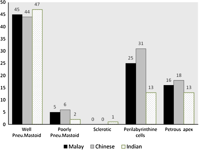
The degree of symmetry noted in the pneumatization of temporal bone parts namely mastoid, perilabyrinthine and petrous apex region was 99.4, 33.3 and 24 % respectively. Moreover, pneumatization extending into other regions of the temporal bone corresponded linearly with squamomastoid pneumatization (Graph 1).
Graph 1.
Histogram showing the correlation between pneumatization of the mastoid and other temporal bone parts (perilabyrinthine and petrous apex) in all races
Pneumatization of Sphenoid Sinus
The post-sellar type with its two subdivisions complete and incomplete was the most predominant type 83.3 %. The presellar type was found in 16.7 %. No conchal type was recorded in our studied subjects. (P = 0.4) showing no statistical relationship between the race and type of sphenoid sinuses.
Pneumatization of Certain Anatomical Variants
Comparison of common anatomical variants in Malay, Chinese and Indian are presented in Table 1.
Table 1.
Comparison of common anatomical variants among the three different races
| Race | Agger nasi cells | Haller’s cells | Onodi cells | Concha bullosa | Pneu. crista galli |
|---|---|---|---|---|---|
| Malay | 24 | 17 | 17 | 17 | 4 |
| Chinese | 29 | 32 | 24 | 16 | 17 |
| Indian | 17 | 15 | 14 | 22 | 16 |
| Total | 70 | 64 | 55 | 55 | 37 |
Agger nasi cells were the most frequently observed variant in Malaysian population, 70 patients (46.6 %). The Chinese group has the highest incidence of these cells (29 cases), unilaterally recorded in 14 patients, seven on the right side and seven on the left. Significant relationship was noted between racial group and the incidence of Agger nasi cells (P = 0.036), with no statistical significance of these cells with the age or gender.
Haller’s cell was the next most common variation seen in 64 patients (42.6 %) of Malaysian population. It was also highly recorded among Chinese individuals 32 cases (21.3 %) which is more than Malay (11.3 %) and Indian (10 %). Of the 64 cases recorded, 53 displayed Haller’s cells unilaterally, 16 on the right and 37 on the left. This variant was also significant regarding race but not for age or gender (P = 0.003).
Of the other anatomical variants that had a higher incidence in Chinese group rather than the other is the Onodi cell variant. It was found in 24 Chinese patients out of 55 cases of Malaysian population who presented with these cells. (P = 0.008). These variant were noted to be unilateral in 46 cases, while only 9 cases seen bilaterally.
True middle turbinate concha bullosa was found in 36.7 % of the Malaysian skulls scanned, 18 on the right and 15 on the left. Our recorded data for Malay, Chinese and Indian proved that there was little difference in the prevalence of concha bullosa between all groups (P = 0.301). The present study found a pneumatized crista galli in 37 (24.7 %) cases out of 150, most frequently among Chinese and Indian 17 (34 %) and 16 (32 %) respectively (P = 0.004).
Discussion
Temporal Bone Pneumatisation
Temporal bone aeration has been found to be symmetrical in 72–99 % of the general population [11], similar to our study. Our finding of petrous-apex pneumatization was 31.3 % of all studied scans correspond with other studies [11, 12]. In our study the probability of petrous-apex and peri-labyrinthine pneumatization corresponds directly with the degree of mastoid pneumatization, as in another study [13].
Sphenoid Sinus Pneumatization
Our study was similar to those in that the post-sellar sphenoid was the predominant type 83.3 % while presellar type was 16.7 % only which is similar to other study [14]. A positive correlation noted between the pneumatization of mastoid air cells and that of the sphenoid sinus in our study similar to another study [15]. This correlation may be explained by the close proximity of the openings of these structures into the nasal cavity, thus they might expose to the same driving forces that control pneumatization process or other pathological events. Furthermore, during the development of these structures, the growth of the mastoid air cells and sphenoid sinus takes longer duration than other sinuses, increasing the possibility of exposing these areas to various pathological events that might affect these structures separately or together [16].
Ethmoidal Group of Cells Pneumatization
The mean incidence of Agger nasi cell in other studies [8, 17] was 50.6 % as compared to 46.6 % in our study. Racial group comparison revealed that the prevalence in Chinese was significantly higher than in Malay and Indian groups.
When the pneumatization process of the ethmoidal air cells progresses inferolaterally into an infraorbital location, HaIler’s cells are created. It can be clinically significant as a causal factor in maxillary sinusitis and the potential risk of injury to the orbit [18]. In our study, Haller’s cells were present in 42.6 % as compared to other studies of 45 % [7]. Malaysian Chinese were also noted to have higher percentage of infraorbital cells at 64 %.
Extension of the middle ethmoidal air cells in an inferomedial direction into the middle turbinate formulate a commonly encountered normal variant on coronal sinus CT scans called concha bullosa, implicated in the pathogenesis of rhinosinusitis because of its tendency to obstruct the ventilation and reduce mucociliary clearance of the middle meatus. However, the prevalence of concha bullosa is reported to be the same in patients with and without symptoms of sinus disease [19]. In our study, a true concha bullosa was observed in 36.7 % as compared to 48 % in Thailand [20].
Potential damage to the optic nerve and ICA can occur in the presence of an Onodi cell [21] when attempts to enter the sphenoid sinus endoscopically by passing through what is thought to be the posterior most ethmoidal air cells is instead an Onodi cell. Our study revealed a prevalence of 36.6 % for Onodi cell as compared to 51 % in Singapore [22]. Chinese also showed a significantly higher rate of Onodi cells 48 % in our study.
Pneumatization of the Crista Galli
Pneumatization of crista galli has not been implicated in sinus disease. Our result with 24.7 % was similar to one American study [23] with the prevalence of 24 %. Malay population had the lowest incidence (8 %) compared to the other two races.
Computerized tomography is the most precise imaging technique for measurement of air space volume. It has many advantages for obtaining quantitative volumetric information about the sinuses or mastoid cells. It is cost effective, more automated and allow repeated measurement over time in the same individual to evaluate the improvement by observing any changes in the bony walls and in the sinonasal drainage pathway. Furthermore, 3D reconstruction using multidetector CT provides an accurate assessment of the sinonasal cavities. Although axial view may be useful for describing the anatomical landmarks of the sinonasal cavity, coronal and sagittal reconstructed images give more information required for an endoscopic clearance as they show progressively the deeper structures that might be encountered by the surgeon during the operation (posterior ethmoidal air cells and sphenoid sinus) [24].
A study of correlation between the volumes of the air-containing structures showed no correlation between the areas of the PNS and the mastoids, but good correlation between the sizes of the frontal and maxillary sinuses on the same side, suggesting that the PNS develop by a common process of pneumatization, whereas the pair of mastoid air cell systems may develop from a different process [1]. Similar results have been shown in another study [25]. The possibility that the incidence of certain anatomical variants in the PNS and nasal cavity vary significantly in certain populations highlights our aim in this study to compare the prevalence of these findings among different ethnic groups of Malaysian population which is representative of this region.
Conclusion
There was a positive correlation between the pneumatization of mastoid air cells and that of the sphenoid sinus. The prevalence of certain anatomical variants of PNS (Agger nasi, Haller’s and Onodi cells) were observed to be significantly higher in certain races (Chinese) than the others, however no relationship was detected between the pneumatization of these ethmoid air cells and that of the temporal bone.
Preoperative assessment of the temporal bones and PNS with CT scan may be helpful in the evaluation of their anatomical landmark and decreases the possibility of surgical complications related to 3D structures. Coronal reconstructed images of CT scan should always be obtained prior to any surgery in the sphenoid sinus area to identify the limits of dissection and prevent iatrogenic injury to the surrounding neurovascular structures. In the current study, Chinese had a higher incidence of certain anatomical variants (Agger nasi, Hallers’ and Onodi cells) makes them more prone to the risk of operative complication, therefore extra precaution should be taken by surgeons during operating on such individuals.
Summary
The PNS and other parts of temporal bone like mastoid air cells system have been found to have multiple functions including humidification, adding resonance during phonation, decreasing the weight of the skull, control of inspired air, and the moderation of air flow in respiration.
The process of pneumatisation of these structures might affected by several factors like environmental conditions, genetic diseases, and past infections.
Temporal bone pneumatization process normally passes into three stages: the infantile, transitional and adult stage with the development of the last air cells in the petrous apex.
There are many observable commonly occurring variants of the PNS and nasal cavity, most being harmless, while others are of surgical importance if they obstruct normal operating procedures.
Particular attention should be paid when evaluating the extent of pneumatisation of the sphenoid sinus as far as its anatomic variations and relationships with the adjacent structures, such as internal carotid artery and optic nerve, are concerned.
The development and refinement of (CT) scan imaging has allowed detailed assessment of each individual, thus providing a map that assist the sinus surgeon to operate safely.
The aims of the present study were to compare the pneumatization of the temporal bone, ethmoid and sphenoid sinuses, among the three major races in Malaysia, namely the Malays, Chinese and Indians based on analysing the conventional two-dimensional CT scan images with Osirix software station.
There was a high degree of symmetry noted in the pneumatization of temporal bone parts namely mastoid, perilabyrinthine and petrous apex region (99.4, 33.3 and 24 % respectively). Moreover, Pneumatization extending into other regions of the temporal bone corresponded linearly with squamomastoid pneumatization.
A positive correlation between the pneumatization of mastoid air cells and that of the sphenoid sinus was noted.
Significant relationship was noted between racial group and the incidence of Agger nasi, Haller’s and Onodi cells with a higher incidence in Chinese group rather than the others.
References
- 1.Thomas A, Raman R. A comparative study of the pneumatization of the mastoid air cells and the frontal and maxillary sinuses. AJNR Am J Neuroradiol. 1989;10:S88. [PMC free article] [PubMed] [Google Scholar]
- 2.Rubensohn G. Mastoid pneumatization in children at various ages. Acta Otolaryngol. 1965;60:11–14. doi: 10.3109/00016486509126983. [DOI] [PubMed] [Google Scholar]
- 3.Andreasson L, Mortensson W. Comparison between the area and the volume of the air filled ear space. Acta Radiol Diagn. 1975;16:347–352. doi: 10.1177/028418517501600405. [DOI] [PubMed] [Google Scholar]
- 4.Luntz M, Malatskey S, Tan M, Bar-Meir E, Ruimi D. Volume of mastoid pneumatization: three-dimensional reconstruction with ultrahigh-resolution computed tomography. Ann Otol Rhinol Laryngol. 2001;110:486–490. doi: 10.1177/000348940111000516. [DOI] [PubMed] [Google Scholar]
- 5.Schuknect HF, Gulya AJ. Anatomy of the temporal bone with surgical implications. Philadelphia: Lea & Febiger; 1986. [Google Scholar]
- 6.Kinnman J. Surgical aspects of the anatomy of the sphenoidal sinuses and the sellaturcica. J Anat. 1977;124:541–553. [PMC free article] [PubMed] [Google Scholar]
- 7.Bolger W, Butzin C, Parsons D. Paranasal sinus bony anatomic variations and mucosal abnormalities: CT analysis for endoscopic sinus surgery. Laryngoscope. 1991;101:56–64. doi: 10.1288/00005537-199101000-00010. [DOI] [PubMed] [Google Scholar]
- 8.Kantarci M, Karasen MR, Alpe F, Onbas O, Okur A, Karaman A. Remarkable anatomic variations in paranasal sinus region and their clinical importance. Eur J Radiol. 2004;50:296–302. doi: 10.1016/j.ejrad.2003.08.012. [DOI] [PubMed] [Google Scholar]
- 9.Driben JS, Bolger WE, Robles HA, Cable B, Zinreich SJ. The reliability of computerized tomographic detection of the Onodi (sphenoethmoid) cell. Am J Rhinol. 1998;12:105–111. doi: 10.2500/105065898781390325. [DOI] [PubMed] [Google Scholar]
- 10.Earwaker J. Anatomic variants in sinonasal CT. Radiographics. 1993;13:381–415. doi: 10.1148/radiographics.13.2.8460226. [DOI] [PubMed] [Google Scholar]
- 11.Hagens EW. Anatomy and pathology of the petrous bone based on a study of 50 temporal bones. Arch Otolaryngol. 1934;19:556–573. doi: 10.1001/archotol.1934.03790050021003. [DOI] [Google Scholar]
- 12.Glick HN. Microscopic observations of the petrous apex. Ann Otol Rhinol Laryngol. 1933;42:175–185. doi: 10.1177/000348943304200115. [DOI] [Google Scholar]
- 13.Kraus L. Die Pyramidenspitzen-pneumatisation im Aoentgenbild. Arch OhrenNasen Kehlkopfh. 1931;128:307–338. doi: 10.1007/BF01623419. [DOI] [Google Scholar]
- 14.Sethi DS, Stanley RE, Pillay PK. Endoscopic anatomy of sphenoid sinus and sella turcica. J Laryngol Otol. 1995;109:951–955. doi: 10.1017/s0022215100131743. [DOI] [PubMed] [Google Scholar]
- 15.Kim J, Song SW, Cho JH, Chang KH, Jun BC. Comparative study of the pneumatization of the mastoid air cells and paranasal sinuses using three-dimensional reconstruction of computed tomography scans. Surg Radiol Anat. 2010;32:593–599. doi: 10.1007/s00276-009-0618-4. [DOI] [PubMed] [Google Scholar]
- 16.Tan HK, Ong YK, Teo MS, Fook-Chong SM. The development of sphenoid sinus in Asian children. Int J Pediatr Otorhinolaryngol. 2003;67:1295–1302. doi: 10.1016/j.ijporl.2003.07.012. [DOI] [PubMed] [Google Scholar]
- 17.Badia L, Lund VJ, Wei W, Ho WK. Ethnic variation in sinonasal anatomy on CT-scanning. Rhinology. 2005;43:210–214. [PubMed] [Google Scholar]
- 18.Arslan H, Aydinlioglu A, Bozkurt M, Egeli E. Anatomical variations of the paranasal sinuses: CT examination for endoscopic sinus surgery. Auris Nasus Larynx. 1999;26:39–48. doi: 10.1016/S0385-8146(98)00024-8. [DOI] [PubMed] [Google Scholar]
- 19.Jones NS, Strobl A, Holland I. A study of the CT findings in 100 patients with rhinosinusitis and 100 controls. Clin Otolaryngol. 1997;22:47–51. doi: 10.1046/j.1365-2273.1997.00862.x. [DOI] [PubMed] [Google Scholar]
- 20.Nitinavakarn B, Thanaviratananich S, Sangsilp N. Anatomical variations of the lateral nasal wall and paranasal sinuses: a CT study for endoscopic sinus surgery (ESS) in Thai patients. J Med Assoc Thai. 2005;88(6):763–768. [PubMed] [Google Scholar]
- 21.Weinberger DG, Anand VK, Al-Rawi M, Cheng HJ, Messina AV. Surgical anatomy and variations of the Onodi cell. Am J Rhinol. 1996;10:365–370. doi: 10.2500/105065896781794851. [DOI] [Google Scholar]
- 22.Yeoh KH, Tan KK. The optic nerve in the posterior ethmoid in Asians. Acta Otolaryngol. 1994;114:329–336. doi: 10.3109/00016489409126065. [DOI] [PubMed] [Google Scholar]
- 23.Van Alyea OE. Nasal sinuses. An anatomic and clinical consideration. Baltimore: Williams and Wilkins Company; 1951. [Google Scholar]
- 24.Kaluskar SK, Patil NP, Sharkey AN. The role of CT in functional endoscopic sinus surgery. Rhinology. 1993;31(2):49–52. [PubMed] [Google Scholar]
- 25.Karakas S, Kavakli A. Morphometric examination of the paranasal sinuses and mastoid air cells using computed tomography. Ann Saudi Med. 2005;25:41–45. doi: 10.5144/0256-4947.2005.41. [DOI] [PMC free article] [PubMed] [Google Scholar]


