Abstract
Retrograde urethrogram is employed for adequate demonstration of anterior urethral stricture and is commonly performed by trainee residents. Not uncommonly, contrast is injected under pressure to overcome the resistance of a stricture which can lead to extravasation or intravasation exposing the patient to risk of bacteremia, sepsis, contrast reactions, and worsening of stricture. We report two such cases of extensive intravasation delineating the “venogram” of peno-pelvic venous arcade. Such rare occurrences highlight the importance of eliciting history of various allergies and asthma, urethral instrumentation, obtaining sterile urine before the study, and performing the study under dynamic fluoroscopy.
Keywords: Urethrogram, Intravasation, Stricture
Case Details
Case 1
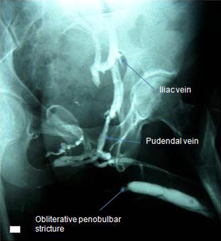
A 25-year-old male presented with progressively deteriorating urinary stream for the past 6 months. Uroflowmetry revealed a peak flow of 4 mL/s. He underwent a retrograde urethrogram (RUG) using Lapides and Stone technique [1] in 45° oblique position under antibiotic cover using 60 % meglumine–sodium–diatrizoate without fluoroscopic guidance. After initial 3–4 mL of instillation, resistance was felt; however, the instillation was continued for another 4–5 mL in the hope of delineating the stricture well. RGU revealed a tight stricture at the penobulbar junction and gross intravasation of contrast delineating the entire penile and pelvic venous system (Fig. 1).
Fig. 1.
Retrograde urethrogram showing tight stricture at penobulbar junction and gross intravasation delineating dorsal vein of penis, circumflex veins, main trunks, and tributaries of internal pudendal, internal iliac and common iliac veins, and inferior vena cava (not shown)
Case 2
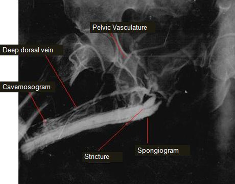
A 50-year-old male presented with complaint of straining and poor stream. He came with an RGU that was done outside. It showed a stricture in proximal bulbar urethra. Apart from delineating just the urethra, the RUG showed dense intravasation with spongiogram, cavernosogram, and deep dorsal vein of penis along with penile as well as pelvic vasculature (Fig. 2).
Fig. 2.
Retrograde urethrogram demonstrating stricture in midbulbar urethra along with dense intravasation with extensive visualization of corpus spongiosum, periurethral veins, dorsal vein of the penis, pudendal venous plexus and internal pudendal veins
Discussion
RUG is used for adequate demonstration of anterior urethral stricture. Instilling contrast under pressure can lead to extravasation or intravasation of contrast material exposing the patient to risk of bacteremia, sepsis, contrast reactions, and embolism especially if oil-based contrast is used [1–3]. Dense contrast intravasation has potential to induce fibrosis in the erectile tissue of penis. Reported incidence of contrast intravasation is 1 % [4]. It occurs due to the breach in the urethral mucosa. It is essential to perform the study under dynamic fluoroscopy without undue pressure while contrast instillation.
References
- 1.Hertz M. Cystourethrography. In: Pollack HM, editor. Clinical urography, vol 1. Philadelphia: Saunders; 1990. pp. 256–295. [Google Scholar]
- 2.Mukherjee MG, Deshon GE, Brackman JA, Mittemeyee BT, Borski AA. Urethrovascular reflux and its significance in urology. J Urol. 1974;112:608–609. doi: 10.1016/s0022-5347(17)59808-0. [DOI] [PubMed] [Google Scholar]
- 3.Ulma AH, Waghshul EC. Pulmonary embolism following urethrography with oily medium. New Eng J Med. 1960;263:137–139. doi: 10.1056/NEJM196007212630307. [DOI] [PubMed] [Google Scholar]
- 4.Gupta SK, Kaur B, Shukla RC. Urethrovenous intravasation during retrograde urethrography. J Postgrad Med. 1991;37:102–104. [PubMed] [Google Scholar]


