Abstract
AIM: Bicyclol, 4,4’-dimethoxy-5,6,5’,6’-dimethylene-dioxy-2-hydroxymethyl-2’-carbonyl biphenyl, is a new anti-hepatitis drug. The aim of the present study was to investigate the protective effect of bicyclol on concanavalin A (Con A)-induced immunological liver injury in mice and its mechanism.
METHODS: Liver injury was induced by injection of Con A via tail vein of mice and assessed biochemically and histologically. Serum transaminase and tumor necrosis factor alpha (TNF-α) were determined. Liver lesions were observed by light microscope. Expressions of TNF-α, interferon gamma (IFN-γ), Fas and Fas ligand (FasL) mRNA in the livers were measured by RT-PCR.
RESULTS: Serum transaminase level and liver lesions in Con A-induced mice were markedly reduced by oral administration of 100, 200 mg/kg of bicyclol. TNF-α level in serum was also reduced by bicyclol. Con A injection induced up-regulation of TNF-α, IFN-γ, Fas and FasL mRNA expression in liver tissues. Bicyclol significantly down-regulated the expression of IFN-γ, Fas and FasL mRNA, but only slightly affected TNF-α mRNA expression in liver tissues.
CONCLUSION: Bicyclol protects against Con A-induced liver injury mainly through inhibition of Fas/FasL mRNA expression in liver tissues and TNF-α release in mice.
INTRODUCTION
Viral hepatitis is a serious health problem worldwide, as more than two billion people alive today have been infected by hepatitis B virus, and 170 million people have been infected by hepatitis C virus[1]. Although a number of drugs such as interferon-2α and lamivudine have been used in the treatment of viral hepatitis, the efficacy of these drugs is still limited and some side effect are severe[2,3]. Therefore, it is necessary to develop new anti-hepatitis drugs. Bicyclol is new synthesized anti-hepatitis drug, chemically named 4, 4’-dimethoxy-5, 6, 5’, 6’-dimethylene-dioxy-2-hydroxymethyl-2’-carbonyl biphenyl. Our previous studies demonstrated that bicyclol has anti-liver injury, anti-liver fibrosis and anti-hepatitis virus activities[4]. Clinical trials indicated that bicyclol markedly normalized the elevated level of serum transaminase in patient with viral hepatitis B, and also inhibited HBV-DNA replication[5]. It is known that the liver injury induced by CCl4, acetaminophen and D-galactosamine is mainly caused by free radicals and oxidative stress[6,7]. The protection of bicyclol against CCl4-, acetaminophen-induced liver injury, is closely related to its elimination of free radicals and inhibition of oxidative stress. However, the pathogenesis of human viral hepatitis is different from hepatotoxicants induced liver injury. Human viral hepatitis is a disease resulting from destruction of virus-infected hepatocytes through immune-mediated mechanism[8,9]. Thus, it is necessary to further investigate if there are other mechanisms involved in the protective effect of bicyclol against liver injury in addition to its anti-oxidative activity. Recently, a model of immune-mediated liver injury in mice was established by injection of T cell mitogen Con A, which is an available animal model relevant to human hepatitis and hepatocellular damage[10]. Previous studies demonstrated that activated T cell-mediated cellular immunity is responsible for the liver damages in Con A model. TNF-α released from activated T lymphocytes and Kupffer cells plays a critical role in the process of liver damage and hepatocyte apoptosis[11-13]. In addition, Fas and Fas ligand (FasL) pathway is involved in the pathogenesis of hepatocytes apoptosis and necrosis[14]. Therefore, the present study was to investigate the effect of bicyclol on Con A induced immune-mediated liver injury and its mechanisms. Figure 1 shows the chemical structure of bicyclol.
Figure 1.
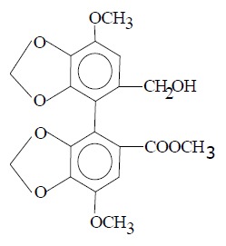
Chemical structure of bicyclol.
MATERIALS AND METHODS
Animals
Male C57BL/6 mice weighing 21-25 g were obtained from the Animal Center of Chinese Academy of Medical Sciences. They were fed standard maintenance diet and water throughout the experiments. All animals received care in compliance with the guidelines of China Ministry of Health.
Reagents
Bicyclol was kindly provided by Professor Zhang CZ in the Department of Pharmaceutical Chemistry of our institute. The purity of bicyclol was over 99% and not dissolved in water. Type IV Con A was purchased from Sigma Chemical Co. (St. Louis, MO, USA). ALT kit was purchased from BHKT Chemical Reagent Co., Ltd (Beijing, China). ELISA kit for TNF-α determination was provided by Jingmei Biotechnology Co, Ltd (Beijing, China).
Con A-induced hepatitis in mice and drug administration
Control mice were injected pyrogen-free phosphate-buffered saline (PBS). A dose of 23 mg/kg of Con A was injected via tail vein. Bicyclol was suspended in 5 g/L sodium carboxymethylcellulose (Na-CMC) just before use. The 200 mg/kg and 100 mg/kg of bicyclol were administered orally three times, 24, 12, 1 h before Con A injection.
Assay for serum ALT activity and TNF-α levels
Serum from individual mouse was obtained at various intervals after Con A injection. Alanine aminotransferase (ALT) level was determined by a biochemical kit according to the manufacturer’s instruction. The assay of serum TNF-α level was performed with specific ELISA kit.
Histopathological examination of liver tissue
Liver samples from individual mouse were fixed in 40 g/L neutral formaldehyde solution, embedded in paraffin, sliced into sections 5-μm of thickness and stained with hematoxylin and eosin for histological examination. The degrees of liver injury were graded based on a score of 0-3[15] and expressed as the mean of 10 different fields of each slide.
Detection of Fas, Fas ligand (FasL), TNF-α, and IFN-γ expression by semiquantitative RT-PCR
Total RNA from mouse liver was extracted by TRIzol reagent (Gibco USA)[16]. RT-PCR was conducted to detect Fas, FasL, TNF-α and IFN-γ gene expression. The primers for Fas were as follows: 5’-TGCACAGAAGGGAAGGAGTA-3’ and 5’-ATG GTTTCACGACTGGAGGT-3’; for FasL: 5’-GACAGCAGTG CCACTTCATC-3’ and 5’- TTAAGGCTTTGGTTG GTGAA-3’; for IFN-γ: 5’-CTCAAGTGGCATAGATGTGG-3’ and 5’-ACT CCTTTTCCGCTTCCTGA-3’; for TNF-α: 5’-GGCGGTGCCT ATGTCTCAG-3’ and 5’-GGGCAGCCTTGTC CCTTGA-3’; and for Cu/Zn SOD: 5’-TCTGCGTGCTGAAG GGCGAC-3’; and 5’-CTCCTGAGAGTGAGATCACA-3’. mRNA was reversely transcribed with AMV reverse transcriptase and then amplified using Tfl DNA polymerase for 35 cycles as follows: at 94 °C for 30 s (dissociation), at 60 °C for 60 s (annealing), and at 72 °C for 90 s (primer extension). PCR amplification products were visualized in 2% agarose gels after staining with ethidium bromide. The density of the PCR products was compared with Cu/Zn SOD, which was used as an internal control. Briefly, the spot density of each band in the gel was measured with the Kodak digital imaging system (Kodak DC120, Digital science 1D system, USA). The percentage of gene expression was calculated by the following equation: (density of cytokine product/density of SOD product) ×100.
Statistical analysis
All data are expressed as mean ± SD and analyzed by Student’s t-test.
RESULTS
Effect of bicyclol on Con A-induced aminotransferase elevation in serum
Injection of Con A to mice caused a time dependent increase in serum ALT levels (Figure 2). ALT levels increased significantly and reached a maximum at 6 h and maintained at plateau for 16 h after Con A injection. Administration of 200 mg/kg and 100 mg/kg of bicyclol to mice for three times significantly reduced the elevated serum ALT levels at 12 h and 16 h after Con A administration (Table 1).
Figure 2.
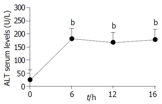
Time-course of Con A-induced liver serum ALT el-evation in mice. bP < 0.01 vs other time.
Table 1.
Protection of bicyclol against Con A induced liver injury in mice as determined by ALT(U/L)
| Time | Con A alone | Con A+bicyclol | Con A+bicyclol |
| 200 mg/kg | 100 mg/kg | ||
| 12 h | 161.5 ± 63.11 | 27.1 ± 10.8b | 50.1 ± 15.9a |
| 16 h | 165.8 ± 8.76 | 10.6 ± 12.8b | 30.7 ± 27.4b |
Data are mean ± SD of 8 mice per group.
P < 0.05,
P < 0.01. vs Con A alone. Serum ALT levels were measured 12 h and 16 h after Con A injection, respectively.
Effect of bicyclol on liver histology
Twelve hours after Con A injection, histological analysis of the liver sections of Con A-treated mice showed widespread areas of necrosis and inflammation within the liver lobules and around the central veins and portal tracts (Figure 3A,B). Extensive lesions were characterized by massive hepatocytes coagulative necrosis, and cytoplasmic swelling of most living hepatocytes and nuclear chromatin condensation were found frequently, which indicated hepatocytes apoptosis. A moderate infiltration of lymphocytes and mononuclear cells in the portal area and around the central vein were also observed. As shown in Figure 3C, bicyclol reduced the areas of lesions and the extent of liver injury induced by Con A. The extent of lymphocyte infiltration in bicyclol treated mice also reduced significantly. The extent of liver necrosis was classified using a four degree score as shown in the footnote of Table 2.
Figure 3.
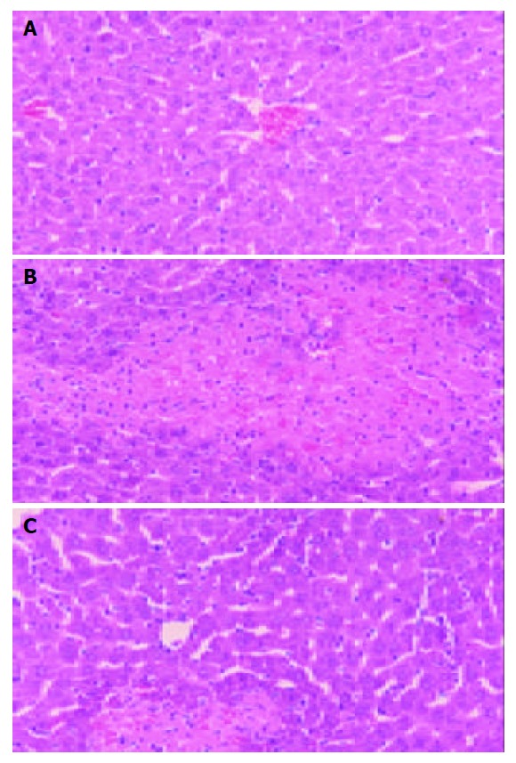
Light micrographs of livers from mice treated with Con A alone or in combination with bicyclol. Original magnification×200. A: normal control mouse. B: Con A injected mouse; C, mouse treated with bicyclol 200 mg/kg.
Table 2.
Effect of bicyclol on liver histology by evaluation the degree of liver injury 12 h after Con A injection
| Group |
Grade of liver injury |
|||
| 0 | 1 | 2 | 3 | |
| Normal (n = 7) | 7 | 0 | 0 | 0 |
| Con A alone (n = 7) | 0 | 0 | 0 | 7 |
| ConA+bicyclol 200 mg/kg (n = 8) | 3 | 2 | 1 | 2 |
| ConA+bicyclol 100 mg/kg (n = 7) | 1 | 2 | 3 | 1 |
Histological degree of liver injury is classified according to the area of coagulative necrosis in hepatic lobules as follows: grade 0, no coagulative necrosis; grade 1, coagulative necrosis area < 10%; grade 2, coagulative necrosis area between 10% and 25%; grade 3, coagulative necrosis area > 25%.
Lowering effect of bicyclol on serum TNF-α level in Con A-injected mice
Mice were killed at the indicated time points after injection of 23 mg/kg Con A. As shown in Figure 4, serum TNF-α level increased significantly 1-2 h after Con A injection, and then showed a marked decline at 4 h. At 24 h, the level of TNF-α level was still slightly higher than the normal mice but not significantly (Figure 4).
Figure 4.
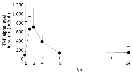
Dynamic changes of serum TNF-α level after Con A injection in mice. Each point represents mean ± SD of six mice.
The treatments of mice with bicyclol 200 mg/kg caused a decrease of the elevated TNF-α level by about 50% two hours after Con A injection (P < 0.05). Bicyclol 100 mg/kg also decreased serum TNF-α concentration about 25% but not significantly statistically (Figure 5).
Figure 5.
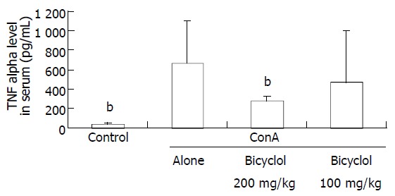
Lowering effect of bicyclol on the elevated serum TNF-α Level induced by Con A injection in mice. Each bar represents mean ± SD of 6 mice per group bP < 0.01 vs Con A alone.
Effect of bicyclol on expressions of liver TNF-α, IFN-γ, Fas and FasL mRNA in Con A-treated mice
One hour after the last administration of bicyclol, mice were injected 23 mg/kg of Con A. The livers were removed 2 h later. Total liver RNA was isolated, reversely transcribed, and amplified by PCR using primers for TNF-α, IFN-γ, Fas and FasL, respectively. Cu/Zn SOD was used as an internal control. As shown in Figure 6, TNF-α and IFN-γ mRNA were undetectable in livers from normal control mice, while the mice injected with Con A showed TNF-α and IFN-γ mRNA expression apparently. Prior administration of bicyclol 200 mg/kg reduced about 40% IFN-γ mRNA expression induced by Con A, but had slight influence on up-regulation of TNF-α mRNA in Con A injected mice.
Figure 6.
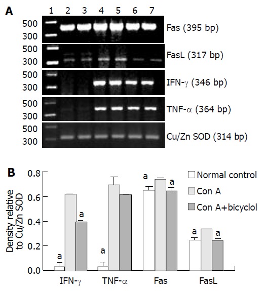
Effect of bicyclol on Con A induced TNF-α, IFN-γ, Fas and FasL mRNA expression determined by RT-PCR. A: Pho-tograph of ethidium bromide-stained amplification products. Lane 1, molecular mass markers; lane 2 and lane 3, control liver; lane 4 and lane 5, liver 2 h after Con A administration alone; lane 6 and lane 7, liver 2 h after injection of Con A in combination with 200 mg/kg bicyclol. B: Relative band intensity of amplification products compared with Cu/Zn SOD mRNA. Data represent the mean of two lanes. Each lane corresponds to the pooled livers from two mice. aP < 0.05 vs Con A alone.
Although Fas and FasL mRNA levels were detectable in normal control mice, significant increase of both Fas and FasL mRNA levels was demonstrated in Con A challenged mice. Mouse liver Fas mRNA level was more than that of FasL both in normal control and Con A injected groups. Administration of bicyclol down-regulated Fas and FasL mRNA expression to approximately normal level.
DISCUSSION
The immune cell-mediated liver injury is a critical event in the pathogenesis of human viral hepatitis[17-19]. Cell immunity is involved in the immune clearance of virus-infected hepatocytes and at the same time damage virus-infected hepatocytes. This conclusion came from the findings that infiltrating T cells were observed in the liver tissue of hepatitis patients[20,21]. Con A induced liver injury is a T-cell-dependent hepatitis model, which situation is relevant to human liver injury[22]. In Con A induced hepatitis, T cell activation plays a crucial role in the process of liver injury, because lymphocytes depleted mice or severe combined immunodeficiency disease (SCID) mice can not develop liver injury after Con A injection, and pretreatment of mice with a monoclonal antibody which in vivo destroys Thy1.2 antigen-bearing T cells abrogated the susceptibility of these animals toward the hepatotoxicity induced by Con A[10]. In the present study lymphocyte infiltration was observed in the portal area and around the central vein of damaged liver tissue. Biochemical and histological data showed that prior administration of bicyclol, a new anti-hepatitis drug has protective effect against Con A induced liver injury in mice. In order to elucidate how bicyclol exerts its protective effect against Con A induced liver injury, mice were pretreated with bicyclol. The results showed that bicyclol markedly decreased lymphocyte accumulation into liver.
Cytotoxic T lymphocytes play an important role in the pathogenesis of viral hepatitis by inducing hepatocyte apoptosis[23,8]. In the study of Moriyama et al[24] transfer of HBV-specific cytotoxic T lymphocytes can mediate liver cell necrosis in the HBV transgenic murine. As viral hepatitis, hepatocytes apoptosis is a dominant feature of Con A induced hepatitis as measured by DNA fragmentation and morphological observation. The apoptosis cascade can be initiated by a ligand, which binds to its receptors on the cell surface. Ligands such as FasL and TNF-α play an important role in Con A induced hepatitis. Interaction of Fas and FasL is an important pathway that mediates the target cytolysis by cytotoxic T lymphocytes[25]. In gld/gld mice, in which FasL is defective, the development of the disease is completely suppressed and in lpr/lpr mice, the suppression of Fas mRNA is incomplete, the hepatitis is partially suppressed. Antibody against Fas can block the development of hepatitis[14]. When FasL binds to Fas, the receptor molecules trimerize, leading to clustering of death domains, activation of a series of caspases and the final apoptosis of liver cells[26]. In chronic hepatitis B (CHB), the intensity of FasL mRNA showed a positive correlation with the histological activity and serum alanine aminotransferase level[27]. Immunohistochemical study suggested that Fas antigen expression plays an important role in inflammation in the hepatitis C virus-infected liver[28]. In our previous study, we found that bicyclol inhibited Con A induced DNA fragmentation[29]. To further investigate the mechanism of bicyclol in protection against liver injury, we measured the expression of Fas/FasL in Con A-induced liver injury.
In our study, we found that both FasL and Fas mRNA expressions were up-regulated significantly after Con A injection. Bicyclol down-regulated the expression of FasL and Fas close to normal level. The results explained why bicyclol could inhibit apoptosis of hepatocytes in Con A hepatitis model in our previous experiment[29]. It is known that Fas expression can be activated by IFN-γ to enhance apoptosis. Fas mRNA expression was reduced by 50% in the liver of IFN-γ-/- mice compared with that in IFN-γ+/- mice [30]. Because IFN-γ can affect the expression of Fas, we further investigated the effect of bicyclol on IFN-γ mRNA expression. Our results indicated that bicyclol reduced the expression of IFN-γ mRNA by about 40% in the livers of Con A treated mice, which was associated with the inhibition of Fas.
TNF-α is another important ligand to induce hepatocyte apoptosis. Activated lymphocytes stimulated the macrophages to release TNF-α and the activated lymphocyte itself is the potential source of TNF-α[31]. This proinflammatory cytokine can produce a direct cytotoxic effect to transcription-inhibited hepatocytes in vitro when bound to TNF-R[32]. In addition to the effect on Fas/FasL pathway, bicyclol significantly inhibited the release of TNF-α. Con A significantly increased the release of serum TNF-α and mRNA expression in liver tissue. Bicyclol reduced the serum level of TNF-α by about 50%. It, however, showed a slight effect (10%) on the mRNA expression in liver. It seems that bicyclol inhibited TNF-α release mostly at posttranscriptional level, which needs further investigation.
In conclusion, bicyclol protects mice against Con A-induced liver injury through at least two mechanisms: down-regulation the expression of Fas/FasL mRNA, and prevention of proinflammatory cytokine TNF-α release.
ACKNOWLEDGEMENTS
We thank Ms. Yong-Rong Zhang and Ms. Yan Liu from the Department of Pathology of our institute for their support in the examination of liver morphology by light microscopy. We also thank Ling-Hong Yu for her helpful advice and excellent technical assistance.
Footnotes
Supported by the Grant from Ministry of Science and Technology of China, No. 94-ZD-02
Edited by Zhu LH and Wang XL Proofread by Xu FM
References
- 1.Liu J, McIntosh H, Lin H. Chinese medicinal herbs for chronic hepatitis B: a systematic review. Liver. 2001;21:280–286. doi: 10.1034/j.1600-0676.2001.021004280.x. [DOI] [PubMed] [Google Scholar]
- 2.Haria M, Benfield P. Interferon-alpha-2a. A review of its pharmacological properties and therapeutic use in the management of viral hepatitis. Drugs. 1995;50:873–896. doi: 10.2165/00003495-199550050-00007. [DOI] [PubMed] [Google Scholar]
- 3.Heathcote J. Treatment of HBe antigen-positive chronic hepatitis B. Semin Liver Dis. 2003;23:69–80. doi: 10.1055/s-2003-37588. [DOI] [PubMed] [Google Scholar]
- 4.Liu GT. The anti-virus and hepatoprotective effect of bicyclol and its mechanism of action. Chin J New Drug Clin. 2001;10:325–327. [Google Scholar]
- 5.Yao GB, Ji YY, Wang QH, Zhou XQ, Xu DZ, Chen XY. A ran-domized double-blind controlled trial of bicyclol in treatment of chronic hepatitis B. Chin J New Drug Clin. 2002;21:457–461. [Google Scholar]
- 6.Suematsu T, Matsumura T, Sato N, Miyamoto T, Ooka T, Kamada T, Abe H. Lipid peroxidation in alcoholic liver disease in humans. Alcohol Clin Exp Res. 1981;5:427–430. doi: 10.1111/j.1530-0277.1981.tb04926.x. [DOI] [PubMed] [Google Scholar]
- 7.Slater TF. Necrogenic action of carbon tetrachloride in the rat: a speculative mechanism based on activation. Nature. 1966;209:36–40. doi: 10.1038/209036a0. [DOI] [PubMed] [Google Scholar]
- 8.Chisari FV. Cytotoxic T cells and viral hepatitis. J Clin Invest. 1997;99:1472–1477. doi: 10.1172/JCI119308. [DOI] [PMC free article] [PubMed] [Google Scholar]
- 9.Jacobson Brown PM, Neuman MG. Immunopathogenesis of hepatitis C viral infection: Th1/Th2 responses and the role of cytokines. Clin Biochem. 2001;34:167–171. doi: 10.1016/s0009-9120(01)00210-7. [DOI] [PubMed] [Google Scholar]
- 10.Tiegs G, Hentschel J, Wendel A. A T cell-dependent experimental liver injury in mice inducible by concanavalin A. J Clin Invest. 1992;90:196–203. doi: 10.1172/JCI115836. [DOI] [PMC free article] [PubMed] [Google Scholar]
- 11.Gantner F, Leist M, Lohse AW, Germann PG, Tiegs G. Concanavalin A-induced T-cell-mediated hepatic injury in mice: the role of tumor necrosis factor. Hepatology. 1995;21:190–198. doi: 10.1016/0270-9139(95)90428-x. [DOI] [PubMed] [Google Scholar]
- 12.Ksontini R, Colagiovanni DB, Josephs MD, Edwards CK, Tannahill CL, Solorzano CC, Norman J, Denham W, Clare-Salzler M, MacKay SL, et al. Disparate roles for TNF-alpha and Fas ligand in concanavalin A-induced hepatitis. J Immunol. 1998;160:4082–4089. [PubMed] [Google Scholar]
- 13.Zang GQ, Zhou XQ, Yu H, Xie Q, Zhao GM, Wang B, Guo Q, Xiang YQ, Liao D. Effect of hepatocyte apoptosis induced by TNF-alpha on acute severe hepatitis in mouse models. World J Gastroenterol. 2000;6:688–692. doi: 10.3748/wjg.v6.i5.688. [DOI] [PMC free article] [PubMed] [Google Scholar]
- 14.Tagawa Y, Kakuta S, Iwakura Y. Involvement of Fas/Fas ligand system-mediated apoptosis in the development of concanavalin A-induced hepatitis. Eur J Immunol. 1998;28:4105–4113. doi: 10.1002/(SICI)1521-4141(199812)28:12<4105::AID-IMMU4105>3.0.CO;2-8. [DOI] [PubMed] [Google Scholar]
- 15.Mochida S, Ohno A, Arai M, Tamatani T, Miyasaka M, Fujiwara K. Role of adhesion molecules in the development of massive hepatic necrosis in rats. Hepatology. 1996;23:320–328. doi: 10.1053/jhep.1996.v23.pm0008591859. [DOI] [PubMed] [Google Scholar]
- 16.Chomczynski P, Sacchi N. Single-step method of RNA isolation by acid guanidinium thiocyanate-phenol-chloroform extraction. Anal Biochem. 1987;162:156–159. doi: 10.1006/abio.1987.9999. [DOI] [PubMed] [Google Scholar]
- 17.Koziel MJ. The immunopathogenesis of HBV infection. Antivir Ther. 1998;3:13–24. [PubMed] [Google Scholar]
- 18.Chisari FV, Ferrari C. Hepatitis B virus immunopathogenesis. Annu Rev Immunol. 1995;13:29–60. doi: 10.1146/annurev.iy.13.040195.000333. [DOI] [PubMed] [Google Scholar]
- 19.Koziel MJ. Cytokines in viral hepatitis. Semin Liver Dis. 1999;19:157–169. doi: 10.1055/s-2007-1007107. [DOI] [PubMed] [Google Scholar]
- 20.Dienes HP, Hütteroth T, Hess G, Meuer SC. Immunoelectron microscopic observations on the inflammatory infiltrates and HLA antigens in hepatitis B and non-A, non-B. Hepatology. 1987;7:1317–1325. doi: 10.1002/hep.1840070623. [DOI] [PubMed] [Google Scholar]
- 21.Volpes R, van den Oord JJ, Desmet VJ. Memory T cells represent the predominant lymphocyte subset in acute and chronic liver inflammation. Hepatology. 1991;13:826–829. [PubMed] [Google Scholar]
- 22.Tiegs G, Gantner F. Immunotoxicology of T cell-dependent experimental liver injury. Exp Toxicol Pathol. 1996;48:471–476. doi: 10.1016/S0940-2993(96)80058-3. [DOI] [PubMed] [Google Scholar]
- 23.Koziel MJ, Dudley D, Wong JT, Dienstag J, Houghton M, Ralston R, Walker BD. Intrahepatic cytotoxic T lymphocytes specific for hepatitis C virus in persons with chronic hepatitis. J Immunol. 1992;149:3339–3344. [PubMed] [Google Scholar]
- 24.Moriyama T, Guilhot S, Klopchin K, Moss B, Pinkert CA, Palmiter RD, Brinster RL, Kanagawa O, Chisari FV. Immunobiology and pathogenesis of hepatocellular injury in hepatitis B virus transgenic mice. Science. 1990;248:361–364. doi: 10.1126/science.1691527. [DOI] [PubMed] [Google Scholar]
- 25.Kägi D, Vignaux F, Ledermann B, Bürki K, Depraetere V, Nagata S, Hengartner H, Golstein P. Fas and perforin pathways as major mechanisms of T cell-mediated cytotoxicity. Science. 1994;265:528–530. doi: 10.1126/science.7518614. [DOI] [PubMed] [Google Scholar]
- 26.Kaplowitz N. Mechanisms of liver cell injury. J Hepatol. 2000;32:39–47. doi: 10.1016/s0168-8278(00)80414-6. [DOI] [PubMed] [Google Scholar]
- 27.Seino K, Kayagaki N, Takeda K, Fukao K, Okumura K, Yagita H. Contribution of Fas ligand to T cell-mediated hepatic injury in mice. Gastroenterology. 1997;113:1315–1322. doi: 10.1053/gast.1997.v113.pm9322527. [DOI] [PubMed] [Google Scholar]
- 28.Hiramatsu N, Hayashi N, Katayama K, Mochizuki K, Kawanishi Y, Kasahara A, Fusamoto H, Kamada T. Immunohistochemical detection of Fas antigen in liver tissue of patients with chronic hepatitis C. Hepatology. 1994;19:1354–1359. [PubMed] [Google Scholar]
- 29.Zhao D, Liu G. [Protective effect of bicyclol on concanavalin A-induced liver nuclear DNA injury in mice] Zhonghua Yi Xue Za Zhi. 2001;81:844–848. [PubMed] [Google Scholar]
- 30.Tagawa Y, Sekikawa K, Iwakura Y. Suppression of concanavalin A-induced hepatitis in IFN-γ(-/-) mice, but not in TNF-α(-/-) mice: role for IFN-γ in activating apoptosis of hepatocytes. J Immunol. 1997;159:1418–1428. [PubMed] [Google Scholar]
- 31.Gantner F, Leist M, Küsters S, Vogt K, Volk HD, Tiegs G. T cell stimulus-induced crosstalk between lymphocytes and liver macrophages results in augmented cytokine release. Exp Cell Res. 1996;229:137–146. doi: 10.1006/excr.1996.0351. [DOI] [PubMed] [Google Scholar]
- 32.Leist M, Gantner F, Jilg S, Wendel A. Activation of the 55 kDa TNF receptor is necessary and sufficient for TNF-induced liver failure, hepatocyte apoptosis, and nitrite release. J Immunol. 1995;154:1307–1316. [PubMed] [Google Scholar]


