INTRODUCTION
Hepatocellular carcinoma (HCC) is one of the most frequent malignancies in the world. It is more common in far eastern countries and relatively rare in the United States and western European countries where at autopsy it accounts for only 1%-2% of malignant tumors. The disease is usually manifested in the the 6th and 7th decade of life. HCC is one of the highly malignant neoplasms. Extrahepatic metastases are seen in 64% of patients with HCC. The lungs, regional lymph nodes, kidney, bone marrow and adrenals are common sites of HCC metastasis[1-3]. But, metastasis to brain and skull is extremely rare. Table 1 shows some of the reported cases of HCC with brain metastasis. These case reports reaffirms the complex and multidisciplinary care of these patients[4-15].
Table 1.
Some of the previous case presentations with HCC and brain metastasis in the literature
| Author | Distinctive presentation |
| Moriya et al[4] | Brain metastasis seen in 1 year interval after hepatectomy for HCC |
| Endo et al[6] | Subgaleal and epidural metastasis presenting as epidural hemorrhage and died from hepatic failure |
| Peres et al[7] | Cerebral metastasis presenting as initial finding of HCC |
| Tanabe et al[11] | 15-year-old boy and the other case presenting as headache |
| Loo et al[12] | Two cases with cerebral metastasis presenting as initial finding of HCC |
| Salvati et al[9] | Cerebral metastasis with stroke-like presentation |
| Asahara et al[10] | Brain metastasis seen after hepatectomy for HCC in 5 cases |
| Kim et al[8] | Seven patients with brain metastasis |
| Yen et al[16] | Eighteen cases with brain metastasis |
| Shuangshoti et al[14] | Nine cases with brain metastasis |
| Friedman et al[13] | A rare case with no identifiable risk factor for primary liver cancer |
The interval between diagnosis of primary cancer and detection of brain metastasis ranged from 2 to 54 mo. The mean survival period was only 3 mo after diagnosis of brain metastasis. The patients with HCC metastasized to brain died of neurologic causes rather than hepatic failure. Although no treatment is clearly defined to increase survival in patients with unresectable tumors, early diagnosis could improve the chance of curative surgical resection[9-12].
We describe a case of HCC presenting with the initial manifestations of an intracranial mass lesion without any symptoms or signs suggestive of the primary hepatic site of the tumor. The diagnosis could not be made until he was admitted to hospital with unilateral weakness and numbness.
CASE REPORT
The patient was a 55-year-old man admitted to our hospital due to numbness and weakness on his right side. The patient’s medical history was significant for chronic HBV-related hepatitis and insulin dependent-diabetes mellitus. The patient was oriented and did not have pathologic reflexes. His initial laboratory examination revealed Hb: 12.6 g/dL, Hct: 36.8, white blood cell count 3560/mL, plt: 54000/mL, prothrombin time (INR) : 1.9, erythrocyte sedimentation rate: 28 mm/h, blood glucose: 196 mg/dL, urea: 39 mg/dL, creatinine: 0.8 mg/dL, AST: 160 U/L, ALT: 88 U/L, GGT: 55 U/L, alkalene phospatase: 288 mg/dL, albumin: 2.59, globulin: 3.7, total bilirubin: 1.6 mg/dL. Serum electrolyte levels, urinalysis were within normal range.
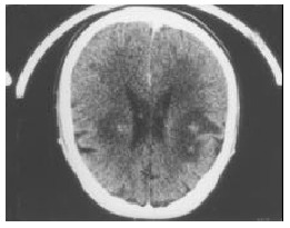
Computed tomography (CT) of the patient’s head revealed multiple intracranial masses and homogenous enhancement by post-contrast CT (Figure 1).
Figure 1.

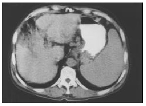
CT image of the lesion in the liver.
Abdominal sonography revealed findings consistent with chronic hepatitis. Thorax-abdomen-pelvic CT scan showed a hypodense mass lesion with irregular margins and 2.8 cm in diameter in the left lobe of the liver (Figure 2). Serum alpha-feto-protein level was higher than 400 U/L. Fine needle biopsy from the mass in the liver was performed. Pathological examination was consistent with the HCC.
Figure 2.

CT image of the metastatic lesions in brain.
He was given glucocorticoid therapy and referred to radiation oncology division for cranial radiotherapy.
DISCUSSION
Although brain metastasis may arise from primary sites in various organs and tissues, they are frequently seen with bronchogenic, breast, and prostate cancers. HCC commonly metastasizes to the lung, regional lymph nodes, peritoneum, and adrenal glands, but rarely to brain. Shuangshoti et al[14] reported that the secondary intracranial hepatic carcinomas were 1.3%-2.9% among intracranial metastatic tumors.
Most extrahepatic HCC occurs in patients at advanced intrahepatic tumor stage. Incidental extrahepatic lesions found at CT in patients having HCC of intrahepatic stage I or II are unlikely to represent metastatic HCC[2]. Our case had single nodule 2.8 cm in diameter. His bilirubin level was near normal. He did not have ascites. He had neither ascites nor history of hepatic encephalopathy. He had liver cirrhosis in Child A stage. If he had not had brain metastasis, he could have been candidate for curative hepatectomy. We believe that brain metastasis from HCC is relatively early in the disease progression.
Yen et al[16] reported a well documented group of 33 patients. Eighteen had brain parenchymal metastasis without skull involvement, the other 15 cases disclosed skull metastasis with brain invasion. The underlying HCC are mainly of expanding 39.4% and multifocal 39.4% types, and 54.5% had mental changes not related to hypoglycemia or hepatic encephalopathy. Nevertheless, our case had unifocal HCC.
Yen et al[16] reported that 90% of 15 cases had hyperdense mass lesion by non-contrast computed tomography scan and 17 cases showed homogenous enhancement (77.3%) by post-contrast CT images. In the non-skull involved group, 41.7% disclosed ring-shape enhancement and 87.5% had perifocal edema, and 24.2% presented as intracerebral hemorrhage. Our patient had perifocal edema in the brain but without hemorrhage.
Because no effective treatment for brain metastasis from HCC is available, further study is needed. Yen et al[16] reported 36.4% death of brain herniation.
Most of non-skull involved cases had simultaneous lung metastasis without bony metastasis, while the skull involved group (66.7%) disclosed extracranial bony metastasis without lung metastasis[15,16]. However, no other simultaneous metastasis site was found of our patient. HCC with intracranial metastasis is symptomatic and life threatening. Half the cases may come from pulmonary metastasis and the other half may be from bony metastasis even though our patient had neither of them. Surgery of the brain or skull metastasis is of no particular technical problem as long as they are located in accessible areas. Brain irradiation or surgery can prolong the survival. Radiotherapy seems to improve the quality and quantity of residual life, although the number of patients describes in the literature is not large enough to draw any definite conclusion.
Loo et al[12] reported that light microscopic examination of the metastatic tumor from HCC revealed a trabecular HCC with focal hemorrhage and necrosis. Their immunohistochemical profile was identical to that described in primary HCC.
Salvati et al[9] suggested that the stroke-like presentation of the cerebral localization of the disease can be explained by both the important vascularization of the tumor and the frequent hemocoagulative alterations caused by the cirrhosis.
In conclusion, the rarity of this type of case gives the clinician the suspicion of such associations when confronted with a patient with liver dysfunction and neurologic findings.
Footnotes
Edited by Ma JY Proofread by Xu FM
References
- 1.Katyal S, Oliver JH, Peterson MS, Ferris JV, Carr BS, Baron RL. Extrahepatic metastases of hepatocellular carcinoma. Radiology. 2000;216:698–703. doi: 10.1148/radiology.216.3.r00se24698. [DOI] [PubMed] [Google Scholar]
- 2.Tang ZY. Hepatocellular carcinoma--cause, treatment and metastasis. World J Gastroenterol. 2001;7:445–454. doi: 10.3748/wjg.v7.i4.445. [DOI] [PMC free article] [PubMed] [Google Scholar]
- 3.Sithinamsuwan P, Piratvisuth T, Tanomkiat W, Apakupakul N, Tongyoo S. Review of 336 patients with hepatocellular carcinoma at Songklanagarind Hospital. World J Gastroenterol. 2000;6:339–343. doi: 10.3748/wjg.v6.i3.339. [DOI] [PMC free article] [PubMed] [Google Scholar]
- 4.Moriya H, Ohtani Y, Tsukui M, Tanaka Y, Tajima T, Makuuchi H, Tanaka Y, Itou K. A case report: tumorectomy for brain metastasis of hepatocellular carcinoma. Tokai J Exp Clin Med. 1999;24:105–110. [PubMed] [Google Scholar]
- 5.Frati A, Salvati M, Giarnieri E, Santoro A, Rocchi G, Frati L. Brain metastasis from hepatocellular carcinoma associated with hepatitis B virus. J Exp Clin Cancer Res. 2002;21:321–327. [PubMed] [Google Scholar]
- 6.Endo M, Hamano M, Watanabe K, Wakai S. [Combined chronic subdural and acute epidural hematoma secondary to metastatic hepatocellular cancer: case report] No Shinkei Geka. 1999;27:331–334. [PubMed] [Google Scholar]
- 7.Peres MF, Forones NM, Malheiros SM, Ferraz HB, Stávale JN, Gabbai AA. Hemorrhagic cerebral metastasis as a first manifestation of a hepatocellular carcinoma. Case report. Arq Neuropsiquiatr. 1998;56:658–660. doi: 10.1590/s0004-282x1998000400023. [DOI] [PubMed] [Google Scholar]
- 8.Kim M, Na DL, Park SH, Jeon BS, Roh JK. Nervous system involvement by metastatic hepatocellular carcinoma. J Neurooncol. 1998;36:85–90. doi: 10.1023/a:1005716408970. [DOI] [PubMed] [Google Scholar]
- 9.Salvati M, Cimatti M, Frati A, Santoro A, Gagliardi FM. Brain metastases from hepatocellular carcinoma. A case report. J Neurosurg Sci. 2002;46:77–80; discussion 80. [PubMed] [Google Scholar]
- 10.Asahara T, Yano M, Fukuda S, Fukuda T, Nakahara H, Katayama K, Itamoto T, Dohi K, Nakanishi T, Kitamoto M, et al. Brain metastasis from hepatocellular carcinoma after radical hepatectomy. Hiroshima J Med Sci. 1999;48:91–94. [PubMed] [Google Scholar]
- 11.Tanabe H, Kondo A, Kinuta Y, Matsuura N, Hasegawa K, Chin M, Saiki M. Unusual presentation of brain metastasis from hepatocellular carcinoma--two case reports. Neurol Med Chir (Tokyo) 1994;34:748–753. doi: 10.2176/nmc.34.748. [DOI] [PubMed] [Google Scholar]
- 12.Loo KT, Tsui WM, Chung KH, Ho LC, Tang SK, Tse CH. Hepatocellular carcinoma metastasizing to the brain and orbit: report of three cases. Pathology. 1994;26:119–122. doi: 10.1080/00313029400169321. [DOI] [PubMed] [Google Scholar]
- 13.Friedman HD. Hepatocellular carcinoma with central nervous system metastasis: a case report and literature review. Med Pediatr Oncol. 1991;19:139–144. doi: 10.1002/mpo.2950190215. [DOI] [PubMed] [Google Scholar]
- 14.Shuangshoti S, Rungruxsirivorn S, Panyathanya R. Intracranial metastasis of hepatic carcinomas: a study of 9 cases within 28 years. J Med Assoc Thai. 1989;72:307–313. [PubMed] [Google Scholar]
- 15.Lee JP, Lee ST. Hepatocellular carcinoma presenting as intracranial metastasis. Surg Neurol. 1988;30:316–320. doi: 10.1016/0090-3019(88)90306-0. [DOI] [PubMed] [Google Scholar]
- 16.Yen FS, Wu JC, Lai CR, Sheng WY, Kuo BI, Chen TZ, Tsay SH, Lee SD. Clinical and radiological pictures of hepatocellular carcinoma with intracranial metastasis. J Gastroenterol Hepatol. 1995;10:413–418. doi: 10.1111/j.1440-1746.1995.tb01593.x. [DOI] [PubMed] [Google Scholar]


