Figure 7.
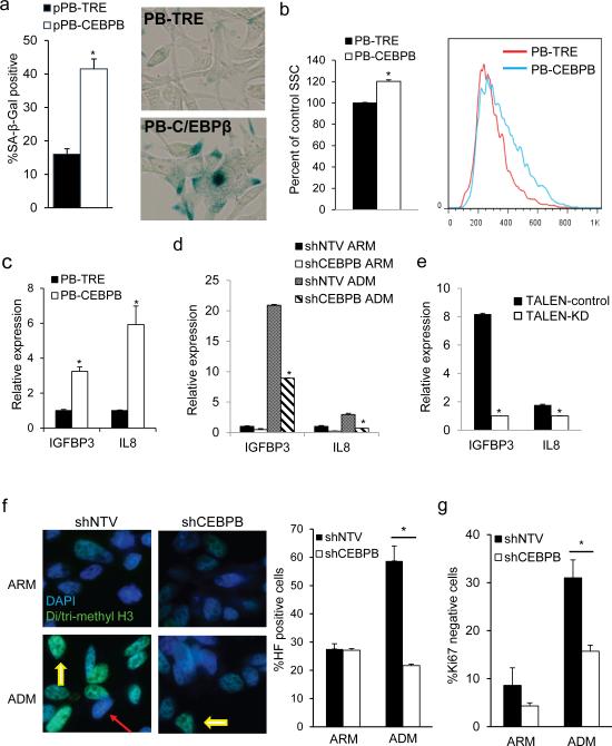
C/EBPβ is required for androgen deprivation induced senescence. (a) The percent of SA-β-gal-positive PB-TRE or PB-CEBPB cells was quantified after 5 days of culture in 0.5 μg/ml doxycycline. An average from 3 experiments (left) and representative images are shown (right). (b) Quantification of cell granularity by the median side scatter value of G1 PB-TRE and PBCEBPB cells 5 days after seeding in doxycycline. A representative histogram is shown on the right. (c) IL8 and IGFBP3 expression were quantified by qRT-PCR in PB-TRE and PB-CEBPB cells cultured under the above conditions. Conversely, total cellular RNA was extracted from shNTV or shCEBPB (d), or TALEN-control or TALEN-KD (e) LNCaP cells after 7 days in culture in ARM or ADM and transcript levels of IL8 and IGFBP3 were assessed by qRTPCR. (f) Expression of an shRNA against CEBPB or non-targeting vector (shNTV) control were induced by doxycycline in LNCaP cells cultured in androgen replete (ARM) or depleted media (ADM). After 7 days cells were stained for senescence associated heterochromatin foci (HF) (yellow arrows). Red arrow points to HF negative cell. Representative photomicrographs are shown (left, 600X magnification). The average and standard error of three independent counts is shown (right panel). *p<0.001. (g) Ki67 was quantified using flow cytometry in shNTV or shCEBPB LNCaP cells cultured in ARM or ADM for 7 days and the average proportion of Ki67 negative cells from 3 experiments is presented. *p<0.01.

