Abstract
Introduction
Gastrocutaneous fistulas remain an uncommon complication of upper gastrointestinal surgery. Less common but equally problematic are gastrocutaneous fistulas secondary to non-healing gastrostomies. Both are associated with considerable morbidity and mortality. Surgical repair remains the gold standard of care. For those unfit for surgical intervention, results from conservative management can be disappointing. We describe a case series of seven patients with gastrocutaneous fistulas who were unfit for surgical intervention. These patients were managed successfully in a minimally invasive manner using the Surgisis® (Cook Surgical, Bloomington, IN, US) anal fistula plug.
Methods
Between September 2008 and January 2009, seven patients with gastrocutaneous fistulas presented to Wishaw General Hospital. Four gastrocutaneous fistulas represented non-healing gastrostomies, two followed an anastomotic leak after an oesophagectomy and one following an anastomotic leak after a distal gastrectomy. All patients had poor nutritional reserve with no other identifiable reason for failure to heal. All were deemed unfit for surgical intervention. Five gastrocutaneous fistulas were closed successfully using the Surgisis® anal fistula plug positioned directly into the fistula tract under local anaesthesia and two gastrocutaneous fistulas were closed successfully using the Surgisis® anal fistula positioned endoscopically using a rendezvous technique.
Results
For the five patients with gastrocutaneous fistulas closed directly under local anaesthesia, oral alimentation was reinstated immediately. Fistula output ceased on day 12 with complete epithelialisation occurring at a median of day 26. For the two gastrocutaneous fistulas closed endoscopically using the rendezvous technique, oral alimentation was reinstated on day 5 with immediate cessation of fistula output. Follow-up upper gastrointestinal endoscopy confirmed re-epithelialisation at eight weeks. In none of the cases has there been fistula recurrence (range of follow-up duration: 30–59 months).
Conclusions
Surgisis® anal fistula plugs can be used safely and effectively to close gastrocutaneous fistulas in a minimally invasive manner in patients unfit for surgical intervention.
Keywords: Gastrocutaneous fistula, Surgisis® anal fistula plug
The incidence of clinically significant anastomotic leaks following upper gastrointestinal surgery is reported as ranging from 4% to 20%.1 The resultant oesophagocutaneous and gastrocutaneous fistulas are associated with significant morbidity and a mortality rate of 80%.1 Less common but equally problematic are gastrocutaneous fistulas secondary to non-healing gastrostomies. Depending on the clinical presentation, treatment varies. Surgery provides definitive repair and remains the gold standard of care for septic patients and for those who have failed conservative management. Conservative management with external drainage can offer success in the absence of sepsis. However, for those with sepsis or for those who have failed conservative management and are not fit for surgery, treatment options remain limited.
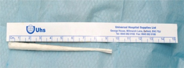
A number of minimally invasive therapies, primarily endoscopic therapies, have been described to date but with limited success. In contrast, we have described the successful use of the Surgisis® (Cook Surgical, Bloomington, IN, US) anal fistula plug (Fig 1) to close three oesophagocutaneous fistulas following minimally invasive endoscopic therapy for the management of Boerhaave’s syndrome.2 In the present paper, we describe a similar success using the Surgisis® anal fistula plug in a minimally invasive manner to close seven gastrocutaneous fistulas.
Figure 1.

Surgisis® anal fistula plug
Methods
Seven patients with gastrocutaneous fistulas presented to Wishaw General Hospital between November 2008 and January 2010 (Table 1). Four of these (at a median of 8 months) following removal of a percutaneous endoscopic gastrostomy (PEG) tube, two following an anastomotic leak from the gastric tube after an oesophagectomy and one following an anastomotic leak after a distal gastrectomy.
Table 1.
Patients with gastrocutaneous fistulas
| Case | Age / sex | Co-morbidity | Morbidity |
|---|---|---|---|
| 1 | 48 M | Chronic pancreatitis Pancreatic insufficiency Choledocholithiasis Ethanol excess Smoker |
Non-healing percutaneous endoscopic gastrostomy |
| 2 | 61 F | Multiple sclerosis Colorectal cancer |
Non-healing percutaneous endoscopic gastrostomy |
| 3 | 42 M | Multiple sclerosis | Non-healing percutaneous endoscopic gastrostomy |
| 4 | 63 M | Chronic obstructive pulmonary disease Ethanol excess Smoker |
Non-healing percutaneous endoscopic gastrostomy |
| 5 | 59 M | Gastric cancer Ethanol excess Smoker |
Anastomotic leak following distal gastrectomy |
| 6 | 52 M | Oesophageal cancer Chronic obstructive pulmonary disease Smoker |
Anastomotic leak following oesophagectomy |
| 7 | 64 F | Oesophageal cancer Ischaemic heart disease Smoker |
Anastomotic leak following oesophagectomy |
In all seven cases, there was no demonstrable endoscopic or radiological evidence of disease in the tissues or of a distal obstruction. All were, however, nutritionally deplete with medical co-morbidity and, in some cases, a history of smoking or alcohol excess. All cases were deemed not fit for formal surgical repair.
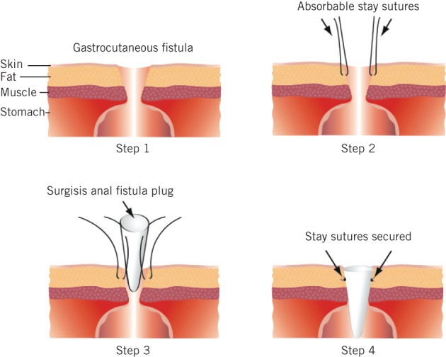
Five gastrocutaneous fistulas were closed directly under local anaesthesia (Fig 2). This cohort included the four non-healing gastrostomies and the resultant gastrocutaneous fistula following distal gastrectomy.
Figure 2.

The four steps used in direct closure of five gastrocutaneous fistulas under local anaesthesia
Step 1: The area of the gastrocutaneous fistula was prepared and draped with povidone-iodine. Local anaesthesia was instilled around the margins of the fistula to produce a field block and an incision was made along the gastrocutaneous junction of the fistula to free the skin edges from the edge of the fistula tract.
Step 2: Using 2/0 PDS® (Ethicon, Somerville, NJ, US), two opposing stay sutures were applied to the internal aspect of the fistula tract.
Step 3: The Surgisis® anal fistula plug was moistened with water as per the manufacturer’s instructions and its tip trimmed to the estimated length of the fistula. The Surgisis® anal fistula plug was incorporated into the stay sutures.
Step 4: The stay sutures were tied, lowering the Surgisis® anal fistula plug down into the gastrocutaneous fistula. The skin edges were left open over the fistula to prevent abscess formation and the wound was dressed accordingly. Antisecretories in the form of proton pump inhibitors were prescribed to reduce gastric output. Oral alimentation was reintroduced immediately in four patients and via one patient’s second PEG tube. In the postgastrectomy patient, feeding was continued via his percutaneous jejunostomy until oral intake became adequate unsupported.
It was not possible to close the two gastrocutaneous fistulas arising after an oesophagectomy directly under local anaesthesia. These fistulas had to be closed by siting the Surgisis® anal fistula plug endoscopically using the rendezvous technique. We have described this technique previously, whereby we successfully closed three oesophagocutaneous fistulas in this manner.2 The demonstration and explanation of this technique will not be repeated in the present paper. However, of note is the fact that the draining chest drains occupying the lateral aspect of the gastrocutaneous fistulas were left in situ for a period of 48 hours to ensure that fistula output had ceased. Feeding was continued via feeding jejunostomies, sited routinely at oesophagectomy. A water soluble oral contrast study was performed to confirm there was no ongoing anastomotic leak, following which oral alimentation was reintroduced on day 5. Jejunal feeding was subsequently reduced and antisecretories were again prescribed to reduce gastric output.
Results
The five patients who underwent direct repair of their gastrocutaneous fistula under local anaesthesia were allowed immediate reintroduction of oral alimentation, which was supported by jejunal feeding in one case and by PEG feeding in another. Ultimately, only the patient who had previously required long-term PEG feeding continued to be fed other than orally without further nutritional support. Fistula output ceased on a median of day 12 with complete epithelialisation occurring on a median of day 26. In none of these five cases has there been fistula recurrence (range of follow-up duration: 30–59 months). No upper gastrointestinal endoscopy was required to confirm mucosal integrity in these short fistulas.
The 2 patients who underwent repair of their gastrocutaneous fistula endoscopically using the rendezvous technique had an immediate cessation of fistula output, enabling the chest drain to be withdrawn at 48 hours. Jejunal feeding continued as per the postoesophagectomy protocol and oral alimentation was reintroduced on day 5 following receipt of a ‘normal’ oral water soluble contrast study. Follow-up upper gastrointestinal endoscopy at 8 weeks confirmed oesophageal integrity and in neither of these two cases has there been fistula recurrence (range of follow-up duration: 30–59 months).
Discussion
The use of both minimally invasive therapies and minimally invasive endoscopic therapies to treat patients with non-healing oesophagocutaneous and gastrocutaneous fistulas continues to expand. It offers patients not fit for surgical intervention an alternative to traditional therapies in a cohort of patients where conservative measures have failed and no other real treatment option exists.
Alternate therapies to date include the use of Vicryl® (Ethicon, Somerville, NJ, US) plugs and fibrin glue,1 over-the-scope clips3,4 and the use of 2-octyl cyanoacrylate.5 All reports are limited primarily to case studies, and carry limited and varied success.
The Surgisis® anal fistula plug is an advanced tissue repair graft made from porcine submucosa. It represents the first minimally invasive alternative to formal fistula-in-ano surgery, recommended as a first line intervention for simple fistula-in-ano.6–8 Innovators from the US have already described the use of the Surgisis® anal fistula plug as a minimally invasive therapy for the management of fistulas not in the perineum. Wood et al described the successful closure of a solitary persistent gastrocutaneous fistula following removal of a PEG tube with a porcine fistula plug9 and Paul et al describe similar success closing a solitary bronchopleural fistula following a partial pneumonectomy.10 We have also described the successful use of the Surgisis® anal fistula plug in closing three oesophagocutaneous fistulas as part of minimally invasive endoscopic therapy for the management of Boerhaave’s syndrome.2 To date, however, these reports remain isolated.
In our cohort, the principal reason for failure to heal a gastrocutaneous fistula was malnutrition, secondary to either the cachexia of chronic disease or catabolism associated with an acute surgical assault. In all seven of our patients, there was no clinical, endoscopic or radiological evidence of disease in the tissues at the site of the fistula, ongoing sepsis or distal obstruction. All had a failed period of conservative management with external drainage and all were regarded as unfit for surgical intervention. The use of the Surgisis® anal fistula plug offered our patients a feasible therapeutic intervention that could be carried out successfully in a minimally invasive manner with low morbidity, where no other described therapy to date has been shown to be consistently successful.
Conclusions
We again demonstrate the successful use of the Surgisis® anal fistula plug in closing fistulas out with of the perineum and, in particular, the repeat success of the endoscopic rendezvous technique. We describe a durable, minimally invasive therapy that can be performed successfully with low morbidity and mortality to close gastrocutaneous fistulas in a cohort of patients unfit for surgical intervention, where other described techniques have proven to be inconsistent.
References
- 1.Böhm G, Mossdorf A, Klink C et al. Treatment algorithm for postoperative upper gastrointestinal fistulas and leaks using combined Vicryl plug and fibrin glue. Endoscopy 2010; 42: 599–602. [DOI] [PubMed] [Google Scholar]
- 2.Darrien JH, Kasem H. Minimally invasive endoscopic therapy for the management of Boerhaave’s syndrome. Ann R Coll Surg Engl 2013; 95: 552–556. [DOI] [PMC free article] [PubMed] [Google Scholar]
- 3.Turner JK, Hurley JJ, Ketchell I, Dolwani S. Over-the-scope clip to close a fistula after removing a percutaneous endoscopic gastrostomy tube. Endoscopy 2010; 42: E197–E198. [DOI] [PubMed] [Google Scholar]
- 4.Conio M, Blanchi S, Repici A et al. Use of an over-the-scope clip for endoscopic sealing of a gastric fistula after sleeve gastrectomy. Endoscopy 2010; 42: E71–E72. [DOI] [PubMed] [Google Scholar]
- 5.Lukish J, Marmon L, Burns C. Nonoperative closure of persistent gastrocutaneous fistulas in children with 2-octylcyanoacrylate. J Laparoendosc Adv Surg Tech A 2010; 20: 565–567. [DOI] [PubMed] [Google Scholar]
- 6.Schwandner T, Roblick MH, Kierer W et al. Surgical treatment of complex anal fistulas with the anal fistula plug: a prospective, multicenter study. Dis Colon Rectum 2009; 52: 1,578–1,583. [DOI] [PubMed] [Google Scholar]
- 7.Ky AJ, Sylla P, Steinhagen R et al. Collagen fistula plug for the treatment of anal fistulas. Dis Colon Rectum 2008; 51: 838–843. [DOI] [PubMed] [Google Scholar]
- 8.Christoforidis D, Etzioni DA, Goldberg SM et al. Treatment of complex anal fistulas with the collagen fistula plug. Dis Colon Rectum 2008; 51: 1,482–1,487. [DOI] [PubMed] [Google Scholar]
- 9.Wood J, Leong S, McCarter M et al. Endoscopic-assisted closure of persistent gastrocutaneous fistula with a porcine fistula plug: report of a new technique. Surg. Innov 2010; 17: 53–56. [DOI] [PubMed] [Google Scholar]
- 10.Paul S, Talbot SG, Carty M et al. Bronchopleural fistula repair during Clagett closure utilizing a collagen matrix plug. Ann Thorac Surg 2007; 83: 1,519–1,521. [DOI] [PubMed] [Google Scholar]


