Fig. 1.
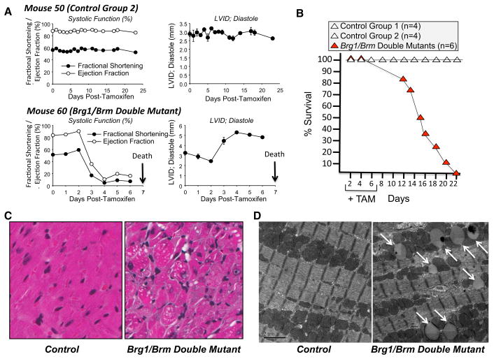
The phenotype of Brg1/Brm double-mutant mice. a Kaplan–Meier survival curve of mice following the administration of tamoxifen (+TAM) on days 1 through 7. b Longitudinal echo time course of an example control group 2 (Brg1floxed/floxed; αMHC-Cre-ERT0/0(no transgene); Brm−/− mice plus tamoxifen treatment) and a Brg1/Brm double mutant (Brg1floxed/floxed; αMHC-Cre-ERT+/0; Brm−/− mice plus tamoxifen treatment) illustrating the rapid decline in systolic function and dilation upon deletion of Brg1 with tamoxifen induction (via chow). c Representative H&E-stained heart sections from control (left) and double mutant (right) mice at ×200 magnification. d Representative transmission electron micrographs of cardiomyocytes from control (left) and double mutant (right) mice at ×5000 magnification. Illustrating accumulation of vacuoles in the mitochondria interspersed in the sarcomere (white arrows) that were found adjacent to fragmented mitochondria

