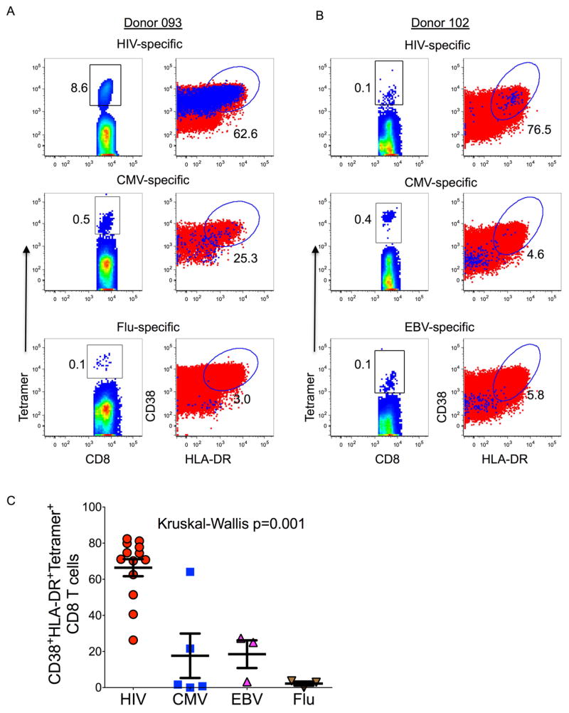
Figure 6. Assessment of bystander CD8+ T cell activation during hyperacute HIV infection.
Intra-donor activation (CD8+ CD38+ HLA-DR+ cells) profiles of HIV, CMV, EBV and influenza virus specific (tetramer+) CD8+ T cells. Data for two donors with detectable tetramer+ cells specific for three different pathogens are shown in panels (a) and (b). The first columns show flow plots gated on tetramer+ cells, the second column shows flow plots gated on tetramer+ cells that are double positive for CD38 and HLA-DR (blue dots) overlaid on total CD8+ T cells (red dots). (c) Activation data for 13 HIV, 5 CMV, 3 EBV and 3 influenza specific tetramer+ cells are graphed. Statistical significance was determined using Kruskal Wallis test.

