

Abstract
To gain insights into the diversification trajectories of qnrB genes, a phylogenetic and comparative genomics analysis of these genes and their surrounding genetic sequences was performed. For this purpose, Citrobacter sp. isolates (n = 21) and genome or plasmid sequences (n = 56) available in public databases harboring complete or truncated qnrB genes were analyzed. Citrobacter species identification was performed by phylogenetic analysis of different genotypic markers. The clonal relatedness among isolates, the location of qnrB genes, and the genetic surroundings of qnrB genes were investigated by pulsed-field gel electrophoresis (PFGE), S1-/I-CeuI-PFGE and hybridization, and PCR mapping and sequencing, respectively. Identification of Citrobacter isolates was achieved using leuS and recN gene sequences, and isolates characterized in this study were diverse and harbored chromosomal qnrB genes. Phylogenetic analysis of all known qnrB genes revealed seven main clusters and two branches, with most of them included in two clusters. Specific platforms (comprising pspF and sapA and varying in synteny and/or identity of other genes and intergenic regions) were associated with each one of these qnrB clusters, and the reliable identification of all Citrobacter isolates revealed that each platform evolved in different recognizable (Citrobacter freundii, C. braakii, C. werkmanii, and C. pasteurii) and putatively new species. A high identity was observed between some of the platforms identified in the chromosome of Citrobacter spp. and in different plasmids of Enterobacteriaceae. Our data corroborate Citrobacter as the origin of qnrB and further suggest divergent evolution of closely related qnrB genes/platforms in particular Citrobacter spp., which were delineated using particular genotypic markers.
INTRODUCTION
The qnrB genes constitute the most prevalent and diverse (>70 allelic variants; see http://www.lahey.org/qnrStudies/) group within the qnr family, encoding proteins responsible for decreased susceptibility to fluoroquinolones (1–4).
Some authors have proposed Citrobacter spp. as the origin of qnrB genes, mainly based on species distribution (>60% in Citrobacter spp., including isolates from the preantibiotic era), location (mostly on the chromosome), and the apparent absence of mobile genetic elements in the immediate genetic environment of qnrB genes, mostly by characterization of clinical Citrobacter sp. isolates (3–5). Nevertheless, the absence of correlation of qnrB genes with particular Citrobacter species, together with the lack of detailed characterization of qnrB platforms, hinders a clear establishment of the origin of qnrB. In fact, most of the methods conventionally used to identify Citrobacter spp. (biochemical or phenotypic features, matrix-assisted laser desorption ionization–time of flight mass spectrometry [MALDI-TOF MS], or 16S rRNA gene sequencing) (6–8) have low discriminatory power, hindering the accurate discrimination of these species.
Recently, Clermont et al. described a multilocus sequence analysis (MLSA) based on partial sequences of rpoB (β subunit of RNA polymerase gene), pyrG (CTP synthetase gene), fusA (protein synthesis elongation factor-G gene), and leuS (leucine tRNA synthetase gene) that allowed the discrimination of the 12 recognized Citrobacter species, namely, Citrobacter freundii, C. amalonaticus, C. braakii, C. farmeri, C. gillenii, C. koseri, C. murliniae, C. rodentium, C. sedlakii, C. werkmanii, C. youngae, and C. pasteurii (6).
In this work, we aim to gain insights into the diversification trajectories of qnrB within Citrobacter species and to unveil qnrB surroundings possibly involved in the dissemination of this gene to other Enterobacteriaceae. For that purpose, we performed an affiliation of Citrobacter species and qnrB genes described to date and a comparative analysis of genetic sequences surrounding qnrB using nonclinical Citrobacter sp. isolates and genome and plasmid sequences deposited in public databases.
MATERIALS AND METHODS
Bacterial isolates.
Twenty-one nonclinical Citrobacter sp. isolates harboring qnrB genes recovered from different nonclinical origins, including untreated waters used for human consumption (n = 12; 2006 to 2008), ready-to-eat salads (n = 3; 2010), and trout aquaculture samples (trout, feed, and sediments from a river located upstream of the trout farm) (n = 6; 2010 to 2012) from different geographic regions in Portugal, were included in this study (see Table S1 in the supplemental material). The isolates carried qnrB6 (n = 1), qnrB9 (n = 1), qnrB10 (n = 3), qnrB17 (n = 1), qnrB18 (n = 1), qnrB56 (n = 3), qnrB57 (n = 2), qnrB58 (n = 1), qnrB59 (n = 3), qnrB72 (n = 2), qnrB73 (n = 1), or truncated qnrB (ΔqnrB; n = 2) genes (9; P. Antunes, E. Machado, and L. Peixe, unpublished data) (see Table S1 in the supplemental material).
In addition, 40 Citrobacter sp. genomes and 16 qnrB-carrying plasmid sequences available from the Pathosystems Resource Integration Center (PATRIC) and/or the National Center for Biotechnology Information (NCBI) database were used for phylogenetic analysis and/or qnrB genetic surrounding comparisons.
Bacterial identification and phylogenetic analysis.
Isolates included in this study were identified by biochemical methods (7), mass spectrometry (MALDI-TOF MS; Bruker Daltonik, Germany), and sequencing of 16S rRNA (8), leuS (leucine tRNA synthetase) (6), and recN (DNA repair protein) genes. PCR amplification and further sequencing of recN genes were performed by using primers recN-Fw (5′-ATTGCCATTGATGCTCTCGG-3′) and recN-Rv (5′-ANCGAGTCGGCCTGATCGT-3′) to amplify a 637-bp internal fragment and the following amplification conditions: one cycle of 3 min at 95°C; 35 cycles of 1 min at 95°C, 1 min at 56°C, and 1 min at 72°C; and 1 cycle of 1 min at 72°C.
Individual nucleotide sequences of genes included in the MLSA scheme of the Citrobacter genus (rpoB, pyrG, fusA, and leuS) (6) and recN were aligned and the average rates of similarity calculated using MEGA version 5.2.2 (http://www.megasoftware.net/) (10). The leuS gene sequences from Clermont et al. were included in this analysis (6). Similarity scores of the leuS and recN genes were calculated and individual phylogenetic trees were constructed in MEGA using the neighbor-joining (NJ) method (11), and genetic distances were calculated using the Kimura two-parameter model (12) in the case of nucleotide sequences and using the Jones-Taylor-Thornton (JTT) model (13) for LeuS and RecN protein sequences. The reliability of internal branches was assessed from bootstrap based on 1,000 resamplings (14). Pantoea ananatis strain LMG 2665T was used as the outgroup.
Clonal relatedness.
Clonal relationships among isolates belonging to the same species were established by pulsed-field gel electrophoresis (PFGE), using XbaI as a restriction enzyme and the following electrophoresis conditions: 10 to 40 s for 21 h at 14°C and 6 V/cm2 (15). The criteria of Tenover et al. were used for comparison of band patterns obtained by PFGE, and isolates representing different PFGE-types were selected for the following studies (16).
Location, transferability, and phylogenetic analysis of qnrB genes.
Location of qnrB genes was assessed by S1-/I-CeuI-PFGE and further hybridization with qnrB and 16S rRNA probes (17, 18). Conjugative transfer of qnrB was evaluated by broth and filter mating assays using Escherichia coli HB101 (streptomycin and azide resistant) as the recipient at a 1:2 donor-to-recipient ratio and selection plates containing ciprofloxacin (0.06 to 0.5 μg/ml) plus sodium azide (130 μm/ml) (19).
Affiliation within all qnrB genes described at the time of study design (n = 74; http://www.lahey.org/qnrStudies/) was generated as specified above for leuS and recN phylogenetic analysis.
Characterization of genetic surroundings of the qnrB genes.
The genetic context of qnrB genes was characterized by PCR mapping (pspF, sapA, intI1, intI2, intI3, ISEcp1, IS3000, ISCR1, IS26) and sequencing based on previously described sequences (3, 20–22). Sequences surrounding qnrB were further aligned and compared in silico with those deposited in the GenBank database using BLAST (http://blast.ncbi.nlm.nih.gov.sci-hub.org/Blast.cgi) and ClustalW2 (http://www.ebi.ac.uk/Tools/msa/clustalw2/).
Nucleotide sequence accession numbers.
The sequences of the genetic platforms associated with the different qnrB alleles characterized in this study have been deposited in the GenBank database under the accession numbers KP339254 (qnrB6), KP339255 (qnrB9), KP339256 (qnrB10), KP339257 (qnrB17), KP339258 (qnrB18), KP339259 (qnrB56), KP339260 (qnrB57), KP339261 (qnrB58), KP339262 (qnrB59), KP339263 (qnrB72), and KP339264 (qnrB73). recN and leuS nucleotide sequence data from the different Citrobacter sp. isolates identified in this study are available in the GenBank database under accession numbers KR998019 (Citrobacter sp. I), KR998020 (Citrobacter sp. I), KR998021 (Citrobacter sp. III), KR998022 (Citrobacter sp. I), KR998023 (C. braakii), KR998024 (Citrobacter sp. I), KR998025 (Citrobacter sp. I), KR998026 (C. freundii), KR998027 (C. freundii), KR998028 (Citrobacter sp. I), KR998029 (Citrobacter sp. I), KR998030 (C. braakii), KR998031 (C. braakii), KR998032 (Citrobacter sp. I), KR998033 (Citrobacter sp. I), KR998034 (Citrobacter sp. III), KR998035 (Citrobacter sp. I), KR998036 (C. braakii), KR998037 (Citrobacter sp. I), KR998038 (Citrobacter sp. I), KR998039 (C. freundii), KR998040 (C. freundii), KR998041 (Citrobacter sp. I), KR998042 (Citrobacter sp. I), KR998043 (C. braakii), and KR998044 (C. braakii).
RESULTS AND DISCUSSION
Citrobacter species identification and clonality.
The identification at the species level of all the Citrobacter sp. isolates included in this study was not possible by biochemical methods, MALDI-TOF MS, or sequencing of the 16S rRNA gene (data not shown), as previously recognized (6, 23). In contrast, analysis of leuS and recN gene sequences provided an accurate discrimination of the currently recognized Citrobacter species, as explained below.
The leuS gene presented the highest discriminatory power (average rate of similarity close to 88.5%, statistically supported) of the genes included in the MLSA scheme proposed by Clermont et al. (6). Therefore, the leuS-based phylogenetic tree allowed the delineation of 12 distinct clusters (Fig. 1), each one supported by a type strain from each Citrobacter species, corroborating the topology obtained by the concatenated affiliation of the MLSA scheme (6). These clusters were defined with a cutoff value of <97.5%, supported by bootstrap values greater than 92% (Fig. 1).
FIG 1.
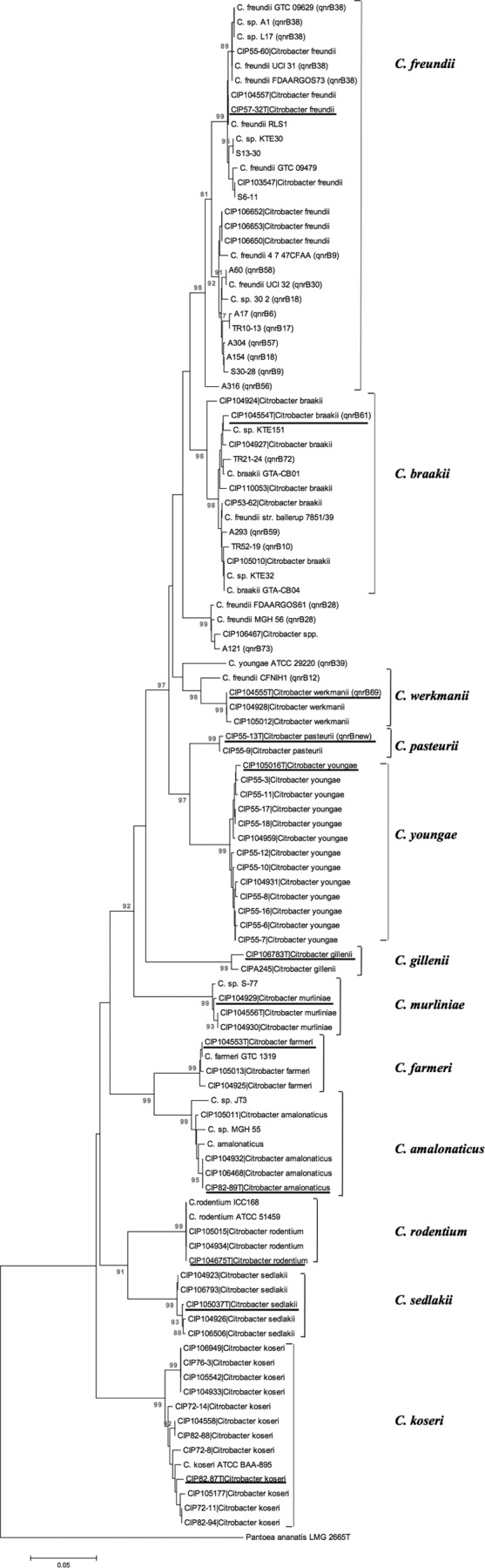
Neighbor-joining (NJ) tree based on the comparison of leuS gene sequences of Citrobacter species analyzed in this study. Genetic distances were constructed using Kimura's two-parameter model. Numbers at branch points indicate bootstrap percentages (1,000 replications) from NJ analysis, and only values greater than 80% are shown. Horizontal bar, genetic distance of 0.05. Citrobacter species type strains are underlined, and the qnrB alleles are shown in parentheses. Pantoea ananatis strain LMG 2665T was used as the outgroup (PATRIC fig|1378093.3.peg.2577). Please refer to Table S2 in the supplemental material for accession numbers of the sequences used.
The recN gene provided a greater resolution than leuS, presenting an average rate of similarity close to 85.6%. The recN tree topology was overall congruent with that obtained for leuS sequences (Fig. 2), with the presence of the same 12 clusters observed (cutoff values of <96.1% statistically supported by bootstrap values greater than 94%) supported by sequences from the available type strains. Interestingly, 3 new clusters were observed, namely, Citrobacter sp. I (n = 10), Citrobacter sp. II (n = 1), and Citrobacter sp. III (n = 3), which might correspond to isolates from novel species (Fig. 2). Citrobacter sp. I presented a genetic distance of 0.071 (bootstrap value of 97%) with its closest related species C. freundii, whereas Citrobacter sp. II and Citrobacter sp. III presented genetic distances of 0.081 (bootstrap value of 99%) and 0.073 (bootstrap value of 100%) with the closest related species C. werkmanii and C. braakii, respectively. Further studies are in progress to clearly establish the identity of the isolates included in these clusters.
FIG 2.

Neighbor-joining (NJ) tree based on the comparison of recN gene sequences of all Citrobacter species analyzed in this study. Genetic distances were constructed using Kimura's two-parameter model. Numbers at branch points indicate bootstrap percentages (1,000 replications) from NJ analysis, and only values greater than 80% are shown. Horizontal bar, genetic distance of 0.05. Citrobacter species type strains are underlined, and qnrB alleles are shown in parentheses. *, Citrobacter sp. I, Citrobacter sp. II, and Citrobacter sp. III correspond to putative novel species. Pantoea ananatis strain LMG 2665T was used as the outgroup (PATRIC fig|1378093.3.peg.2577). Please refer to Table S2 in the supplemental material for accession numbers of the sequences used.
Phylogenetic trees constructed based on amino acid sequences of LeuS and RecN showed that most nucleotide substitutions were synonymous, despite resulting in a less clear delineation between species due to the higher conservative character of amino acid sequences (see Fig. S1 and S2 in the supplemental material).
According to our phylogenetic analysis, Citrobacter sp. isolates characterized in this study were identified as C. braakii (n = 83 PFGE types), C. freundii (n = 22 PFGE types), and putatively two novel species (Citrobacter sp. I [n = 107 PFGE types] or Citrobacter sp. III [n = 11 PFGE types]) (see Table S1 in the supplemental material).
Location and affiliation of qnrB genes.
No plasmids were detected in any of the Citrobacter isolates included in this study, and in all cases, qnrB was chromosomally located and not transferable by conjugation, further supporting the natural occurrence of this gene in the chromosome of Citrobacter spp. (3–5). The qnrB gene diversity found was in accordance with previous data (24, 25), probably driven by the interplay of different selective events (natural recombination events and/or alternative selective forces) (1, 26–28).
The phylogenetic tree constructed based on qnrB gene sequences (Fig. 3A) revealed seven distinct clusters (I to VII) and two branches comprising qnrB39 and a new qnrB (C. pasteurii strain CIP 55-13T), supported by bootstraps of ≥92% and sharing ≤92.83% identity between them. The corresponding affiliation based on amino acid sequences of QnrB showed that most nucleotide substitutions were synonymous, which resulted in a similar tree topology (see Fig. S3 in the supplemental material), with some exceptions consisting of genes showing a higher degree of nucleotide divergence (qnrB31, qnrB53, or qnrB39), as observed by other authors for blaCTX-M genes (29). Our phylogenetic analysis also showed that most of the qnrB genes, including those characterized in this study, belonged to cluster I (n = 33, including qnrB6, qnrB9, qnrB17, qnrB18, qnrB57, and qnrB58) or to cluster III (n = 18, including qnrB10, qnrB56, qnrB59, and qnrB72), whose diversification might be favored by their association with particular host species and/or niches (see below). Few qnrB genes were enclosed in cluster II (n = 2), IV (n = 4, including qnrB73), V (n = 6), VI (n = 4), or VII (n = 5).
FIG 3.

Affiliation of qnrB genes and qnrB genetic platforms from Citrobacter spp. (A) Neighbor-joining tree based on 74 qnrB gene sequences (http://www.lahey.org/qnrStudies/). Genetic distances were constructed using the Kimura 2-parameter model. Numbers at branch points indicate bootstrap percentages (1,000 replications) from NJ analysis, and only values greater than 80% are shown. Horizontal bar, genetic distance of 0.05. The nucleotide sequence of qnrD1 (GenBank accession number FJ228229) was used as the outgroup. The qnrB genes for which the genetic environment was first characterized in this study are surrounded by circles, whereas those available in the GenBank database are underlined. pl, plasmid-borne qnrB; cr, chromosomally located qnrB; *, qnrB location not assessed. (B) Schematic representation of the genetic platforms (GP) carrying chromosomally located qnrB genes. Numbers between ORFs indicate the size of the intergenic region in base pairs (bp). Vertical black bars represent IRR2. Genes identified in qnrB platforms are pspF (encoding a phage shock protein), orf2 (open reading frame of a gene of unknown function), sdr (encoding a short-chain dehydrogenase/reductase protein), cinA (encoding competence/damage-inducible domain protein), HP (encoding a hypothetical protein), ppp (encoding putative periplasmic protein), and/or sapA (encoding a protein involved in antimicrobial peptide resistance). Genetic platforms have been deposited in the GenBank database under accession numbers KP339254 (qnrB6), KP339255 (qnrB9), ADLG01000026.1 (qnrB9), CP007557 (qnrB12), KP339256 (qnrB10), KP339257 (qnrB17), KP339258 (qnrB18), ACDJ02000027.1 (qnrB18), JMUJ01000007.1 (qnrB28), JTBV01000001.1 (qnrB28), JAPA01000008.1 (qnrB30), JN173057 (qnrB35), JN173060 (qnrB38), NZ_AMPE01000004.1 (qnrB38), NZ_AKTT01000018.1 (qnrB38), NZ_AOUE01000004.1 (qnrB38), JTBJ01000001.1 (qnrB38), JAPB01000002.1 (qnrB38), ABWL02000005.1 (qnrB39), KP339259 (qnrB56), KP339260 (qnrB57), KP339261 (qnrB58), KP339262 (qnrB59), AB734055 (qnrB60), AB734053 (qnrB61), BBMW01000005.1 (qnrB69), KP339263 (qnrB72), KP339264 (qnrB73), and CDHL01000019 (new qnrB from CIP 55-13T).
Detailed characterization of qnrB genetic platforms.
Analysis of the genetic surroundings of complete qnrB genes revealed eight different qnrB genetic platforms (GP1 to GP8) (Fig. 3B). pspF (encoding a phage shock protein) and sapA (encoding a protein involved in antimicrobial peptide resistance) genes were consistently found upstream and downstream of qnrB genes, respectively. A high variability was observed mostly downstream of qnrB, with differences in the presence of other genes (orf2, cinA, HP, and/or ppp) and in the size and identity of intergenic regions (IGRs) upstream and downstream of qnrB (Fig. 3B). Interestingly, we observed conserved genetic platforms (gene content and sequence identity) for closely related qnrB genes (i.e., those grouped in the same cluster), with an exception in cluster I, possibly explained by a recombination event (Fig. 3).
As the characterization of IGRs was important to elucidate the origin and evolutionary routes of other antibiotic resistance genes (29, 30), we performed a detailed analysis of IGRs located in the qnrB genetic environment. In fact, the intergenic regions upstream of qnrB (IGR-1) were closely related (in size and in nucleotide sequence) among qnrB alleles that were grouped in the same cluster (identity, >96%) (Fig. 4), including those from cluster I (see above), whereas they exhibited a loss of identity between clusters (identity, 60% to 85%). This IGR-1 encompassed a LexA box consensus sequence located upstream of qnrB and downstream −35 and −10 promoter sequences (Fig. 4), which might directly regulate the expression of qnrB genes, as previously suggested (31, 32).
FIG 4.

Nucleotide sequence alignment of intergenic regions upstream of chromosomally located qnrB. The −35 and −10 promoters are indicated by gray shading, and the sequence of the LexA box is boxed. Sequences were aligned using ClustalW2 software (http://www.ebi.ac.uk/Tools/msa/clustalw2/). The IGR-1 sequences represented in this figure are found in the GenBank database through the accession numbers KP339254 (qnrB6), KP339255 (qnrB9), KP339256 (qnrB10), CP007557 (qnrB12), KP339257 (qnrB17), KP339258 (qnrB18), JMUJ01000007.1 (qnrB28), JAPA01000008.1 (qnrB30), JN173057 (qnrB35), JN173060 (qnrB38), ABWL02000005.1 (qnrB39), KP339259 (qnrB56), KP339260 (qnrB57), KP339261 (qnrB58), KP339262 (qnrB59), AB734055 (qnrB60), AB734053 (qnrB61), BBMW01000005.1 (qnrB69), KP339263 (qnrB72), KP339264 (qnrB73), and CDHL01000019 (new qnrB from CIP 55-13T).
Interestingly, taking into consideration the similarity of the platforms carrying closely related qnrB genes and the identification of Citrobacter isolates carrying each qnrB, an association was found between each particular qnrB platform and specific Citrobacter species. The qnrB cluster I was associated with Citrobacter sp. I, qnrB cluster III with C. braakii, qnrB cluster IV with Citrobacter sp. III, qnrB cluster V with C. freundii, qnrB cluster VII with C. werkmanii, the branch comprising qnrB39 with Citrobacter sp. II, and finally the branch comprising the new qnrB allele with C. pasteurii. One unique exception was detected (an isolate carrying qnrB56 from cluster III belonged to Citrobacter sp. I instead of C. braakii), which may be explained by a genomic recombination event. This relationship was not established for qnrB alleles included in clusters II and VI due to the lack of genomic information from the corresponding strains in available databases. Thus, our findings provide additional data to support the acquisition of qnrB between pspF and sapA by a progenitor of at least some Citrobacter species prior to platform diversification. This hypothesis is further supported by the observation that 89% of isolates from particular species (C. freundii, C. braakii, C. werkmanii, C. pasteurii, Citrobacter sp. I, Citrobacter sp. II, and Citrobacter sp. III) carry a complete or truncated qnrB gene, suggesting species adaptation to variable ecological niches (see Table S2 in the supplemental material).
Analysis of the genetic environment surrounding the truncated qnrB genes (ΔqnrB) identified in this study revealed that the end of the pspF-qnrB intergenic region (encompassing promoter regions) and the first 360 bp of the qnrB gene were truncated (pspF-[47/49 bp]-ΔqnrB-[643 bp]-sapA). This genetic environment was identical (97% to 100%) with those described in the chromosome of other Citrobacter spp., including C. freundii strain ATCC 8090T (GenBank accession numbers AB734052, AB734052, and AB734054), which suggests pseudogenization or deletion processes driven by insertion sequences (ISs) and eventually prophages (33, 34).
In silico analysis of qnrB-carrying plasmid platforms.
Our in silico analysis revealed that some of the qnrB genetic platforms identified in the chromosome of Citrobacter sp. I and C. braakii have already been detected in plasmids of different Enterobacteriaceae species (Fig. 1). This is the case for the genetic platforms containing qnrB2, qnrB1, or qnrB6 (qnrB cluster I), previously identified in IncN, IncL/M, or IncFII plasmids in different Enterobacteriaceae species (GenBank accession numbers JX193301, JX101693, EU715254, KF193607, JX424423, JF775514, GU723682, and GU723680). Also, an identity was observed between the qnrB10 platform detected in the chromosome of C. braakii and that in IncR plasmids (GenBank accession numbers EU052800, EU091084, and CP006662).
Some possibilities of mobilization of qnrB and/or regions surrounding qnrB were investigated. We did not find insertion sequences (ISs) or integrons in the qnrB genetic environment of the isolates characterized in this study, but an inverted repeat region (IRR; CTGAATTACTGGGT) was detected within the coding sequence of the pspF gene (including those associated with ΔqnrB). The IRR is also found in the same position in the chromosome of Citrobacter spp. (GenBank accession numbers AB734055, JN173060, AB734055, and AB734054) and in plasmids of different Enterobacteriaceae species (GenBank accession numbers EU523120, JN995611, JX101693, GU295957, JX424423, JX298080, and EU643617). This IRR is similar (0- to 5-bp mismatches) to IRR2, which was previously implicated in the mobilization of qnrB19 after recognition by ISEcp1C (35) and which might have been involved in the mobilization of other qnrB genes to plasmids. Nevertheless, different ISs (e.g., IS26, ISCR1, ISEcp1, IS3000, IS6100) have been identified in the vicinity of diverse plasmid-mediated qnrB genes deposited in the GenBank database, suggesting the involvement of multiple mechanisms in the mobilization and/or assembly of the plasmid-associated qnrB genetic surroundings.
In conclusion, this study provides a comprehensive and extensive analysis of all qnrB genes and surrounding genetic platforms described to date and contributes to delineating the taxonomic positions of the different species within the Citrobacter genus. Our data corroborate Citrobacter as the origin of qnrB and further suggest independent diversification trajectories of specific qnrB genes/platforms in particular Citrobacter species (C. freundii, C. braakii, C. werkmanii, C. pasteurii, and in three putatively new Citrobacter species). Moreover, we unveil a potential route for mobilization of qnrB genes to plasmids, potentiating the dissemination of particular qnrB alleles in the clinical setting.
Supplementary Material
ACKNOWLEDGMENTS
This work received financial support from the European Union (FEDER funds through COMPETE), National Funds (Fundação para a Ciência e Tecnologia [FCT]) through projects Pest-C/EQB/LA0006/2013, PTDC/AAC-AMB/103386/2008, and UID/Multi/04378/2013, and Fundação Ensino e Cultura Fernando Pessoa. T.G.R. and R.B. were supported by Ph.D. fellowships from FCT (SFRH/BD/75752/2011 and SFRH/BD/61410/2009, respectively). A.N. was supported by funds from the European Union under the framework of QREN through Project NORTE-07-0124-FEDER-000066.
Footnotes
Supplemental material for this article may be found at http://dx.doi.org/10.1128/AAC.00027-15.
REFERENCES
- 1.Baquirin MH, Barlow M. 2008. Evolution and recombination of the plasmidic qnr alleles. J Mol Evol 67:103–110. doi: 10.1007/s00239-008-9131-3. [DOI] [PubMed] [Google Scholar]
- 2.Strahilevitz J, Jacoby GA, Hooper DC, Robicsek A. 2009. Plasmid-mediated quinolone resistance: a multifaceted threat. Clin Microbiol Rev 22:664–689. doi: 10.1128/CMR.00016-09. [DOI] [PMC free article] [PubMed] [Google Scholar]
- 3.Jacoby GA, Griffin CM, Hooper C. 2011. Citrobacter spp. as a source of qnrB alleles. Antimicrob Agents Chemother 55:4979–4984. doi: 10.1128/AAC.05187-11. [DOI] [PMC free article] [PubMed] [Google Scholar]
- 4.Saga T, Sabtcheva S, Mitsutake K, Ishii Y, Tateda K, Yamaguchi K, Kaku M. 2013. Characterization of qnrB-like genes in Citrobacter species of the American Type Culture Collection. Antimicrob Agents Chemother 57:2863–2866. doi: 10.1128/AAC.02396-12. [DOI] [PMC free article] [PubMed] [Google Scholar]
- 5.Kehrenberg C, Friederichs S, de Jong A, Schwarz S. 2008. Novel variant of the qnrB gene, qnrB12, in Citrobacter werkmanii. Antimicrob Agents Chemother 52:1206–1207. doi: 10.1128/AAC.01042-07. [DOI] [PMC free article] [PubMed] [Google Scholar]
- 6.Clermont D, Motreff L, Passet V, Fernandez JC, Bizet C, Brisse S. 2015. Multilocus sequence analysis of the genus Citrobacter and description of Citrobacter pasteurii sp. nov. Int J Syst Evol Microbiol 65:1486–1490. doi: 10.1099/ijs.0.000122. [DOI] [PubMed] [Google Scholar]
- 7.O'Hara CM, Roman SB, Miller JM. 1995. Ability of commercial identification systems to identify newly recognized species of Citrobacter. J Clin Microbiol 33:242–245. [DOI] [PMC free article] [PubMed] [Google Scholar]
- 8.Héritier C, Poirel L, Aubert D, Nordmann P. 2003. Genetic and functional analysis of the chromosome-encoded carbapenem-hydrolyzing oxacillinase OXA-40 of Acinetobacter baumannii. Antimicrob Agents Chemother 47:268–273. doi: 10.1128/AAC.47.1.268-273.2003. [DOI] [PMC free article] [PubMed] [Google Scholar]
- 9.Campos J, Mourão J, Pestana N, Peixe L, Novais C, Antunes P. 2013. Microbiological quality of ready-to-eat salads: an underestimated vehicle of bacteria and clinically relevant antibiotic resistance genes. Int J Food Microbiol 166:464–470. doi: 10.1016/j.ijfoodmicro.2013.08.005. [DOI] [PubMed] [Google Scholar]
- 10.Tamura K, Peterson D, Peterson N, Stecher G, Nei M, Kumar S. 2011. MEGA5: molecular evolutionary genetics analysis using maximum likelihood, evolutionary distance, and maximum parsimony methods. Mol Biol Evol 28:2731–2739. doi: 10.1093/molbev/msr121. [DOI] [PMC free article] [PubMed] [Google Scholar]
- 11.Saitou N, Nei M. 1987. The neighbor-joining method: a new method for reconstructing phylogenetic trees. Mol Biol Evol 4:406–425. [DOI] [PubMed] [Google Scholar]
- 12.Kimura M. 1980. A simple method for estimating evolutionary rate of base substitutions through comparative studies of nucleotide sequences. J Mol Evol 16:111–120. doi: 10.1007/BF01731581. [DOI] [PubMed] [Google Scholar]
- 13.Jones DT, Taylor WR, Thornton JM. 1992. The rapid generation of mutation data matrices from protein sequences. Comput Appl Biosci 8:275–282. [DOI] [PubMed] [Google Scholar]
- 14.Felsenstein J. 1985. Confidence limits on phylogenies: an approach using the bootstrap. Evolution 39:783–791. doi: 10.2307/2408678. [DOI] [PubMed] [Google Scholar]
- 15.Novais Â, Rodrigues C, Branquinho R, Antunes P, Grosso F, Boaventura L, Ribeiro G, Peixe L. 2012. Spread of an OmpK36-modified ST15 Klebsiella pneumoniae variant during an outbreak involving multiple carbapenem-resistant Enterobacteriaceae species and clones. Eur J Clin Microbiol Infect Dis 31:3057–3063. doi: 10.1007/s10096-012-1665-z. [DOI] [PubMed] [Google Scholar]
- 16.Tenover FC, Arbeit RD, Goering RV, Mickelsen PA, Murray BE, Persing DH, Swaminathan B. 1995. Interpreting chromosomal DNA restriction patterns produced by pulsed-field gel electrophoresis: criteria for bacterial strain typing. J Clin Microbiol 33:2233–2239. [DOI] [PMC free article] [PubMed] [Google Scholar]
- 17.Barton BM, Harding GP, Zuccarelli AJ. 1995. A general method for detecting and sizing large plasmids. Anal Biochem 226:235–240. doi: 10.1006/abio.1995.1220. [DOI] [PubMed] [Google Scholar]
- 18.Liu SL, Hessel A, Sanderson KE. 1993. Genomic mapping with I-Ceu I, an intron-encoded endonuclease specific for genes for ribosomal RNA, in Salmonella spp., Escherichia coli, and other bacteria. Proc Natl Acad Sci U S A 90:6874–6878. doi: 10.1073/pnas.90.14.6874. [DOI] [PMC free article] [PubMed] [Google Scholar]
- 19.Antunes P, Mourão J, Machado J, Peixe L. 2011. First description of qnrS1-IncN plasmid in a ST11 Salmonella Enteritidis clinical isolate from Portugal. Diagn Microbiol Infect Dis 69:463–465. doi: 10.1016/j.diagmicrobio.2010.11.004. [DOI] [PubMed] [Google Scholar]
- 20.Machado E, Cantón R, Baquero F, Galán JC, Rollán A, Peixe L, Coque TM. 2005. Integron content of extended-spectrum-beta-lactamase-producing Escherichia coli strains over 12 years in a single hospital in Madrid, Spain. Antimicrob Agents Chemother 49:1823–1829. doi: 10.1128/AAC.49.5.1823-1829.2005. [DOI] [PMC free article] [PubMed] [Google Scholar]
- 21.Novais Â, Cantón R, Valverde A, Machado E, Galán JC, Peixe L, Carattoli A, Baquero F, Coque TM. 2006. Dissemination and persistence of blaCTX-M-9 are linked to class 1 integrons containing CR1 associated with defective transposon derivatives from Tn402 located in early antibiotic resistance plasmids of IncHI2, IncP1-α, and IncFI groups. Antimicrob Agents Chemother 50:2741–2750. doi: 10.1128/AAC.00274-06. [DOI] [PMC free article] [PubMed] [Google Scholar]
- 22.Quiroga MP, Andres P, Petroni A, Soler Bistué AJ, Guerriero L, Vargas LJ, Zorreguieta A, Tokumoto M, Quiroga C, Tolmasky ME, Galas M, Centrón D. 2007. Complex class 1 integrons with diverse variable regions, including aac(6′)-Ib-cr, and a novel allele, qnrB10, associated with ISCR1 in clinical enterobacterial isolates from Argentina. Antimicrob Agents Chemother 51:4466–4470. doi: 10.1128/AAC.00726-07. [DOI] [PMC free article] [PubMed] [Google Scholar]
- 23.Brady C, Cleenwerck I, Venter S, Coutinho T, De Vos P. 2013. Taxonomic evaluation of the genus Enterobacter based on multilocus sequence analysis (MLSA): proposal to reclassify E. nimipressuralis and E. amnigenus into Lelliottia gen. nov. as Lelliottia nimipressuralis comb. nov. and Lelliottia amnigena comb. nov., respectively, E. gergoviae and E. pyrinus into Pluralibacter gen. nov. as Pluralibacter gergoviae comb. nov. and Pluralibacter pyrinus comb. nov., respectively, E. cowanii, E. radicincitans, E. oryzae and E. arachidis into Kosakonia gen. nov. as Kosakonia cowanii comb. nov., Kosakonia radicincitans comb. nov., Kosakonia oryzae comb. nov. and Kosakonia arachidis comb. nov., respectively, and E. turicensis, E. helveticus and E. pulveris into Cronobacter as Cronobacter zurichensis nom. nov., Cronobacter helveticus comb. nov. and Cronobacter pulveris comb. nov., respectively, and emended description of the genera Enterobacter and Cronobacter. Syst Appl Microbiol 36:309–319. doi: 10.1016/j.syapm.2013.03.005. [DOI] [PubMed] [Google Scholar]
- 24.Poirel L, Cattoir V, Nordmann P. 2012. Plasmid-mediated quinolone resistance; interactions between human, animal, and environmental ecologies. Front Microbiol 3:24. doi: 10.3389/fmicb.2012.00024. [DOI] [PMC free article] [PubMed] [Google Scholar]
- 25.Halová D, Papousek I, Jamborova I, Masarikova M, Cizek A, Janecko N, Oravcova V, Zurek L, Clark AB, Townsend A, Ellis JC, Literak I. 2014. Plasmid-mediated quinolone resistance genes in Enterobacteriaceae from American crows: high prevalence of bacteria with variable qnrB genes. Antimicrob Agents Chemother 58:1257–1258. doi: 10.1128/AAC.01849-13. [DOI] [PMC free article] [PubMed] [Google Scholar]
- 26.Matic I, Radman M, Taddei F, Picard B, Doit C, Bingen E, Denamur E, Elion J. 1997. Highly variable mutation rates in commensal and pathogenic Escherichia coli. Science 277:1833–1834. doi: 10.1126/science.277.5333.1833. [DOI] [PubMed] [Google Scholar]
- 27.Saint-Ruf C, Matic I. 2006. Environmental tuning of mutation rates. Environ Microbiol 8:193–199. doi: 10.1046/j.1462-2920.2003.00397.x-i1. [DOI] [PubMed] [Google Scholar]
- 28.Rodríguez-Martínez JM, Briales A, Velasco C, Conejo MC, Martínez-Martínez L, Pascual A. 2009. Mutational analysis of quinolone resistance in the plasmid-encoded pentapeptide repeat proteins QnrA, QnrB and QnrS. J Antimicrob Chemother 63:1128–1134. doi: 10.1093/jac/dkp111. [DOI] [PubMed] [Google Scholar]
- 29.Cantón R, González-Alba JM, Galán JC. 2012. CTX-M enzymes: origin and diffusion. Front Microbiol 3:110. doi: 10.3389/fmicb.2012.00110. [DOI] [PMC free article] [PubMed] [Google Scholar]
- 30.Gil R, Latorre A. 2012. Factors behind junk DNA in bacteria. Genes 3:634–650. doi: 10.3390/genes3040634. [DOI] [PMC free article] [PubMed] [Google Scholar]
- 31.Wang M, Jacoby GA, Mills DM, Hooper DC. 2009. SOS regulation of qnrB expression. Antimicrob Agents Chemother 53:821–823. doi: 10.1128/AAC.00132-08. [DOI] [PMC free article] [PubMed] [Google Scholar]
- 32.Briales A, Rodriguez-Martinez JM, Velasco C, Machuca J, Díaz de Alba P, Blazquez J, Pascual A. 2012. Exposure to diverse antimicrobials induces the expression of qnrB1, qnrD and smaqnr genes by SOS-dependent regulation. J Antimicrob Chemother 67:2854–2859. doi: 10.1093/jac/dks326. [DOI] [PubMed] [Google Scholar]
- 33.Petty NK, Feltwell T, Pickard D, Clare S, Toribio AL, Fookes M, Roberts K, Monson R, Nair S, Kingsley RA, Bulgin R, Wiles S, Goulding D, Keane T, Corton C, Lennard N, Harris D, Willey D, Rance R, Yu L, Choudhary JS, Churcher C, Quail MA, Parkhill J, Frankel G, Dougan G, Salmond GP, Thomson NR. 2011. Citrobacter rodentium is an unstable pathogen showing evidence of significant genomic flux. PLoS Pathog 7:e1002018. doi: 10.1371/journal.ppat.1002018. [DOI] [PMC free article] [PubMed] [Google Scholar]
- 34.Siguier P, Gourbeyre E, Chandler M. 2014. Bacterial insertion sequences: their genomic impact and diversity. FEMS Microbiol Rev 38:865–891. doi: 10.1111/1574-6976.12067. [DOI] [PMC free article] [PubMed] [Google Scholar]
- 35.Cattoir V, Nordmann P, Silva-Sanchez J, Espinal P, Poirel L. 2008. ISEcp1-mediated transposition of qnrB-like gene in Escherichia coli. Antimicrob Agents Chemother 52:2929–2932. doi: 10.1128/AAC.00349-08. [DOI] [PMC free article] [PubMed] [Google Scholar]
Associated Data
This section collects any data citations, data availability statements, or supplementary materials included in this article.