Figure 2.
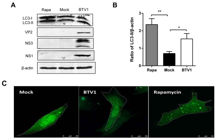
Autophagy was induced in primary lamb lingual epithelial cells by BTV1 infection. (A) The change from LC3-I to LC3-II in primary cells after BTV1 infection for 48 h was detected by Western blotting using an anti-LC3B antibody. Rapamycin group was positive control; (B) Ratios of LC3-II to β-actin of panel (A). The values represent the mean ± SD of three independent experiments. Statistical significance was analyzed with Student’s t-test (* p < 0.05); (C) The primary lamb lingual epithelial cells were transfected with pEGFP-LC3. At 24 h post-transfection, cells were treated with mock-infection (left) or BTV1-infection at an MOI of 1 for 48 h (middle); or rapamycin treatment for 36 h (right). The fluorescence signals were visualized by confocal microscopy. Scale bar: 7.5 μm.

