Abstract
Introduction: Guillain-Barré syndrome (GBS) is a fulminant polyradiculoneuropathy that is acute, frequently severe and autoimmune in nature. Etiology of GBS is incompletely understood, prognosis is usually good with early detection and prompt treatment. This retrospective study was done to evaluate clinical profile, epidemiological, laboratory, and electrodiagnostic features of patients with GBS and mode of management, complications and prognostic factors.
Methods: Data of 1,166 patients admitted with GBS or presented to outpatient department (previous medical records) with GBS between January 2003 and January 2014 were analyzed.
Results: No difference in genders noted. Around 35% of patients are above 50 years of age. Poor control of diabetes with mean HbA1c of 8.1 ± 2.11 is found on analysis. Seasonal occurrence in GBS is prominent in winter 484 (41.50%) and mechanically ventilated were 449 (38.50%) patients. 48 (4.11%) deaths were attributed to GBS. Neurological analysis revealed cranial nerve involvement in 407 (34.90%) patients, facial palsy in 401 (34.39%) and ataxia in 88 (7.54%) patients. Most patients in plasma exchange group belonged to the lower socio-economic status. Mean cerebrospinal fluid (CSF) protein levels was (n=962) 113.8 ± 11.8 mg/dl. Conduction block determined indirectly by absent H-reflex was noted in 891 (90.64%) patients. No difference in complications and outcome is found in treatment regimens of intravenous immunoglobulin (IVIG) and plasma exchange.
Conclusion: Seasonal occurrence predominantly in winter is noted. Peak flow test may be a predictor of assessing requirement of mechanical ventilation and prognosis. Conduction block is the major abnormality noted in electrophysiological studies and proximal nerve segment assessing with Erb’s point stimulation has high predictive value. IVIG treatment is more expensive but is associated with less duration of hospital stay.
Keywords: Guillain-Barré syndrome, autoimmune, acute inflammatory demyelinating polyradiculoneuropathy, conduction motor action potential, IVIG, immunoglobulin, electromyography studies, plasma exchange
Zusammenfassung
Einführung: Das Guillain-Barré-Syndrom (GBS) ist eine fulminant verlaufende Polyradiculoneuropathie, die akut, meist schwer verlaufend, auf der Basis eines Autoimmunprozesses auftritt. Die Ätiologie der Erkrankung wird nicht ganz verstanden, die Prognose ist bei früher Diagnose und Therapie gewöhnlich gut. Eine retrospektive Studie wurde durchgeführt, um das klinische Profil, die Epidemiologie, die Laborwerte, die Elektrodiagnose, die Behandlungsarten und die Prognose von Patienten mit GBS auszuwerten.
Methode: Die klinischen Daten von 1.166 Patienten, die zwischen Januar 2003 und Januar 2014 mit GBS überwiesen oder in den Ambulanzen vorgestellt wurden, wurden ausgewertet.
Ergebnisse: Geschlechtsspezifische Unterschiede wurden nicht beobachtet. Etwa 35% der Patienten waren älter als 50 Jahre. Die Analyse zeigte schlecht eingestellten Diabetes mellitus (HBA1c = 8,1 ± 2,11%). Die saisonale Abhängigkeit der GBS ist deutlich, im Winter wurden 484 (41,5%) gefunden und 449 (38,5%) Patienten mit GBS wurden künstlich beatmet. 48 (4,11%) der Patienten verstarben an GBS. Die neurologischen Untersuchungen ergaben bei 407 (34,9%) der Patienten Beteiligung der cranialen Nerven, faciale Lähmungen bei 401 (34,39%) und Ataxien bei 88 (7,54%) der Patienten. Die meisten Patienten, die mit Plasmapherese behandelt wurden, hatten einen niedrigeren sozioökonomischen Status. Die mittlere Proteinkonzentration im Liquor war 113,8 ± 11,8 mg/dl. Störungen der Nervenleitung, indirekt bestimmt über den H-Reflex, wurden bei 891 (90,64%) der Patienten beobachtet. Keine Unterschiede hinsichtlich Komplikationen und Endergebnis wurden festgestellt zwischen den Behandlungen mit intravenöser Gabe von Immunglobulinen und Plasmaaustausch.
Schlussfolgerung: GBS tritt vorwiegend in den Wintermonaten auf, der „Peak-Flow-Test“ kann ein Indikator für eine erforderliche künstliche Beatmung und für die Prognose sein. Eine Verminderung oder Blockierung der Nervenleitung ist die schwerwiegendste Veränderung, die bei elektrophysiologischen Untersuchungen beobachtet wird. Die Prüfung des proximalen Nervensegments mit der Stimulierung am Erb-Punkt hat einen hohen prädiktiven Wert. Intravenöse Immunglobulin-Behandlung ist teurer, aber verkürzt den Krankenhausaufenthalt.
Introduction
Guillain-Barré syndrome (GBS) is a fulminant polyradiculoneuropathy that is acute, frequently severe and autoimmune in nature. GBS is the most common cause of acute or subacute generalized paralysis which at one time rivaled polio in frequency [1]. GBS is also known as Landry-Guillain-Barré-Strohl syndrome and acute inflammatory demyelinating polyneuropathy (AIDP). Global annual incidence is reported to be 0.6–2.4 cases per 100,000 per year [2], [3], [4]. Men are more commonly affected by approximately 1.5 times than women [5]. Acute inflammatory demyelinating polyradiculoneuropathy (AIDP) is the most commonly occurring subtype in North America and Europe accounting for about 90% of all cases [6]. However, in other parts of the world (Asia, Central and South America) axonal variants of GBS i.e. acute motor axonopathy (AMAN) and acute motor sensory axonopathy (AMSAN) are found to represent 30% to 47% of cases [6], [7].
The earliest description of GBS dates to 19th century regarding an afebrile generalized paralysis by Wardrop and Ollivier in 1834. Other important landmarks are Landry’s report in 1859 about an acute, ascending, predominantly motor paralysis with respiratory failure, leading to death [8] and Osler’s (1892) description of afebrile polyneuritis [9]. Guillain, Barré, and Strohl (1916) described a benign polyneuritis with albumino-cytological dissociation in the cerebrospinal fluid (CSF) [10] and the first report regarding pathology of GBS was by Haymaker and Kernohan in 1949 who reported that edema of the nerve roots was an important change in the early stages of the disease [11]. Asbury, Arnason and Adams (1969) established that the essential lesion is due to perivascular mononuclear inflammatory infiltration of the roots and nerves [12].
Etiology of GBS is not completely understood but believed to be due to autoimmune cause where majority of cases are triggered by infection stimulating anti-ganglioside antibodies production. Approximately 70% of cases of GBS occur 1–3 weeks after an acute infectious process. The organisms thought to be involved are Campylobacter jejuni (diarrhea), Mycoplasma pneumonia, Haemophilus influenzae, cytomegalovirus, Epstein-Barr virus and influenza [13]. Administration of outmoded anti-rabies vaccines and A/New Jersey (swine) influenza vaccine, given in 1976, was associated with a slight increase in GBS incidence. New influenza vaccines appear to confer risk of <1 per million and are relatively safe.
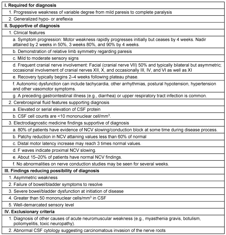
Clinical features include areflexia, limb weakness and uncommonly sensory loss proceeding to neuromuscular paralysis involving bulbar, facial and respiratory function with maximum severity of symptoms in 2–4 weeks [13]. Common clinical pattern is an ascending paralysis first noticed as rubbery legs which evolves over hours to few days with tingling and dysesthesias in the extremities frequently accompanying. Lower limbs are affected more than upper limbs, and facial diparesis occurs in 50% of affected individuals. Usually prognosis is good with 90% of patients having complete functional recovery or with minimal deficits in 1 year after the onset of GBS [3], [14]. Mortality rate is between 1–18% [15]. Diagnosis of AIDP is made by recognizing the pattern of paralysis that is rapidly evolving along with areflexia, absence of fever or other systemic symptoms (Table 1 (Tab. 1)) [16].
Table 1. Diagnostic features of acute inflammatory demyelinating polyneuropathy (AIDP).
The seasons in the Indian subcontinent were divided as: summer/pre-monsoon: March to May; monsoon/rainy: June to September; post-monsoon/autumn: October to November; winter: December to February according to the seasonal classification of the Indian Meteorological Department. This retrospective study which evaluated 1,166 patients was conducted to study clinical profile, epidemiological, laboratory, and electrodiagnostic features of patients with GBS and mode of management, complications and prognostic factors.
Methods
The retrospective study analyzed data of patients admitted with GBS or presented to outpatient department (previous medical records) with GBS between January 2003 and January 2014. Data was pooled from 7 hospitals in which 4 are specialized neurological centers. The medical records were analyzed for the demographic data (age, sex), clinical features, co-morbid conditions, investigations, electrophysiological data, mode and results of the treatment and complications of the procedures. 1,166 patients fulfilled the levels 1, 2 or 3 described by Sejvar et al. which are of diagnostic certainty for GBS/MFS [17].
Medical Research Council (MRC) sum score was used for valuing the muscle strength from 0 to 5 in proximal and distal muscles in upper and lower limbs bilaterally; score ranged from 40 (normal) to 0 (quadriplegic) and by Hughes et al. disability score for GBS [18]. Cranial nerve involvement was noted along with respiratory muscle weakness which was assessed by need of mechanical ventilation, oxygen administration, non-invasive ventilation and spirometer record. Sensory system and autonomic abnormalities were analyzed.
Classification of patients as axonal or demyelinating subtype was based on electrodiagnostic criteria of Hadden et al. [19]. AMSAN diagnosis was based on criteria of Rees et al. [20]. CSF examination was available in 884 (75.81%) patients. Even though 1,166 patient records were analyzed for demographic data, specific treatment protocols with plasma exchange or intravenous immunoglobulin were available in 962 patients and were analyzed for mode of treatment and complications.
Records indicated that serological testing was done for cytomegalovirus (CMV), herpes simplex virus (HSV), Epstein-Barr virus (EBV), varicella-zoster (VZV), Mycoplasma pneumoniae, hepatitis B and C, Haemophilus influenzae and Campylobacter jejuni. GBS disability score [18] (Table 2 (Tab. 2)) was used for evaluation of functional impact during discharge of patients.
Table 2. Hughes grade scale for assessing functional motor deficits.
962 patients received treatment with either IVIG or plasma exchange, of which 494 (51.35%) received IVIG and 468 (48.64%) plasma exchange. Plasma exchange regimen is considered as removal of 200–250 mL/kg of plasma (total) over 5 to 8 cycles on a daily or alternate day basis. Plasma exchange was performed using a peripheral or central venous catheter. Replacement fluids used were fresh frozen plasma (FFP), lactated Ringer’s solution (RL), substitute fluid (SF) and 3% hydroxylethyl starch (HES). TPE was performed every alternate day for majority of patients and 1 plasma volume was exchanged during each procedure. IVIG was administered as 0.4 g/kg daily for 5 consecutive days.
The common indigenous IVIG preparations available in India are Bharat Serum: Ivigama, VHB Pharma: Iviglob, Intas: Globucel, Reliance: Immunorel, Claris: Norglobin, Synergy: MeGlob, and Nirlife Healthcare: IVIG. FDA or EMA preparations of IVIG available in India are Biotest: Intratect, Intraglobin, and Pentaglobin, Bharat Serum/Talecris: Gamunex, Baxter: Kiovig, Gammagard, Novartis: Sandoglobulin, Octapharma: Octagam, and Alpha: Venoglobulin. The average cost per 5 grams of indigenous preparations is around INR 7,500 (US $ 138) while FDA or EMA approved preparations cost around INR 25,000 (US $ 462).
Statistical analyses were performed using SPSS 16 program. Chi-square test for dichotomic variables was used for univariate analysis. Student t-test in parametric variables was used for continuous variables. Mean, standard deviation was performed. P-value was considered significant if <0.05. For calculation of treatment costs, Indian rupee (INR) to US dollar (USD) conversion is done as per standard conversion rates on January 1st 2015.
Results
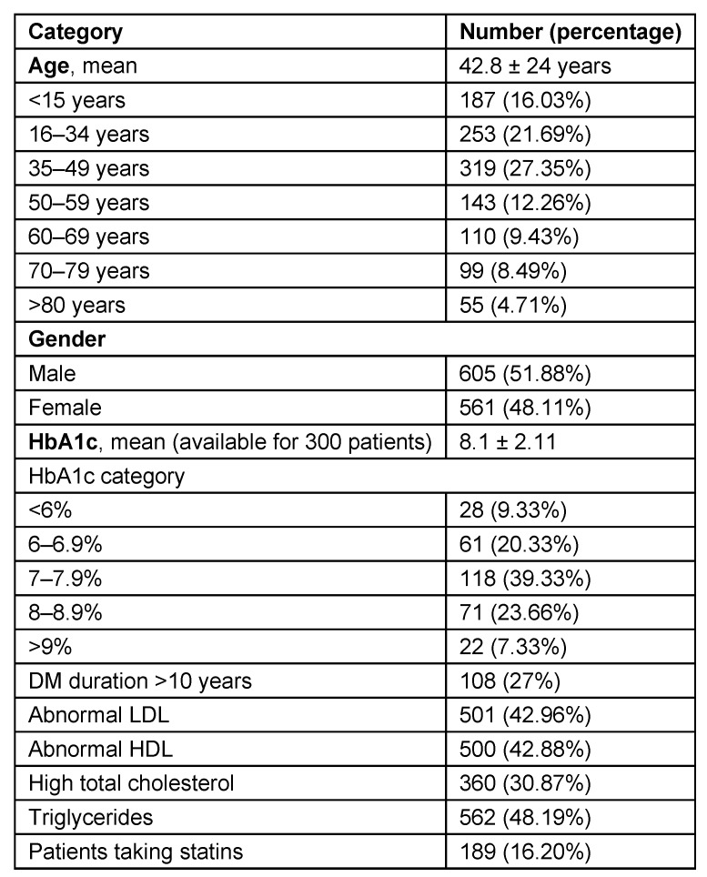
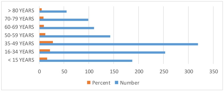
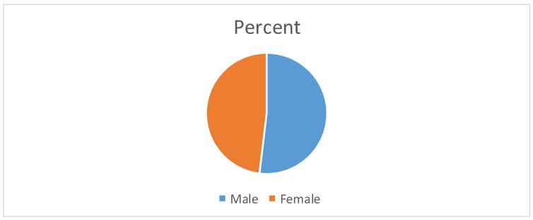
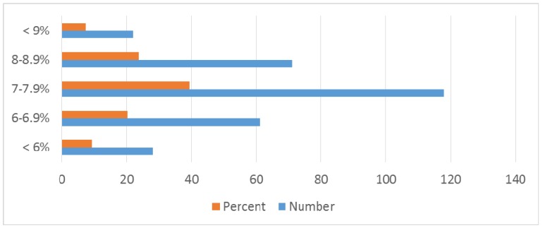
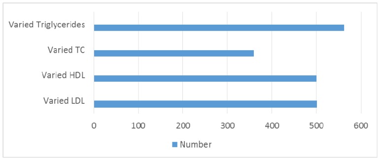
Study group of 1,166 patients consisted of 605 (51.88%) males and 561 females (48.11%). Mean age of onset was 42.8 ± 24 years (range from 0–85). Demographic and clinical data are illustrated in Table 3 (Tab. 3) and in Figure 1 (Fig. 1), Figure 2 (Fig. 2), Figure 3 (Fig. 3), Figure 4 (Fig. 4). There is no difference in genders. Around 35% of patients are above 50 years of age. Diabetic patients constituted 400 out of 1,166 patients. Poor control of diabetes with mean HbA1c of 8.1±2.11 is found on analysis. 70.33% of diabetic patients had HbA1c >7%. Even though dyslipidemia is found in 70% of patients only 16% patients are taking statins which is alarming (Table 3 (Tab. 3)).
Table 3. Epidemiological data of 1,166 patients.
Figure 1. Bar diagram showing age wise distribution.
Figure 2. Pie chart showing gender distribution.
Figure 3. Bar diagram showing HbA1c distribution.
Figure 4. Bar diagram showing lipid parameters .
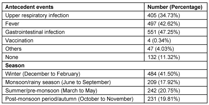
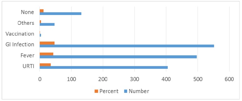
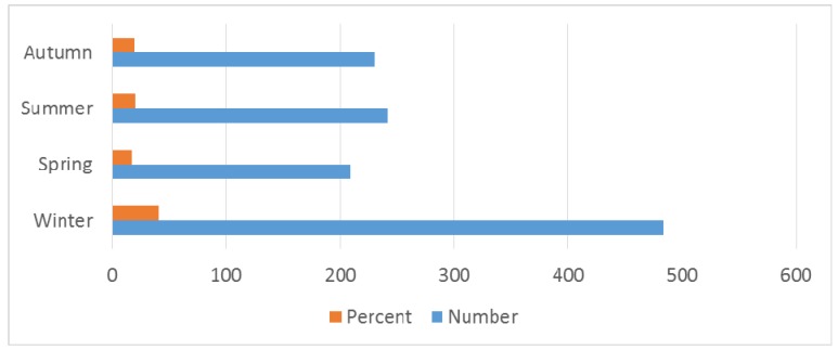
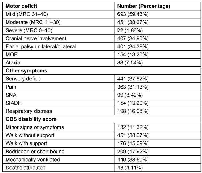
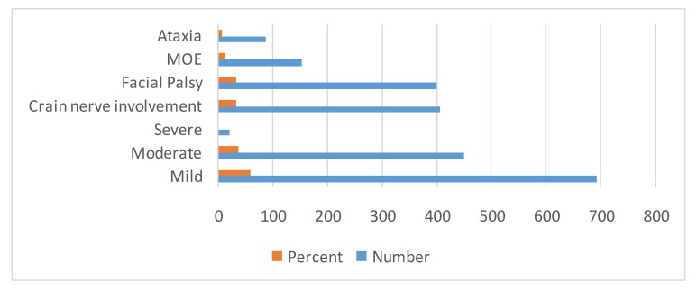
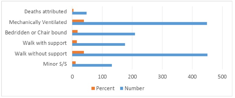
Among antecedent events that were associated with GBS, fever was present in 497 (42.62%) patients and gastrointestinal infection in 551 (47.25%) patients. Seasonal occurrence in GBS is prominent in winter 484 (41.50%), monsoon/rainy season 209 (17.92%) and summer/pre-monsoon 242 (20.75%) seasons (Table 4 (Tab. 4), Figure 5 (Fig. 5), Figure 6 (Fig. 6)). Neurological deficits and GBS disability score are illustrated in Table 5 (Tab. 5) (Figure 7 (Fig. 7), Figure 8 (Fig. 8)). According to GBS disability score, minor signs or symptoms were found in 132 (11.32%) patients, 451 (38.67%) patients walked without support, 176 (15.09%) walked with support, 209 (17.92%) were bedridden or chair bound and 449 (38.50%) patients were mechanically ventilated. 48 (4.11%) deaths were attributed to GBS.
Table 4. Seasonal occurrence and antecedent events in GBS.
Figure 5. Bar diagram showing antecedent events.
Figure 6. Bar diagram showing seasonal occurrence.
Table 5. Neurological deficits and GBS disability score (1,166 patients).
Figure 7. Bar diagram showing motor deficits.
Figure 8. Bar diagram showing GBS disability score.
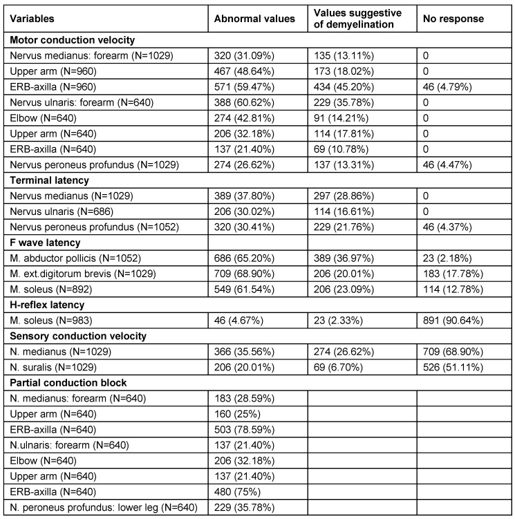
Neurological analysis revealed cranial nerve involvement in 407 (34.90%) patients, facial palsy in 401 (34.39%) and ataxia in 88 (7.54%) patients (Table 5 (Tab. 5)). Electrodiagnostic features (abnormal frequencies) in Guillain-Barré syndrome patients are illustrated in Table 6 (Tab. 6). The highest frequency of affection in patients was observed in the sixth (36.7%) and fifth (27.9%) decade of life. In patients classified as axonal variety, electrophysiology showed low distal CMAP amplitudes, normal motor, sensory conduction velocities and normal distal latencies. F waves were absent or of normal latency and normal sensory neurography. 891 (90.64%) patients had absent H-reflex in soleus muscle. Conduction block (CB) in Erb-to-axilla segment was noted in 503 (78.59%) of 962 patients.
Table 6. Electrodiagnostic features (abnormal frequencies) in Guillain-Barré syndrome patients (1,166 patients).
CMAP amplitude less than 3 mV was noted in the thumb abductor muscle in 183 (28.59%) of 640 patients and in little finger abductor muscle in 137 (21.40%) of 640 patients. But, amplitude reductions observed were not accompanied by CMAP duration prolongation. In the Erb to axilla segment compared to reduction of distal CMAP amplitude and conduction block in forearm (p=0.0004) and upper arm (p=0.0002), median nerve conduction block was more commonly involved and is statistically significant (p=0.0003). In the Erb-to-axilla segment compared to upper arm (p=0.0001), forearm (p=0.0001) and elbow (p=0.0031) segments, ulnar nerve conduction block involvement was common and statistically significant.
Motor conduction velocity delay is observed more commonly in Erb-to-axilla segment than in the upper arm (p=0.005) and forearm (p=0.0002) segments. Statistically significant difference was not observed between slowing of motor conduction velocity in the Erb-to-axilla segment and prolongation of F wave latency (p>0.05). The difference between patients who had normal sural nerve neurography and had normal median nerve sensory neurography was not statistically significant (p>0.05). There is statistically significant difference observed between reduction of sural nerve and median nerve sensory nerve action potential (SNAP) amplitudes compared to sensory conduction slowing down in both nerves (p=0.0001).
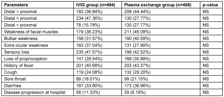
In the 962 patients where treatment records were available, 494 (51.35%) received IVIG and 468 (48.64%) plasma exchange. Males constituted 286 (57.89%) and 312 (66.66%) in IVIG and plasma exchange groups, while females constituted 208 (42.10%) and 156 (33.33%) in IVIG and plasma exchange groups. Mean and range of age were comparable in both groups. Clinical features at admission were almost similar in both groups. Limb weakness was observed in all patients. Distal weakness was more common than proximal weakness. It is the commonest clinical presentation observed. Epidemiological data of patients treated with IVIG or plasma exchange (962 patients) is illustrated in Table 7 (Tab. 7).
Table 7. Epidemiological data of patients treated with IVIG or plasma exchange (962 patients).
AIDP was observed to be the commonest type followed by AMAN and AMSAN. CSF examination evaluation revealed mean CSF protein level of 113.8 ± 11.8 mg/dl (range: 18–450 mg/ dl). Plasma exchange group patients had statistically significant increase in mean length of stay compared to IVIG group (p=0.001) (Table 7 (Tab. 7)). Clinical features of patients that received treatment (962 patients) are illustrated in Table 8 (Tab. 8). Complications and outcome of GBS are illustrated in Table 9 (Tab. 9). There was no statistically significant difference of complication occurrence in both groups. Clinical outcome analysis showed statistically significant mean MRC sum score at time of admission and at time of discharge in IVIG group, which were 19.62 ± 8.20 and 40.10 ± 14.80 (p<0.0001); and in plasma exchange group were 19.42 ± 11.16 and 40.64 ± 15.80 (p<0.0001) respectively.
Table 8. Clinical features of patients that received treatment (962 patients).
Table 9. Complications and outcome of GBS.
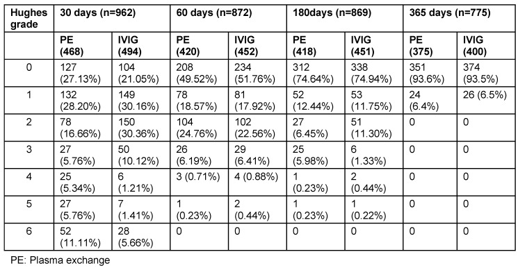
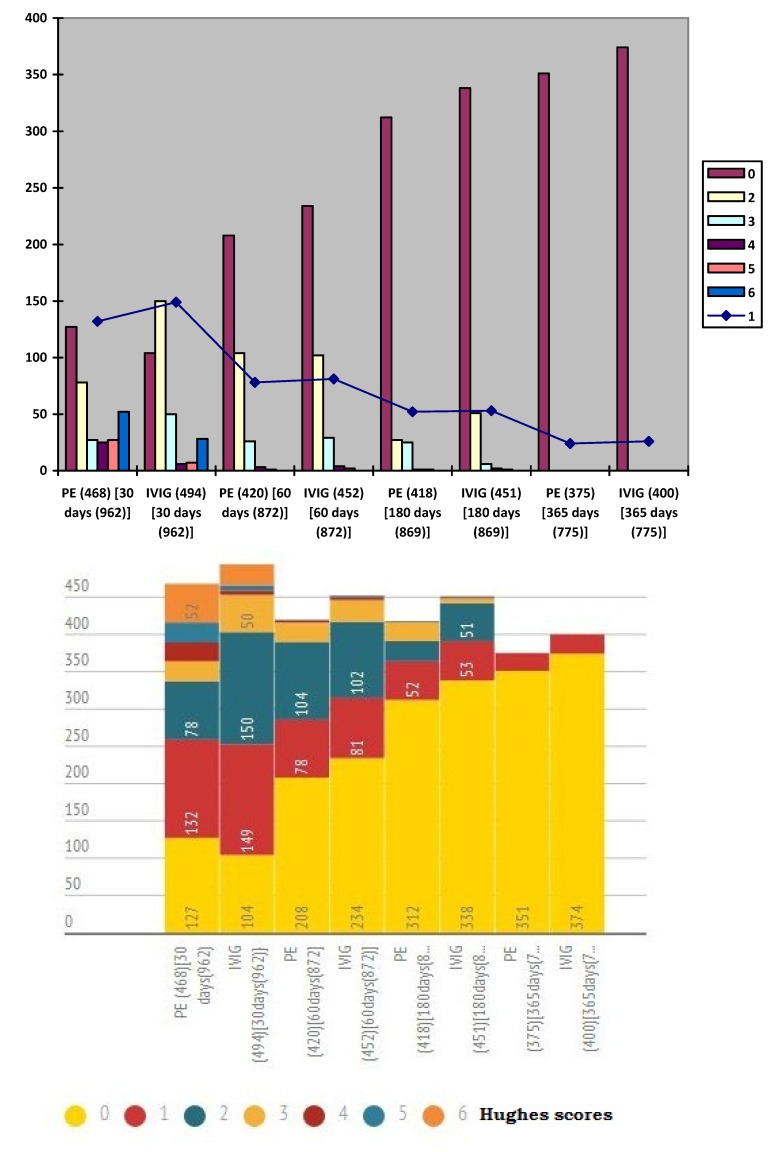
Follow-up of patients with GBS were illustrated in Table 10 (Tab. 10) and Figure 9 (Fig. 9). At 30 days of follow-up, Hughes grade of 0 was observed in 127 (27.13%) patients of plasma exchange group and 104 (21.05%) patients of IVIG groups. At 180 days of follow-up, Hughes grade of 0 was noted in 312 (74.64%) patients of plasma exchange group and 338 (74.94%) patients of IVIG groups. At 365 days of follow-up, progressive improvement in muscle strength is noted. Hughes grade of 0 was noted in 351 (93.6%) patients of plasma exchange group and 374 (93.5%) patients of IVIG groups at 365 days follow-up. No statistically significant difference in follow-up and outcome at discharge was found at 30, 60, 180 and 365 days between both groups.
Table 10. Follow-up of patients with GBS.
Figure 9. Follow-up of patients with GBS with Hughes score.
However, not all patients had financial records showing cost of hospital care available. The cost varied from government and private hospitals significantly. One important observation was that plasma exchange costs were significantly less than IVIG in all the hospitals (p<0.01). The cost of hospital care of the 2 groups is illustrated in Table 11 (Tab. 11).
Table 11. Hospital care cost in plasma exchange and IVIG groups.

Discussion
Our retrospective study shows that there is no difference in gender similar to other studies [21]. Linear increase in incidence with age until 5th decade is noted and patients above 50 years constituted 35% of all patients [22], [23]. Winter predominance (484 (41.50%) patients) of occurrence of illness was noted in our study [23] followed by summer/pre-monsoon (242 (20.75%) patients) even though it was considered that GBS is sporadic without seasonal preference [13], [22]. Our observation differs from studies by Kaur et al. [24], Sharma et al. who reported a peak incidence between June–July and Sept–October [25] and Sriganesh K et al. in 2013, who reported increased occurrence of GBS during the months of June to August and December to February [26]. In many studies, patients admitted with predisposing or associated infection constituted 40–70% of patients [2], [22], [27]. Our study reveals that around 80% of patients had predisposing infection. Gastrointestinal infection was present in 551 (47.25%) followed by upper respiratory infection in 405 (34.73%) patients.
Patients with syndrome of inappropriate antidiuretic hormone hypersecretion (SIADH) constituted 154 (13.20%) patients. In some studies SIADH was reported to be up to 58% in GBS. There is no clear consensus regarding the neurophysiological values defining Guillain-Barré syndrome and its variants [28], [29], [30], [31]. Motor conduction velocity (MCV) decrease, prolongation of motor distal latency, conduction blocks (CB), temporal dispersion and increased F wave latency are the commonly accepted demyelination parameters. First electromyography changes reported are the alteration of F wave and H-reflex response. The important electrophysiological features of peripheral nerve demyelination are conduction block and conduction slowing. In a study reported in 1981 regarding experimental demyelination, conduction block is an early manifestation of demyelination and slow conduction velocity is a feature of remyelination [32].
Many studies revealed that the main cause of acute paralysis and the most common observation in early GBS is conduction block and it may be the only sign in early GBS [33], [34]. This is also found in our study showing that conduction block is the commonest abnormality observed. Conduction block determined indirectly by absent H-reflex was noted in 891 (90.64%) patients, which was the most frequent electrophysiological abnormality observed in GBS patients. Many series of GBS revealed that electrophysiological abnormalities are unevenly distributed with more frequency of occurrence in most proximal and terminal segments of the peripheral nervous system and across entrapment sites [35], [36], [37], [38]. Relative deficiency of blood-nerve barrier may be reason for uneven distribution as per studies [39].
Our study shows concordance with previous studies in the distribution of electrophysiological abnormalities. Conduction block and motor conduction slowing were more frequent in the Erb-to-axilla segment than in distal segments which shows that proximal segment involvement was more common. Mills and Murray reported that the predominant abnormality in early GBS is proximal conduction block, which was measured directly in the nerve segment between axilla and spinal cord [40]. There was difference in conduction block frequency noted in carpal tunnel and elbow segments compared to forearm and upper arm even though it was not significant (p>0.05).
Mean CSF protein levels were increased in all the patients (n=962). 113.8 ± 11.8 mg/dl was the mean noted for CSF and is comparable to studies by Chiò et al. [41] and Corston et al. [42]. Studies reported that abnormal rise of CSF protein in GBS may be due to inflammatory reaction in the choroid plexus or disturbance in process of transport [11], [43], [44], [45], [46] or breakdown of the blood CSF barrier [47], [48], [49], [50], [51], [52].
Outcomes of patients in IVIG and plasma exchange groups were comparable, which were analyzed by muscle strength and Hughes grade. Our study is in concordance with other studies in outcomes [53], [54]. Few studies reported that plasmapheresis is better than IVIG in children with GBS under mechanical ventilation [55]. However, our study did not show any concordance.
Frequencies of occurrence of complications were similar in both groups even though there are reports of increased risk of hypotension and spread of infection in plasma exchange group [56], [57]. 44 patients in plasma exchange group were reported to have breathing difficulty of which 40 of them improved over a period of 3–4 days. One specific incidence of transfusion related acute lung injury (TRALI) was reported in a female patient immediately after plasma exchange. However the patient was also simultaneously diagnosed with scrub typhus infection by Weil-Felix and antibody tests. Scrub typhus is a common infection usually underdiagnosed in rural Bangalore areas. Patient improved over a period of 7 days. The exact reason could not be found from the patient records.
Another significant observation was that most patients in plasma exchange group belonged to the lower socio-economic status as per modified Kuppuswamy’s socio-economic scale used in India [58]. Also statistically significant longer hospital stay was noted in plasma exchange group compared to IVIG group. This may be due to the financial constraints of patients in plasma exchange group which may include delay in getting investigations and treatment. Deaths observed were 24 (4.85%) patients in IVIG group and 20 (4.27%) patients in plasma exchange group, slightly more than in studies reported from Europe and North America [59]. Even though IVIG is comparatively easier to administer and duration of hospital stay is lower compared to plasma exchange, it is more expensive. No difference was noted in the effectiveness of treatment or improvement rate by usage of either treatment.
Conclusion
We put forward the following observations based on our retrospective analysis. This retrospective study has the limitations in accurate calculation of incidence. No gender difference is observed. Increased age is associated with worse prognosis and increased frequency of GBS occurrence. Seasonal occurrence predominantly in winter is noted. Peak flow test may be a predictor of assessing requirement of mechanical ventilation and prognosis. Conduction block is the major abnormality noted in electrophysiological studies and proximal nerve segment assessing with Erb’s point stimulation has high predictive value. Proximal segment involvement is more common than distal segment involvement as per electrophysiology studies.
No difference in complications and outcome is found in treatment regimens of IVIG and plasma exchange. Mortality rate is comparable. IVIG treatment is more expensive but is associated with less duration of hospital stay.
Abbreviations
AIDP: Acute inflammatory demyelinating polyradiculoneuropathy
AMAN: Acute motor axonal neuropathy
AMSAN: Acute motor and sensory axonal neuropathy
CB: Conduction blocks
CMAP: Conduction motor action potential
CMV: Cytomegalovirus
CSF: Cerebrospinal fluid
EBV: Epstein-Barr virus
EMA: European Medicines Agency
EMG: Electromyography studies
GBS: Guillain-Barré syndrome
HSV: Herpes simplex virus
IVIG: Intravenous immunoglobulin
MCV: Motor conduction velocities
MFS: Miller-Fisher syndrome
MRC: Medical Research Council
MV: Mechanical ventilation
NCS: Nerve conduction studies
SIADH: Syndrome of inappropriate antidiuretic hormone hypersecretion
SNAP: Sensory nerve action potentials
VZV: Varicella-zoster virus
Notes
Competing interests
The authors declare that they have no competing interests.
References
- 1.Ropper AH, Brown RH. Adams and Victor's Principles of Neurology. 8th ed. New York: McGraw-Hill; 2005. [Google Scholar]
- 2.McGrogan A, Madle GC, Seaman HE, de Vries CS. The epidemiology of Guillain-Barré syndrome worldwide. A systematic literature review. Neuroepidemiology. 2009;32(2):150–163. doi: 10.1159/000184748. Available from: http://dx.doi.org/10.1159/000184748. [DOI] [PubMed] [Google Scholar]
- 3.Soysal A, Aysal F, Caliskan B, Dogan Ak P, Mutluay B, Sakalli N, Baybas S, Arpaci B. Clinico-electrophysiological findings and prognosis of Guillain-Barré syndrome--10 years' experience. Acta Neurol Scand. 2011 Mar;123(3):181–186. doi: 10.1111/j.1600-0404.2010.01366.x. Available from: http://dx.doi.org/10.1111/j.1600-0404.2010.01366.x. [DOI] [PubMed] [Google Scholar]
- 4.Cuadrado JI, de Pedro-Cuesta J, Ara JR, Cemillán CA, Díaz M, Duarte J, Fernández MD, Fernández O, García-López F, García-Merino A, García-Montero R, Martínez-Matos JA, Palomo F, Pardo J, Tobías A Spanish GBS Epidemiological Study Group. Guillain-Barré syndrome in Spain, 1985-1997: epidemiological and public health views. Eur Neurol. 2001;46(2):83–91. doi: 10.1159/000050769. Available from: http://dx.doi.org/10.1159/000050769. [DOI] [PubMed] [Google Scholar]
- 5.Bogliun G, Beghi E Italian GBS Registry Study Group. Incidence and clinical features of acute inflammatory polyradiculoneuropathy in Lombardy, Italy, 1996. Acta Neurol Scand. 2004 Aug;110(2):100–106. doi: 10.1111/j.1600-0404.2004.00272.x. Available from: http://dx.doi.org/10.1111/j.1600-0404.2004.00272.x. [DOI] [PubMed] [Google Scholar]
- 6.Hughes RA, Cornblath DR. Guillain-Barré syndrome. Lancet. 2005 Nov;366(9497):1653–1666. doi: 10.1016/S0140-6736(05)67665-9. Available from: http://dx.doi.org/10.1016/S0140-6736(05)67665-9. [DOI] [PubMed] [Google Scholar]
- 7.Hung KL, Wang HS, Liou WY, Mak SC, Chi CS, Shen EY, Lin MI, Wang PJ, Shen YZ, Chang KP. Guillain-Barré syndrome in children: a cooperative study in Taiwan. Brain Dev. 1994 May-Jun;16(3):204–208. doi: 10.1016/0387-7604(94)90070-1. Available from: http://dx.doi.org/10.1016/0387-7604(94)90070-1. [DOI] [PubMed] [Google Scholar]
- 8.Landry JB. Note sur la paralysie ascendante aigue. Gaz Hebd Med Chir. 1859;6:472–474, 486–488. [Google Scholar]
- 9.Osler W. The principles and practice of medicine: designed for the use of practitioners and students of medicine. New York: Appleton; 1892. [Google Scholar]
- 10.Guillain G, Barré J, Strohl A. Sur un syndrome de radiculonévrite avec hyperalbuminose du liquide céphalo-rachidien sans réaction cellulaire. Remarques sur les caractères cliniques et graphiques des réflexes tendineux. Bull Mem Soc Med Hop Paris. 1916;40:1462–70. [PubMed] [Google Scholar]
- 11.Haymaker WE, Kernohan JW. The Landry-Guillain-Barré syndrome; a clinicopathologic report of 50 fatal cases and a critique of the literature. Medicine (Baltimore) 1949 Feb;28(1):59–141. doi: 10.1097/00005792-194902010-00003. Available from: http://dx.doi.org/10.1097/00005792-194902010-00003. [DOI] [PubMed] [Google Scholar]
- 12.Asbury AK, Arnason BG, Adams RD. The inflammatory lesion in idiopathic polyneuritis. Its role in pathogenesis. Medicine (Baltimore) 1969 May;48(3):173–215. doi: 10.1097/00005792-196905000-00001. [DOI] [PubMed] [Google Scholar]
- 13.van Doorn PA, Ruts L, Jacobs BC. Clinical features, pathogenesis, and treatment of Guillain-Barré syndrome. Lancet Neurol. 2008 Oct;7(10):939–950. doi: 10.1016/S1474-4422(08)70215-1. Available from: http://dx.doi.org/10.1016/S1474-4422(08)70215-1. [DOI] [PubMed] [Google Scholar]
- 14.Korinthenberg R, Schessl J, Kirschner J. Clinical presentation and course of childhood Guillain-Barré syndrome: a prospective multicentre study. Neuropediatrics. 2007 Feb;38(1):10–17. doi: 10.1055/s-2007-981686. Available from: http://dx.doi.org/10.1055/s-2007-981686. [DOI] [PubMed] [Google Scholar]
- 15.The prognosis and main prognostic indicators of Guillain-Barré syndrome. A multicentre prospective study of 297 patients. The Italian Guillain-Barré Study Group. Brain. 1996 Dec;119(Pt 6):2053–2061. doi: 10.1093/brain/119.6.2053. Available from: http://dx.doi.org/10.1093/brain/119.6.2053. [DOI] [PubMed] [Google Scholar]
- 16.Dumitru D, Amato AA, Zwarts M. Electrodiagnostic medicine. 2nd ed. Philadelphia: Hanley & Belfus; 2002. [Google Scholar]
- 17.Sejvar JJ, Kohl KS, Gidudu J, Amato A, Bakshi N, Baxter R, Burwen DR, Cornblath DR, Cleerbout J, Edwards KM, Heininger U, Hughes R, Khuri-Bulos N, Korinthenberg R, Law BJ, Munro U, Maltezou HC, Nell P, Oleske J, Sparks R, Velentgas P, Vermeer P, Wiznitzer M Brighton Collaboration GBS Working Group. Guillain-Barré syndrome and Fisher syndrome: case definitions and guidelines for collection, analysis, and presentation of immunization safety data. Vaccine. 2011 Jan;29(3):599–612. doi: 10.1016/j.vaccine.2010.06.003. Available from: http://dx.doi.org/10.1016/j.vaccine.2010.06.003. [DOI] [PubMed] [Google Scholar]
- 18.Hughes RA, Newsom-Davis JM, Perkin GD, Pierce JM. Controlled trial prednisolone in acute polyneuropathy. Lancet. 1978 Oct;2(8093):750–753. doi: 10.1016/S0140-6736(78)92644-2. Available from: http://dx.doi.org/10.1016/S0140-6736(78)92644-2. [DOI] [PubMed] [Google Scholar]
- 19.Hadden RD, Cornblath DR, Hughes RA, Zielasek J, Hartung HP, Toyka KV, Swan AV. Electrophysiological classification of Guillain-Barré syndrome: clinical associations and outcome. Plasma Exchange/Sandoglobulin Guillain-Barré Syndrome Trial Group. Ann Neurol. 1998 Nov;44(5):780–788. doi: 10.1002/ana.410440512. Available from: http://dx.doi.org/10.1002/ana.410440512. [DOI] [PubMed] [Google Scholar]
- 20.Rees JH, Gregson NA, Hughes RA. Anti-ganglioside GM1 antibodies in Guillain-Barré syndrome and their relationship to Campylobacter jejuni infection. Ann Neurol. 1995 Nov;38(5):809–816. doi: 10.1002/ana.410380516. Available from: http://dx.doi.org/10.1002/ana.410380516. [DOI] [PubMed] [Google Scholar]
- 21.Vucic S, Kiernan MC, Cornblath DR. Guillain-Barré syndrome: an update. J Clin Neurosci. 2009 Jun;16(6):733–741. doi: 10.1016/j.jocn.2008.08.033. Available from: http://dx.doi.org/10.1016/j.jocn.2008.08.033. [DOI] [PubMed] [Google Scholar]
- 22.A prospective study on the incidence and prognosis of Guillain-Barré syndrome in Emilia-Romagna region, Italy (1992-1993). Emilia-Romagna Study Group on Clinical and Epidemiological Problems in Neurology. Neurology. 1997 Jan;48(1):214–221. doi: 10.1212/WNL.48.1.214. Available from: http://dx.doi.org/10.1212/WNL.48.1.214. [DOI] [PubMed] [Google Scholar]
- 23.Aladro-Benito Y, Conde-Sendin MA, Muñoz-Fernández C, Pérez-Correa S, Alemany-Rodríguez MJ, Fiuza-Pérez MD, Alamo-Santana F. Sindrome de Guillain-Barré en el area norte de Gran Canaria e isla de Lanzarote. [Guillain-Barré syndrome in the northern area of Gran Canaria and the island of Lanzarote]. Rev Neurol. 2002 Oct 16-31;35(8):705–710. [PubMed] [Google Scholar]
- 24.Kaur U, Chopra JS, Prabhakar S, Radhakrishnan K, Rana S. Guillain-Barré syndrome. A clinical electrophysiological and biochemical study. Acta Neurol Scand. 1986 Apr;73(4):394–402. doi: 10.1111/j.1600-0404.1986.tb03295.x. Available from: http://dx.doi.org/10.1111/j.1600-0404.1986.tb03295.x. [DOI] [PubMed] [Google Scholar]
- 25.Sharma A, Lal V, Modi M, Vaishnavi C, Prabhakar S. Campylobacter jejuni infection in Guillain-Barré syndrome: a prospective case control study in a tertiary care hospital. Neurol India. 2011 Sep-Oct;59(5):717–721. doi: 10.4103/0028-3886.86547. Available from: http://dx.doi.org/10.4103/0028-3886.86547. [DOI] [PubMed] [Google Scholar]
- 26.Sriganesh K, Netto A, Kulkarni GB, Taly AB, Umamaheswara Rao GS. Seasonal variation in the clinical recovery of patients with Guillain Barré syndrome requiring mechanical ventilation. Neurol India. 2013 Jul-Aug;61(4):349–354. doi: 10.4103/0028-3886.117582. Available from: http://dx.doi.org/10.4103/0028-3886.117582. [DOI] [PubMed] [Google Scholar]
- 27.Lyu RK, Tang LM, Cheng SY, Hsu WC, Chen ST. Guillain-Barré syndrome in Taiwan: a clinical study of 167 patients. J Neurol Neurosurg Psychiatr. 1997 Oct;63(4):494–500. doi: 10.1136/jnnp.63.4.494. Available from: http://dx.doi.org/10.1136/jnnp.63.4.494. [DOI] [PMC free article] [PubMed] [Google Scholar]
- 28.Saifudheen K, Jose J, Gafoor VA, Musthafa M. Guillain-Barre syndrome and SIADH. Neurology. 2011 Feb;76(8):701–704. doi: 10.1212/WNL.0b013e31820d8b40. Available from: http://dx.doi.org/10.1212/WNL.0b013e31820d8b40. [DOI] [PubMed] [Google Scholar]
- 29.Van den Bergh PY, Piéret F. Electrodiagnostic criteria for acute and chronic inflammatory demyelinating polyradiculoneuropathy. Muscle Nerve. 2004 Apr;29(4):565–574. doi: 10.1002/mus.20022. Available from: http://dx.doi.org/10.1002/mus.20022. [DOI] [PubMed] [Google Scholar]
- 30.Vucic S, Cairns KD, Black KR, Chong PS, Cros D. Neurophysiologic findings in early acute inflammatory demyelinating polyradiculoneuropathy. Clin Neurophysiol. 2004 Oct;115(10):2329–2335. doi: 10.1016/j.clinph.2004.05.009. Available from: http://dx.doi.org/10.1016/j.clinph.2004.05.009. [DOI] [PubMed] [Google Scholar]
- 31.Alam TA, Chaudhry V, Cornblath DR. Electrophysiological studies in the Guillain-Barré syndrome: distinguishing subtypes by published criteria. Muscle Nerve. 1998 Oct;21(10):1275–1279. doi: 10.1002/(SICI)1097-4598(199810)21:10<1275::AID-MUS5>3.0.CO;2-8. Available from: http://dx.doi.org/10.1002/(SICI)1097-4598(199810)21:10<1275::AID-MUS5>3.0.CO;2-8. [DOI] [PubMed] [Google Scholar]
- 32.Sumner AJ. The physiological basis for symptoms in Guillain-Barré syndrome. Ann Neurol. 1981;9 Suppl:28–30. doi: 10.1002/ana.410090706. Available from: http://dx.doi.org/10.1002/ana.410090706. [DOI] [PubMed] [Google Scholar]
- 33.Brown WF, Feasby TE. Conduction block and denervation in Guillain-Barré polyneuropathy. Brain. 1984 Mar;107(Pt 1):219–239. doi: 10.1093/brain/107.1.219. Available from: http://dx.doi.org/10.1093/brain/107.1.219. [DOI] [PubMed] [Google Scholar]
- 34.Atanasova D, Ishpekova B, Muradyan N, Novachkova S, Daskalov M. Conduction block--the diagnostic value in the early stage of Guillain-Barre syndrome. Electromyogr Clin Neurophysiol. 2004 Sep;44(6):361–364. [PubMed] [Google Scholar]
- 35.Albers JW, Donofrio PD, McGonagle TK. Sequential electrodiagnostic abnormalities in acute inflammatory demyelinating polyradiculoneuropathy. Muscle Nerve. 1985 Jul-Aug;8(6):528–539. doi: 10.1002/mus.880080609. Available from: http://dx.doi.org/10.1002/mus.880080609. [DOI] [PubMed] [Google Scholar]
- 36.Kimura J. Proximal versus distal slowing of motor nerve conduction velocity in the Guillain-Barré syndrome. Ann Neurol. 1978 Apr;3(4):344–350. doi: 10.1002/ana.410030412. Available from: http://dx.doi.org/10.1002/ana.410030412. [DOI] [PubMed] [Google Scholar]
- 37.Ropper AH, Wijdicks EF, Shahani BT. Electrodiagnostic abnormalities in 113 consecutive patients with Guillain-Barré syndrome. Arch Neurol. 1990 Aug;47(8):881–887. doi: 10.1001/archneur.1990.00530080065012. Available from: http://dx.doi.org/10.1001/archneur.1990.00530080065012. [DOI] [PubMed] [Google Scholar]
- 38.Brown WF, Snow R. Patterns and severity of conduction abnormalities in Guillain-Barré syndrome. J Neurol Neurosurg Psychiatr. 1991 Sep;54(9):768–774. doi: 10.1136/jnnp.54.9.768. Available from: http://dx.doi.org/10.1136/jnnp.54.9.768. [DOI] [PMC free article] [PubMed] [Google Scholar]
- 39.Olsson Y. Topographical differences in the vascular permeability of the peripheral nervous system. Acta Neuropathol. 1968 Jan;10(1):26–33. doi: 10.1007/BF00690507. Available from: http://dx.doi.org/10.1007/BF00690507. [DOI] [PubMed] [Google Scholar]
- 40.Mills KR, Murray NM. Proximal conduction block in early Guillain-Barré syndrome. Lancet. 1985 Sep;2(8456):659. doi: 10.1016/S0140-6736(85)90017-0. Available from: http://dx.doi.org/10.1016/S0140-6736(85)90017-0. [DOI] [PubMed] [Google Scholar]
- 41.Chiò A, Cocito D, Leone M, Giordana MT, Mora G, Mutani R. Guillain-Barré syndrome: a prospective, population-based incidence and outcome survey. Neurology. 2003 Apr;60(7):1146–1150. doi: 10.1212/01.WNL.0000055091.96905.D0. Available from: http://dx.doi.org/10.1212/01.WNL.0000055091.96905.D0. [DOI] [PubMed] [Google Scholar]
- 42.Corston RN, McGale EH, Stonier C, Aber GM, Hutchinson EC. Abnormalities of cerebrospinal fluid amino acids in patients with the Guillain-Barré syndrome. J Neurol Neurosurg Psychiatr. 1981 Jan;44(1):86–89. doi: 10.1136/jnnp.44.1.86. Available from: http://dx.doi.org/10.1136/jnnp.44.1.86. [DOI] [PMC free article] [PubMed] [Google Scholar]
- 43.Cutler RW, Lorenzo AV. Transport of 1-aminocyclopentanecarboxylic acid from feline cerebrospinal fluid. Science. 1968 Sep;161(3848):1363–1364. doi: 10.1126/science.161.3848.1363. Available from: http://dx.doi.org/10.1126/science.161.3848.1363. [DOI] [PubMed] [Google Scholar]
- 44.Lorenzo AV, Cutler RW. Amino acid transport by choroid plexus in vitro. J Neurochem. 1969 Apr;16(4):577–585. doi: 10.1111/j.1471-4159.1969.tb06857.x. Available from: http://dx.doi.org/10.1111/j.1471-4159.1969.tb06857.x. [DOI] [PubMed] [Google Scholar]
- 45.Snodgrass SR, Cutler RW, Kang ES, Lorenzo AV. Transport of neutral amino acids from feline cerebrospinal fluid. Am J Physiol. 1969 Oct;217(4):974–980. doi: 10.1152/ajplegacy.1969.217.4.974. [DOI] [PubMed] [Google Scholar]
- 46.Cutler RW. Transport of lysine from cerebrospinal fluid of the cat. J Neurochem. 1970 Jul;17(7):1017–1027. doi: 10.1111/j.1471-4159.1970.tb02255.x. Available from: http://dx.doi.org/10.1111/j.1471-4159.1970.tb02255.x. [DOI] [PubMed] [Google Scholar]
- 47.Okuyama H. Mechanism of increase of the cerebrospinal fluid (CSF) protein content in Guillain-Barre syndrome. Study using RISA in evaluation of diffusion within CSF cavity and transport of CSF to circulating plasms. Rinsho Shinkeigaku (Clin Neurol) 1975 Nov;15(11):817–826. [PubMed] [Google Scholar]
- 48.Miyazaki M, Fujita M, Genba H, Shimazaki K, Ogawa M. Study on the blood-cerebrospinal fluid barrir (III). Permeability in the normal state and in polyradiculoneuritis. Rinsho Shinkeigaku (Clin Neurol) 1975 Nov;15(11):843–850. [PubMed] [Google Scholar]
- 49.Schliep G, Felgenhauer K. Serum-CSF protein gradients, the blood-GSF barrier and the local immune response. J Neurol. 1978 May;218(2):77–96. doi: 10.1007/BF02402169. Available from: http://dx.doi.org/10.1007/BF02402169. [DOI] [PubMed] [Google Scholar]
- 50.Jensen K. Cerebrospinal fluid proteins in neurological diseases. Studies on agar gel electrophoresis protein profiles. Acta Neurol Scand, Supplc. 1978;70:1–268. [PubMed] [Google Scholar]
- 51.Snodgrass SR, Lorenzo AV. Transport of GABA from the perfused ventricular system of the cat. J Neurochem. 1973 Mar;20(3):761–769. doi: 10.1111/j.1471-4159.1973.tb00037.x. Available from: http://dx.doi.org/10.1111/j.1471-4159.1973.tb00037.x. [DOI] [PubMed] [Google Scholar]
- 52.Rieder HP, Kaeser HE, Nusselt L. Liquorproteinveränderungen bei Polyneuritis. [Changes in cerebrospinal fluid proteins in polyneuritis]. Schweiz Med Wochenschr. 1972 Jun;102(22):766–772. (Ger). [PubMed] [Google Scholar]
- 53.Bril V, Ilse WK, Pearce R, Dhanani A, Sutton D, Kong K. Pilot trial of immunoglobulin versus plasma exchange in patients with Guillain-Barré syndrome. Neurology. 1996 Jan;46(1):100–103. doi: 10.1212/WNL.46.1.100. Available from: http://dx.doi.org/10.1212/WNL.46.1.100. [DOI] [PubMed] [Google Scholar]
- 54.Nomura K, Hamaguchi K, Hosokawa T, Hattori T, Satou T, Mannen T, et al. A randomized controlled trial comparing intravenous immunoglobulin and plasmapheresis in Guillain-Barré syndrome. Neurol Therapeutics. 2001;18:69–81. [Google Scholar]
- 55.El-Bayoumi MA, El-Refaey AM, Abdelkader AM, El-Assmy MM, Alwakeel AA, El-Tahan HM. Comparison of intravenous immunoglobulin and plasma exchange in treatment of mechanically ventilated children with Guillain Barré syndrome: a randomized study. Crit Care. 2011;15(4):R164. doi: 10.1186/cc10305. Available from: http://dx.doi.org/10.1186/cc10305. [DOI] [PMC free article] [PubMed] [Google Scholar]
- 56.Pohl MA, Lan SP, Berl T. Plasmapheresis does not increase the risk for infection in immunosuppressed patients with severe lupus nephritis. The Lupus Nephritis Collaborative Study Group. Ann Intern Med. 1991 Jun;114(11):924–929. doi: 10.7326/0003-4819-114-11-924. Available from: http://dx.doi.org/10.7326/0003-4819-114-11-924. [DOI] [PubMed] [Google Scholar]
- 57.Chen WH, Yeh JH, Chiu HC. Experience of double filtration plasmapheresis in the treatment of Guillain-Barré syndrome. J Clin Apher. 1999;14(3):126–129. doi: 10.1002/(SICI)1098-1101(1999)14:3<126::AID-JCA4>3.0.CO;2-W. Available from: http://dx.doi.org/10.1002/(SICI)1098-1101(1999)14:3<126::AID-JCA4>3.0.CO;2-W. [DOI] [PubMed] [Google Scholar]
- 58.Bairwa M, Rajput M, Sachdeva S. Modified Kuppuswamy's Socioeconomic Scale: Social Researcher Should Include Updated Income Criteria, 2012. Indian J Community Med. 2013 Jul;38(3):185–186. doi: 10.4103/0970-0218.116358. Available from: http://dx.doi.org/10.4103/0970-0218.116358. [DOI] [PMC free article] [PubMed] [Google Scholar]
- 59.Ramírez-Zamora M, Burgos-Ganuza CR, Alas-Valle DA, Vergara-Galán PE, Ortez-González CI. Síndrome de Guillain-Barré en edad pediátrica. Perfil epidemiológico, clínico y terapéutico en un hospital de El Salvador. [Guillain-Barre syndrome in the paediatric age: epidemiological, clinical and therapeutic profile in a hospital in El Salvador]. Rev Neurol. 2009 Mar 16-31;48(6):292–296. [PubMed] [Google Scholar]


