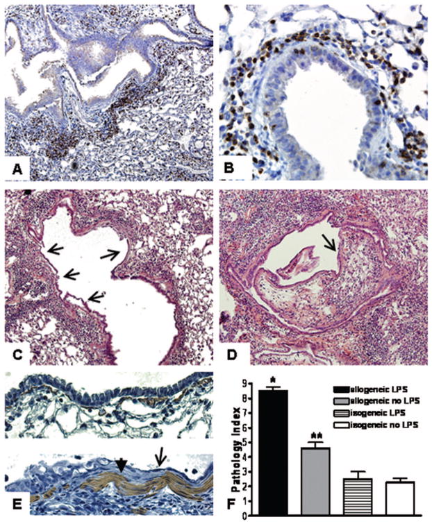
FIGURE 2.
Immunohistochemistry demonstrates peribronchial CD3-positive cells (arrows) in LPS-exposed allogeneic BMT recipient mice (A, 40×, and B, 400× magnification). (C) Epithelial disruption and squamous metaplasia (arrows) are evident throughout a bronchus affected diffusely with lymphocytic bronchiolitis (400×). (D) Intraluminal lesion comprised of loose connective tissue and lymphocytes (arrow) partially obstructs the lumen of a bronchus with disrupted and metaplastic epithelium throughout (400×). (E) α-smooth muscle actin staining demonstrates hyperplasia of myofibroblasts (arrowhead, bottom panel) adjacent to squamous metaplastic epithelium (arrow, bottom panel). Normal epithelium and myofibroblasts in isogeneic mice (top panel). (F) Quantification of perivascular and peribronchial inflammation (*P<0.001 compared to all other groups, **P<0.01 compared to isogeneic mice).

