Abstract
Peste des petits ruminants (PPR) is a viral disease of sheep and goats that is spreading through many countries in the developing world. Work on the virus is often restricted to studies of attenuated vaccine strains or to work in laboratories that have high containment facilities. We have created a helper cell dependent form of PPR virus by removing the entire RNA polymerase gene and complementing it with polymerase made constitutively in a cell line. The resultant L-deleted virus grows efficiently in the L-expressing cell line but not in other cells. Virus made with this system is indistinguishable from normal virus when used in diagnostic assays, and can be grown in normal facilities without the need for high level biocontainment. The L-deleted virus will thus make a positive contribution to the control and study of this important disease.
Introduction
Peste des petits ruminants (PPR) is a severe disease of sheep and goats which has been spreading extensively over the past two decades, and is now found widely distributed through large parts of Africa, the Middle East and Asia; it poses an increasing threat to poor livestock keepers, primarily in developing countries [1–4]. The disease is caused by a virus, PPR virus (PPRV), which is a morbillivirus, related to the human pathogen measles virus (MV), as well as other animal pathogens such as canine distemper virus (CDV) and the now-eradicated rinderpest virus (RPV). Control of this disease has recently become a major international goal, marked by the adoption in 2014 of a resolution by the World Organisation for Animal Health (OIE) to establish a control programme with a view to eventual eradication of the disease [5].
Disease control is mostly achieved through the use of clinical or laboratory-based diagnosis coupled with vaccination. All the vaccines currently in use are attenuated strains of PPRV [6,7]; these vaccines are effective, though they do not provide a DIVA (Distinguishing Infected from Vaccinated Animals) capability, since they provide what is essentially a totally subclinical infection with PPRV, and the antibody signatures of vaccinated and previously-infected animals are identical. Several alternative DIVA vaccines have been proposed based on recombinant viruses [8–13], but none is yet in field use.
The most commonly used laboratory tests are those for anti-PPRV antibodies, partly on cost grounds, and partly because a lot of the effort in infected countries is still on tracking the prevalence of disease through identifying flocks/herd which have been exposed to the virus, rather than acute response diagnostics on animals showing clinical signs. In addition, vaccination programs are increasingly being supported by post-vaccination serum surveillance to measure the effectiveness of the local vaccination process. Although there are still laboratories using agarose gel immunodiffusion (AGID) techniques, the primary method of testing for anti-PPRV antibodies is competition ELISA (cELISA), and the antibodies tested for are either those recognising the viral nucleocapsid protein (N) [14,15], in which the ELISA antigen is a bacterially expressed protein, or those recognising the viral surface glycoprotein H [16,17], where whole virus is used as the ELISA antigen. The latter system presents a problem in the need to grow and purify live virus, even if it is only a vaccine strain of the virus. In many countries, this requires a containment laboratory for the virus culture, and continued application of biosecurity restrictions, including restrictions on the transport of the ELISA kits, even if the virus preparation is subsequently inactivated, e.g. with binary ethylenimine (BEI). For larger scale and simplified production of antigen for ELISA, it would be useful to be able to prepare a suitable antigen without the need for actual virus. The H protein requires mammalian glycosylation for proper folding, so baculovirus-expressed protein is not adequate. We have therefore sought to create a biosafe system to produce virus-like particles (VLPs) which would appear as virus in diagnostic tests and which could be produced in good yield. We have achieved this by deleting an essential gene from the PPRV genome and providing the required protein in trans in a helper cell line.
In our previous studies, it was shown that removing the P gene from a morbillivirus genome and providing the P protein from a helper cell line can lead to the production of suitable VLPs, but in too low yield for practical use, since the P-expressing cell line cannot synthesize the amount of P protein that is required to support normal virus replication and assembly [18]; the P gene is, in any event, a complex system in morbilliviruses, being also required for the production of accessory proteins V and C, both of which are required for optimal virus replication [18–21]. We show here that a PPRV genome lacking the entire gene for the viral polymerase (L) protein grows to near-normal levels in a cell line expressing the L protein, generating VLPs that act as a good antigen in the cELISA, as well as acting as a model virus for detection by PCR-based techniques. The VLPs cannot replicate in other cell lines.
Materials and methods
Cells and viruses
Vero cells expressing the canine version of the morbillivirus receptor SLAM (Signalling Lymphocyte Activation Molecule) (Vero-Dog-SLAM, VDS) were obtained from Dr Paul Duprex, then at Queen’s University Belfast, N. Ireland, and maintained in Dulbecco’s modified Eagle’s medium containing 25 mM HEPES buffer, penicillin (100 U/mL), and streptomycin (100 μg/mL) (DMEM) containing 10% foetal calf serum (FCS). Zeocin was included at 0.1 mg/mL to maintain selection for SLAM expression. PPRV Nigeria 75/1 vaccine strain [6] and recombinant PPRV rPPRV-GFP [22] were propagated and titrated in VDS cells. Titres were determined as the 50% tissue culture infectious dose (TCID50), calculated by the method of Spearman and Kärber [23]. Titres of preparations of PPRV-del-L were determined in VDS-L cells, while titres of complete PPRV were determined in VDS cells.
Transfections and infections of cultured cells
VDS cells or VDS-L cells were plated at 4 × 10⁴ cells per well in 12-well plates or 105 cells per well in 6-well plates. For immunofluorescence studies, the wells contained sterile glass coverslips (10mm in diameter). The cells were allowed to attach and were infected 6-18 h after plating at a multiplicity of infection (moi) as indicated for each experiment. For infection studies, virus inocula were removed after one hour, the cells washed once with phosphate buffered saline and fresh medium added. Cells were transfected using TransIT-LT1 according to the manufacturer’s instructions, using 3 μL transfection reagent per μg DNA. At the indicated time after transfection, cells were fixed for immunofluorescence staining and confocal microscopy [22], or dissolved in SDS-PAGE sample buffer, or subjected to a freeze-thaw cycle to release progeny virus.
Recombinant PPRV del-L
The PPRV genomic clone (pPPRV-GFP) and the procedure for recovering live virus from plasmids was as previously published [22]. The PPRV-del-L genome was constructed by removing bases 10031-16690 from the PPRV-GFP cDNA, i.e. the entire sequence from 26 bases after the H open reading frame (ORF) to 5 bases after the L ORF. The sequences removed include the H gene stop signal, the intergenic trinucleotide, the L gene start signal and the whole L ORF (a total of 6660 bases). This genome manipulation was carried out using Gibson assembly [24] of two large PCR products derived from the PPRV-GFP plasmid. The two fragments were generated using primer pairs Del-L_F1 [TGCAACCATCGCTCGAGCAAGTGATACATCTGCCCCCTTCTC] with Del-L_R1 [GATTCTTGTGTCAACCCCTGGA] and Del-L_F2 [TCCAGGGGTTGACACAAGAATC] with Del-L_R2 [GAGAAGGGGGCAGATGTATCACTTGCTCGAGCGATGGTTGCA]. The entire sequence of the resulting PPRV-del-L genome was checked.
Creation of the helper cell line
To create the cell line expressing PPRV L, the exact L ORF, without any transcription control sequences (i.e. no gene start, gene stop or untranslated region), was fused downstream of the V5 epitope tag and cloned under the control of the promoter in pCAGGS (pCAGSS-V5-L). A large section of this plasmid, consisting of the pCAGGS promoter and intron and the V5-L ORF, was then inserted into plasmid 5’-PTK-3’ [25], upstream of the IRES-driven puromycin resistance marker in that vector. To do this, the En-2 splice acceptor was removed from 5’-PTK-3’ and replaced with a short multiple cloning site (XhoI/SgfI/AflII/SbfI/NheI/MfeI) to give pPB-MCS-IPUR. The CAGGS-V5-L construct from pCAGGS-V5-L was then inserted into pPB-MCS-IPUR to give pPB-V5-PPRV-L. The plasmid pPB-V5-PPRV-L was transfected into VDS by standard techniques along with plasmid pCyL43 encoding the transposase [26] and the cells subjected to puromycin selection through 3 passages to create the VDS-L cell line.
Other assays
RNA was extracted from cells using the Qiagen RNeasy mini kit. cDNA was synthesised using Superscript II reverse transcriptase with oligo(dT)-anch [27] as the primer. Real-time quantitative PCR of PPRV N mRNA or host cell L13A mRNA was carried out essentially as previously described [18,27], using a set of primers recognising the PPRV N gene and optimised for SYBR Green-based qPCR (NF2b:CGGGTTGACCTTTGCATCA and NREVb: CTTTGTTGTGTGTATTTAACCCACCTT). Confocal microscopy was performed as previously described [22]. Mouse anti-V5 tag was obtained from AbD Serotec and AlexaFluor 568 anti-mouse IgG was from Life Technologies. All images were taken by sequential scanning and the resulting separate colour images overlayed in Photoshop. cELISA antigen was prepared as previously described [16]. SDS-PAGE and Western blots were carried out as previously described [28], except that the Western blot transfer was performed using a TransBlot SD Semi Dry Electrophoretic Transfer Cell (Bio-Rad) and Bjerrum and Schafer-Nielsen transfer buffer (48 mM Tris, 39 mM glycine, 37.5 mg/L SDS, 20% methanol, pH 9.2) [29]. Statistical analysis was performed in Minitab v17.
Results
In previous work, it was found that a recombinant rinderpest virus in which the L protein had an amino-terminal HA tag was fully functional (Baron, M.D., unpublished). It seemed likely that insertion of an epitope tag at the amino-terminus of a morbillivirus L protein does not interfere with its function, so a PPRV L protein expression construct was created with the V5 epitope tag at this position. Despite the large size of the L protein, significant amounts of full-length L protein were detected in cells transfected with this construct (Figure 1A). In addition, the V5-L construct was equally effective in supporting the rescue of PPRV from the plasmid copy of the genome as was the original pCAGGS-L (Figure 1B), confirming that it is functional as the viral polymerase.
Figure 1.
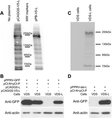
Expression of functionally active PPRV L protein. A VDS cells were transfected with the indicated plasmid (2 μg) in 6-well dishes and cultured for 48 h. After lysis of the transfected cells, expressed proteins were analysed by SDS-PAGE and Western blot using mouse anti-V5 tag. B Cells (VDS or VDS-L) in 6-well dishes were transfected with the indicated plasmids and incubated for 8 days. The transfected cells were subjected to 1 freeze-thaw cycle to release infectious virus and the clarified supernatant used to infect fresh VDS or VDS-L cells. After 7 days, the cells were harvested and the expression of virally-encoded GFP analysed by SDS-PAGE and Western blot. Actin was used as a loading control. C VDS or VDS-L cells (~4 × 10⁵) were lysed directly in SDS-PAGE sample buffer and analysed by SDS-PAGE and Western blot using mouse anti-V5 tag. D As B, except that no plasmid encoding the L protein was included.
Since the V5-L was fully functional, the VDS-L helper cell line was created using this construct. VDS cells were selected as the base for the new cell line as they have been, in our hands, the best cell line for growing PPRV, either wild-type or vaccine strains. The presence of the morbillivirus receptor (SLAM) ensures that virus growth leads to rapid production of large syncytia, which are easy to detect and characteristic of virus replication and viral glycoprotein expression. Normal methods for creating cell lines expressing extra proteins by transfecting with the plasmid of interest, in linear or circular form, along with a plasmid encoding an antibiotic resistance marker, were ineffective. Resistant cells could be isolated, but they either did not express any V5 tag-bearing protein at all or the protein detected was a severely truncated form of the L protein. Since a problem with these methods is that they rely on random recombination events of genomic DNA with the plasmids in question, it appeared that the L sequence was long enough to make it highly probable that such an event would occur within its sequence, or that of the governing promoter. We therefore sought to carry out a directed insertion of a defined expression cassette using the PiggyBac transposase system [30]. This method was very effective. The resultant cell line was isolated at the first attempt and was found to express an L protein that was the same size as the original (Figure 1C), and was able to support rescue of rPPRV-GFP when the cell line was the only source of L protein (Figure 1B), showing that the cell line-expressed L protein was functional.
A corresponding version of pPPRV-GFP was then created which lacked the L gene. In order to preclude any possibility of the full virus being recreated by recombination with L sequences in the cell line, we deleted not only the L ORF, but also all the transcriptional control sequences required for its transcription by the viral RNA-dependent RNA polymerase. Correspondingly, the construct inserted into the cell line genome contains only the L protein ORF, so there is no sequence common to the L protein mRNA and the cut-down PPRV genome. The PPRV-del-L genome could be rescued (as evidenced by expression of GFP and the viral glycoproteins and development of cytopathic effect (cpe)) in VDS-L cells, but not in the normal VDS cells (Figure 1D). The resultant virions could be harvested, titrated and passaged as for the parental virus, but only on VDS-L cells; no replication was seen in VDS cells (Figure 2).
Figure 2.
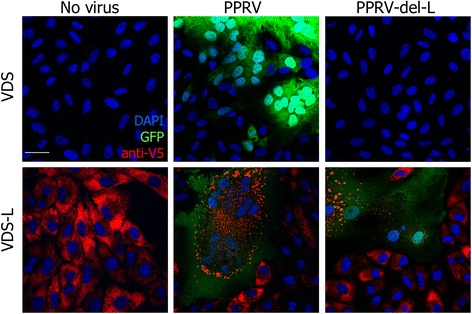
PPRV-del-L requires helper cell line. VDS or VDS-L cells were infected with PPRV-GFP or PPRV-del-L at moi = 0.01 and incubated for 48 h. Cells were fixed and stained with mouse anti-V5 tag and AlexaFluor 568-conjugated goat anti-mouse IgG. Nuclei were stained with DAPI. Confocal images were taken by sequential scanning.
Because it is a normal constituent of the virion, a small amount of the L protein was found in the PPRV-del-L VLPs produced in VDS-L cells. L protein carry-over was sufficient that a low level of GFP expression could be detected in VDS cells infected with PPRV-del-L, though only if the infection was carried out at a relatively high multiplicity of infection (moi) (Figure 3). In order to be sure that no PPRV-del-L was growing in cells without the L protein, serial passage of the VLPs on VDS cells was carried out in order to try to amplify any functional virion. No cpe was seen during these serial passages, nor was there any sign of GFP expression. Quantitative RT-qPCR was used to assay viral mRNA from each one of the passages, and showed only the expected decay of the viral RNA present in the initial inoculum.
Figure 3.
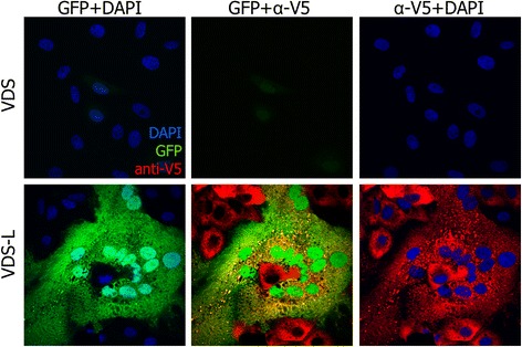
Protein expression from PPRV-del-L after infection at higher moi. Cells were infected with PPRV-del-L at moi = 0.5. At 48 hours post infection, cells were fixed and stained as in Figure 2. Confocal images were taken by sequential scanning. Fields showing infection and protein expression in VDS and VDS-L are shown.
No evidence for replication of the viral genome was seen in VDS cells, even with repeated passage (Figure 4). Note that for PPRV, as for other morbilliviruses, the virus inoculum is not pure virus, and the original inoculum always contains significant non-infectious viral genome and even some viral mRNA; this means that RNA extracted from virus stocks, or RNA extracted from infected cells harvested at t = 0, always gives a positive signal in RT-qPCR (Figure 4).
Figure 4.
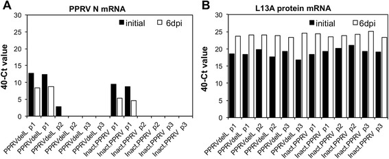
PPRV-del-L behaves as heat-inactivated PPRV. VDS cells were infected with PPRV-del-L (PPRVdelL) or PPRV that had been heat-inactivated (2 h at 58 °C) (inactPPRV) at a nominal moi of 0.5. After removing unattached virus, duplicate wells were harvested immediately for RNA or cultured for 6 days (p1). After 6 days, the infected cells were subjected to 1 freeze-thaw cycle, the cell pellet harvested for RNA, and one third of the supernatant passaged to fresh cells (p2). This procedure was then repeated (p3). RT-qPCR was used to determine the relative amount of viral mRNA in each sample (A); Vero cell L13A mRNA was used as control for the recovery of RNA from each sample (B). Results are expressed as 40-(mean Ct value observed from duplicate PCRs from each of duplicate wells).
Cells infected with live rPPRV-GFP show a steadily increasing amount of viral genome with time, reflecting viral replication (Figure 5). However, if the initial inoculum is heat-inactivated PPRV, the content of viral genome simply decays over time, with no indication of replication (Figure 4). The pattern seen with PPRV-del-L in VDS cells was similar to that seen with inactivated PPRV.
Figure 5.
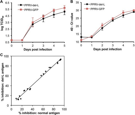
Comparison of growth and antigenicity of PPRV-del-L and full PPRV. VDS-L cells in 6-well plates were infected with PPRV-GFP or PPRV-del-L at moi = 0.01. Duplicate wells were harvested at 0, 1, 2, 3, 4 and 5 days post infection, subjected to one freeze-thaw cycle and the released virus titrated (A). RNA was extracted from the cell pellets and viral (genome + mRNA) assayed by RT-qPCR (B). C VDS-L cells were infected with PPRV-del-L (moi = 0.01) in 175cm2 flasks and cultured for 5 days. Virus antigen was prepared and used in the PPRV-H cELISA (BDSL) according to the manufacturer’s instructions. A set of samples known to be positive (% inhibition >50%) and negative (% inhibition <50%) were assayed in parallel cELISA tests using either the PPRV antigen provided with the kit or the antigen made from PPRV-del-L. All sera were assayed in duplicate in each test and the mean % inhibition obtained with each antigen plotted in the graph. The set of individual mean values for each serum were compared using a paired t-test: the mean difference was not significantly different from zero (p = 0.081). The regression line had a slope of 0.99 ± 0.02 (± S.E.) and an intercept of 1.81 ± 1.34.
Multi-step growth curves were performed to compare the growth kinetics of the parental and del-L viruses. These studies were carried out in VDS-L cells, so that the same cell line was used for the two viruses. It was found that the PPRV del-L virus grew similarly to normal virus in these cells (Figure 5), although still slightly slower than the normal virus (2-way ANOVA, p < 0.05 for growth measured as either virus (Figure 5A) or viral RNA (Figure 5B)). Interestingly, the normal PPRV grew slightly worse in the cells expressing endogenous L protein than in parental VDS cells (compare Figure 5A with Figure 3A of [22] and Figure 2A of [18]). This observation is in accord with the observation that virus-encoded GFP expression from full PPRV is lower in VDS-L cells than in VDS cells (Figure 2). This apparent defect in virus replication in the presence of excess L protein may reflect previous observations that the correct ratio of the P and L proteins is critical in replication/transcription of paramyxovirus genomes [31,32].
Antigen for the PPRV cELISA was prepared from PPRV-del-L grown on VDS-L cells and its performance in the assay compared with that of the antigen distributed with H protein-specific cELISA kit; the results were essentially identical (Figure 5C), showing that the VLPs produce an antigen which behaves exactly like normal antigen in cELISA, while the VLPs can act as a template for PCR tests.
Discussion
In our previous work [18], we showed that deletion of the morbillivirus P protein coding sequence produced a replicon that could replicate only in cell lines expressing the P protein, although even then with greatly reduced replication relative to the intact virus. The viruses lacking the P protein coding sequence were completely dependent on the helper cell line for transcription and replication. In the work presented here, we have shown that a replicon lacking the L (polymerase) gene (PPRV-del-L) can replicate in a cell line expressing the L protein (VDS-L), and with efficiency close to that of the parental virus. This replicon is unable to replicate in any other cell, since it lacks the L gene in its entirety. Repeated blind passage of the VLPs produced by PPRV-del-L in cultured cells showed no replication of the replicon genome (as assessed by RT-qPCR).
A key question in considering the biosafety of the PPRV-del-L is whether there is any possibility of the virus recovering its ability to replicate through the recovery of its polymerase gene or the gene for a polymerase that would fulfil the same function. Replication and transcription of PPRV, as with all nonsegmented negative-strand RNA viruses (NNSRVs), take place exclusively in the cytoplasm of the infected cell. Because the NNSRVs all co-transcriptionally encapsidate their genome and antigenome RNAs, naked viral RNAs (other than mRNAs) are not found in the cytoplasm. In addition, the template for the viral RNA polymerase is encapsidated viral RNA containing specific promoter sequences (the 5’ and 3’ terminal 120 bases), and never naked RNAs, whether viral mRNAs or derived from the host genome. For this reason, recombination is not found in this group of viruses in nature (reviewed in [33]). There is one published example of the creation of a chimeric NNSRV, which was done in culture and under specific selection for recombinant viruses [34]; in that particular case it was a combination of two different strains of respiratory syncytial virus. There is also some indirect evidence from analysis of sequences in the database that recombination between strains of the same virus has occurred in nature [35–41]; there is no evidence either in nature or in culture of recombination between dissimilar NNSRVs or between NNSRV genomes and cellular RNAs.
Given these findings, it is clearly possible for PPRV-del-L to recover its L gene through recombination with a natural PPRV virus. In this case, of course, there is already live PPRV in the system, so this circumstance would not introduce an additional hazard. If the donor is itself a wild type virus, there is already pathogenic virus present. The del-L virus is based on the fully attenuated vaccine strain of PPRV. The stability of this vaccine through several decades of use in the field shows that it has attenuating mutations throughout the genome, as was found for the Plowright vaccine [42,43], and indeed the P, F, H and N proteins of the vaccine strain all show extensive differences from those of wild type viruses isolated at the same time and geographical area, while the promoters in the vaccine show changes to bases that are conserved in all known wild type viruses. Donation of L from a different PPRV vaccine will not therefore result in a chimera that has become virulent. Recombination between the del-L virus and a different PPRV virus would in any event require coinfection of the same cells; since PPRV is restricted to containment laboratories in countries where the virus is not endemic, use of the del-L system in non-containment laboratories does not pose a threat.
Another possibility is that PPRV del-L could gain an L gene from another (non-contained) morbillivirus. This would require recombination between the PPRV genome and that of another morbillivirus (e.g. CDV, measles virus). There is no evidence from nature of recombination having occurred between two different NNSRVs which has given rise to a viable chimeric virus. This may be because such recombination has to occur at very similar sequences, as is known to be the case in pox virus recombination [44]. Alternatively, it may be because co-evolution of the viral proteins has led to incompatibilities between proteins from different, even if closely related, viruses, for example the finding that the surface glycoproteins (F and H) of PPRV will work together, but not in any heterologous pair with the H and F proteins of RPV [45]. More importantly, the N, P and L proteins of morbilliviruses, which together form the transcription/replication complexes, only work effectively when all three come from the same virus [46]. A PPRV that had acquired the L gene of CDV from replicating CDV would therefore have a non-functional N/P/L combination. Whatever the underlying biochemistry, the evidence over hundreds of years of cocirculation of different morbilliviruses (and other paramyxoviruses) is that recombination between genomes of different viruses does not occur, and the PPRV-del-L will not acquire a functional polymerase through recombination with a different morbillivirus.
A third risk might be that the del-L virus could reacquire the L gene from the helper cell line. As mentioned previously, there is no recorded case of a NNSRV picking up genetic material from a host mRNA, making it unlikely that it would happen in this case. In any event, the entire L gene transcription unit is removed from the del-L construct (including the gene transcription start and end sequences), while the helper cell contains only the L protein ORF, so that even if recombination were to occur between viral genome and cell mRNA, vital parts of the virus would be missing.
While we have not carried out this study, it is likely that the PPRV-del-L construct could be replicated by another morbillivirus, though not very well. A related construct based on two defective measles virus genomes has been propagated [47], though the resultant virus did not grow well, presumably because the minimum infectious unit needed to contain one of each genome. It was only maintained because both genomes were defective and had to support each other in trans. Co-infection of a cell by both PPRV-del-L and another morbillivirus (e.g. CDV) would lead to replication of the PPRV-del-L genome and transcription of its genes. However, this is essentially the same as the CDV replicating in the presence of a large deletion DI (defective interfering particle). Replication of the CDV would be decreased, and the “DI” still could not replicate independently. PPRV as such would not be recreated. Such a mixed infection could conceivably give rise to a sort of two-segmented pathogenic virus containing both the full genome of the non-contained morbillivirus and that of the PPRV-del-L replicon, but such a mixture would be under the usual selective pressure to lose the "DI" component.
Our results in morbilliviruses are completely in line with work done with another NNSRV, vesicular stomatitis virus (VSV), which has been developed in several laboratories as a gene delivery tool by removing the gene for the viral G protein; the VSV G protein functions both for viral attachment and fusion with the target cell. The resultant cut down viral genome, with or without the addition of other, heterologous, coding sequences (e.g. influenza HA, luciferase) could be replicated by providing the G protein in a helper cell line, which is used for growing stocks of resultant VLPs, e.g. [48–58]. This type of VSV construct is generally classified as BSL1 e.g. [49], as transmissibility is entirely dependent on the helper cell line’s provision of the G protein. A similar series of constructs has been developed based on Sendai virus (SeV), where either the F or M genes, or both, are deleted e.g. [59–66]. These constructs have been used for various functional studies as well as vaccine vector studies, and are generally recognised as nontransmissible (though risk assessment depends on the nature of the additional gene inserted into the constructs, if any).
In summary, we have created a version of a highly restricted virus which can only replicate in a defined helper cell line. The resultant construct is therefore safe to use and manufacture out of containment. It can be a safe source not only of antigen for diagnostic kits, but for validation of diagnostic assays (PCR, icELISA, VNTs) in laboratories which have restrictions on admitting live PPRV (even if subsequently chemically inactivated), or PPRV from other countries (e.g. international ring trials). In addition, the system described could be used by laboratories wishing to study replication and assembly of PPRV but lacking appropriate high containment facilities. The system has been approved by the UK Health and Safety Executive for use at BSL 1.
Acknowledgements
The work was funded by the BBSRC Institute Strategic Programme on Livestock Viral Diseases at The Pirbright Institute.
Footnotes
Competing interests
The authors declare that they have no competing interests.
Authors’ contributions
JB prepared most of the plasmids, recovered the recombinant viruses, made the cell lines and carried out all the other experimental work. MDB prepared the PPRV del-L genome plasmid, designed the study and prepared the manuscript. Both authors read and approved the manuscript.
Contributor Information
Jana Baron, Email: jana.baron@pirbright.ac.uk.
Michael D. Baron, Email: michael.baron@pirbright.ac.uk
References
- 1.Singh B, Bardhan D, Verma MR, Prasad S, Sinha DK. Estimation of economic losses due to Peste des petits ruminants in small ruminants in India. Vet World. 2014;7:194–199. doi: 10.14202/vetworld.2014.194-199. [DOI] [Google Scholar]
- 2.Albina E, Kwiatek O, Minet C, Lancelot R, Servan de Almeida R, Libeau G. Peste des Petits Ruminants, the next eradicated animal disease? Vet Microbiol. 2013;165:38–44. doi: 10.1016/j.vetmic.2012.12.013. [DOI] [PubMed] [Google Scholar]
- 3.Baron MD, Parida S, Oura CA. Peste des petits ruminants: a suitable candidate for eradication? Vet Rec. 2011;169:16–21. doi: 10.1136/vr.d3947. [DOI] [PubMed] [Google Scholar]
- 4.Banyard AC, Parida S, Batten C, Oura C, Kwiatek O, Libeau G. Global distribution of peste des petits ruminants virus and prospects for improved diagnosis and control. J Gen Virol. 2010;91:2885–2897. doi: 10.1099/vir.0.025841-0. [DOI] [PubMed] [Google Scholar]
- 5.World Organisation for Animal Health (2014) Resolutions Adopted by the World Assembly of Delegates of the OIE during its 82nd General Session 25–30 May 2014. OIE. Paris, France
- 6.Diallo A, Taylor WP, Lefevre PC, Provost A. Attenuation of a strain of rinderpest virus: potential homologous live vaccine. Rev Elev Med Vet Pays Trop. 1989;42:311–319. [PubMed] [Google Scholar]
- 7.Sen A, Saravanan P, Balamurugan V, Rajak KK, Sudhakar SB, Bhanuprakash V, Parida S, Singh RK. Vaccines against peste des petits ruminants virus. Expert Rev Vaccines. 2010;9:785–796. doi: 10.1586/erv.10.74. [DOI] [PubMed] [Google Scholar]
- 8.Rojas JM, Moreno H, Valcarcel F, Pena L, Sevilla N, Martin V. Vaccination with recombinant adenoviruses expressing the peste des petits ruminants virus F or H proteins overcomes viral immunosuppression and induces protective immunity against PPRV challenge in sheep. PLoS One. 2014;9:e101226. doi: 10.1371/journal.pone.0101226. [DOI] [PMC free article] [PubMed] [Google Scholar]
- 9.Herbert R, Baron J, Batten C, Baron M, Taylor G. Recombinant adenovirus expressing the haemagglutinin of peste des petits ruminants virus (PPRV) protects goats against challenge with pathogenic virus; a DIVA vaccine for PPR. Vet Res. 2014;45:24. doi: 10.1186/1297-9716-45-24. [DOI] [PMC free article] [PubMed] [Google Scholar]
- 10.Wang Y, Liu G, Chen Z, Li C, Shi L, Li W, Huang H, Tao C, Cheng C, Xu B, Li G. Recombinant adenovirus expressing F and H fusion proteins of peste des petits ruminants virus induces both humoral and cell-mediated immune responses in goats. Vet Immunol Immunopathol. 2013;154:1–7. doi: 10.1016/j.vetimm.2013.05.002. [DOI] [PubMed] [Google Scholar]
- 11.Qin J, Huang H, Ruan Y, Hou X, Yang S, Wang C, Huang G, Wang T, Feng N, Gao Y, Xia X. A novel recombinant Peste des petits ruminants-canine adenovirus vaccine elicits long-lasting neutralizing antibody response against PPR in goats. PLoS One. 2012;7:e37170. doi: 10.1371/journal.pone.0037170. [DOI] [PMC free article] [PubMed] [Google Scholar]
- 12.Chen W, Hu S, Qu L, Hu Q, Zhang Q, Zhi H, Huang K, Bu Z. A goat poxvirus-vectored peste-des-petits-ruminants vaccine induces long-lasting neutralization antibody to high levels in goats and sheep. Vaccine. 2010;28:4742–4750. doi: 10.1016/j.vaccine.2010.04.102. [DOI] [PubMed] [Google Scholar]
- 13.Berhe G, Minet C, Le Goff C, Barrett T, Ngangnou A, Grillet C, Libeau G, Fleming M, Black DN, Diallo A. Development of a dual recombinant vaccine to protect small ruminants against peste-des-petits-ruminant virus and capripoxvirus infections. J Virol. 2003;77:1571–1577. doi: 10.1128/JVI.77.2.1571-1577.2003. [DOI] [PMC free article] [PubMed] [Google Scholar]
- 14.Libeau G, Prehaud C, Lancelot R, Colas F, Guerre L, Bishop DH, Diallo A. Development of a competitive ELISA for detecting antibodies to the peste des petits ruminants virus using a recombinant nucleoprotein. Res Vet Sci. 1995;58:50–55. doi: 10.1016/0034-5288(95)90088-8. [DOI] [PubMed] [Google Scholar]
- 15.Choi K-S, Nah J-J, Choi C-U, Ko Y-J, Sohn H-J, Libeau G, Kang S-Y, Joo Y-S. Monoclonal antibody-based competitive ELISA for simultaneous detection of rinderpest virus and peste des petits ruminants virus antibodies. Vet Microbiol. 2003;96:1–16. doi: 10.1016/S0378-1135(03)00201-3. [DOI] [PubMed] [Google Scholar]
- 16.Anderson J, McKay JA, Butcher RN. Seromonitoring of Rinderpest throughout Africa: Phase One. Proceedings of the Final Research Coordination Meeting of the IAEA Rinderpest Control Projects, Cote d'Ivoire 19-23 November 1990 IAEA-TECDOC-623. Vienna: International Atomic Energy Agency; 1990. The use of monoclonal antibodies in competitive ELISA for the detection of antibodies to rinderpest and peste des petits ruminants viruses. [Google Scholar]
- 17.Singh RP, Sreenivasa BP, Dhar P, Shah LC, Bandyopadhyay SK. Development of a monoclonal antibody based competitive-ELISA for detection and titration of antibodies to peste des petits ruminants (PPR) virus. Vet Microbiol. 2004;98:3–15. doi: 10.1016/j.vetmic.2003.07.007. [DOI] [PubMed] [Google Scholar]
- 18.Baron J, Baron MD. Creation of a completely helper cell-dependent recombinant morbillivirus. J Gen Virol. 2013;94:1195–1199. doi: 10.1099/vir.0.050872-0. [DOI] [PMC free article] [PubMed] [Google Scholar]
- 19.Baron MD, Barrett T. Rinderpest viruses lacking the C and V proteins show specific defects in growth and transcription of viral RNAs. J Virol. 2000;74:2603–2611. doi: 10.1128/JVI.74.6.2603-2611.2000. [DOI] [PMC free article] [PubMed] [Google Scholar]
- 20.Patterson JB, Thomas D, Lewicki H, Billeter MA, Oldstone MB. V and C proteins of measles virus function as virulence factors in vivo. Virology. 2000;267:80–89. doi: 10.1006/viro.1999.0118. [DOI] [PubMed] [Google Scholar]
- 21.Escoffier C, Manié S, Vincent S, Muller CP, Billeter MA, Gerlier D. Nonstructural C protein is required for efficient measles virus replication in human peripheral blood cells. J Virol. 1999;73:1695–1698. doi: 10.1128/jvi.73.2.1695-1698.1999. [DOI] [PMC free article] [PubMed] [Google Scholar]
- 22.Hu Q, Chen W, Huang K, Baron MD, Bu Z. Rescue of recombinant Peste des petits ruminants virus: creation of a GFP-expressing virus and application in rapid virus neutralization test. Vet Res. 2012;43:48. doi: 10.1186/1297-9716-43-48. [DOI] [PMC free article] [PubMed] [Google Scholar]
- 23.Finney DJ. Statistical Method in Biological Assay. 2. London: Charles Griffen & Co Ltd; 1964. [Google Scholar]
- 24.Gibson DG, Young L, Chuang RY, Venter JC, Hutchison CA, 3rd, Smith HO. Enzymatic assembly of DNA molecules up to several hundred kilobases. Nat Methods. 2009;6:343–345. doi: 10.1038/nmeth.1318. [DOI] [PubMed] [Google Scholar]
- 25.Cadinanos J, Bradley A. Generation of an inducible and optimized piggyBac transposon system. Nucleic Acids Res. 2007;35:e87. doi: 10.1093/nar/gkm446. [DOI] [PMC free article] [PubMed] [Google Scholar]
- 26.Wang W, Lin C, Lu D, Ning Z, Cox T, Melvin D, Wang X, Bradley A, Liu P. Chromosomal transposition of PiggyBac in mouse embryonic stem cells. Proc Natl Acad Sci U S A. 2008;105:9290–9295. doi: 10.1073/pnas.0801017105. [DOI] [PMC free article] [PubMed] [Google Scholar]
- 27.Bin Tarif A, Lasecka L, Holzer B, Baron MD. Ganjam virus/Nairobi sheep disease virus induces a pro-inflammatory response in infected sheep. Vet Res. 2012;43:71. doi: 10.1186/1297-9716-43-71. [DOI] [PMC free article] [PubMed] [Google Scholar]
- 28.Nanda SK, Baron MD. Rinderpest virus blocks type I and type II interferon action: role of structural and nonstructural proteins. J Virol. 2006;80:7555–7568. doi: 10.1128/JVI.02720-05. [DOI] [PMC free article] [PubMed] [Google Scholar]
- 29.Bjerrum OJ, Schafer-Nielsen C. Buffer systems and transfer parameters for semidry electroblotting with a horizontal apparatus. In: Dunn MJ, editor. Analytical Electrophoresis. Weinheim: Wiley-VCH Verlag GmbH; 1986. [Google Scholar]
- 30.Ding S, Wu X, Li G, Han M, Zhuang Y, Xu T. Efficient transposition of the piggyBac (PB) transposon in mammalian cells and mice. Cell. 2005;122:473–483. doi: 10.1016/j.cell.2005.07.013. [DOI] [PubMed] [Google Scholar]
- 31.Baron MD, Barrett T. Rescue of rinderpest virus from cloned cDNA. J Virol. 1997;71:1265–1271. doi: 10.1128/jvi.71.2.1265-1271.1997. [DOI] [PMC free article] [PubMed] [Google Scholar]
- 32.Clarke DK, Sidhu MS, Johnson JE, Udem SA. Rescue of mumps virus from cDNA. J Virol. 2000;74:4831–4838. doi: 10.1128/JVI.74.10.4831-4838.2000. [DOI] [PMC free article] [PubMed] [Google Scholar]
- 33.Collins PL, Bukreyev A, Murphy BR. What are the risks--hypothetical and observed--of recombination involving live vaccines and vaccine vectors based on nonsegmented negative-strain RNA viruses? J Virol. 2008;82:9805–9806. doi: 10.1128/JVI.01336-08. [DOI] [PMC free article] [PubMed] [Google Scholar]
- 34.Spann KM, Collins PL, Teng MN. Genetic recombination during coinfection of two mutants of human respiratory syncytial virus. J Virol. 2003;77:11201–11211. doi: 10.1128/JVI.77.20.11201-11211.2003. [DOI] [PMC free article] [PubMed] [Google Scholar]
- 35.Han GZ, Liu XP, Li SS. Cross-species recombination in the haemagglutinin gene of canine distemper virus. Virus Res. 2008;136:198–201. doi: 10.1016/j.virusres.2008.04.022. [DOI] [PubMed] [Google Scholar]
- 36.Han GZ, He CQ, Ding NZ, Ma LY. Identification of a natural multi-recombinant of Newcastle disease virus. Virology. 2008;371:54–60. doi: 10.1016/j.virol.2007.09.038. [DOI] [PubMed] [Google Scholar]
- 37.Chare ER, Gould EA, Holmes EC. Phylogenetic analysis reveals a low rate of homologous recombination in negative-sense RNA viruses. J Gen Virol. 2003;84:2691–2703. doi: 10.1099/vir.0.19277-0. [DOI] [PubMed] [Google Scholar]
- 38.Qin Z, Sun L, Ma B, Cui Z, Zhu Y, Kitamura Y, Liu W. F gene recombination between genotype II and VII Newcastle disease virus. Virus Res. 2008;131:299–303. doi: 10.1016/j.virusres.2007.10.001. [DOI] [PubMed] [Google Scholar]
- 39.Schierup MH, Mordhorst CH, Muller CP, Christensen LS. Evidence of recombination among early-vaccination era measles virus strains. BMC Evol Biol. 2005;5:52. doi: 10.1186/1471-2148-5-52. [DOI] [PMC free article] [PubMed] [Google Scholar]
- 40.Wittmann TJ, Biek R, Hassanin A, Rouquet P, Reed P, Yaba P, Pourrut X, Real LA, Gonzalez JP, Leroy EM. Isolates of Zaire ebolavirus from wild apes reveal genetic lineage and recombinants. Proc Natl Acad Sci U S A. 2007;104:17123–17127. doi: 10.1073/pnas.0704076104. [DOI] [PMC free article] [PubMed] [Google Scholar]
- 41.Zheng H, Storch GA, Zang C, Peret TC, Park CS, Anderson LJ. Genetic variability in envelope-associated protein genes of closely related group A strains of respiratory syncytial virus. Virus Res. 1999;59:89–99. doi: 10.1016/S0168-1702(98)00132-4. [DOI] [PubMed] [Google Scholar]
- 42.Baron MD, Banyard AC, Parida S, Barrett T. The Plowright vaccine strain of rinderpest virus has attenuating mutations in most genes. J Gen Virol. 2005;86:1093–1101. doi: 10.1099/vir.0.80751-0. [DOI] [PubMed] [Google Scholar]
- 43.Banyard AC, Baron MD, Barrett T. A role for virus promoters in determining the pathogenesis of Rinderpest virus in cattle. J Gen Virol. 2005;86:1083–1092. doi: 10.1099/vir.0.80752-0. [DOI] [PubMed] [Google Scholar]
- 44.Yao XD, Evans DH. Effects of DNA structure and homology length on vaccinia virus recombination. J Virol. 2001;75:6923–6932. doi: 10.1128/JVI.75.15.6923-6932.2001. [DOI] [PMC free article] [PubMed] [Google Scholar]
- 45.Das SC, Baron MD, Barrett T. Recovery and characterization of a chimeric rinderpest virus with the glycoproteins of peste-des-petits-ruminants virus: homologous F and H proteins are required for virus viability. J Virol. 2000;74:9039–9047. doi: 10.1128/JVI.74.19.9039-9047.2000. [DOI] [PMC free article] [PubMed] [Google Scholar]
- 46.Brown DD, Collins FM, Duprex WP, Baron MD, Barrett T, Rima BK. "Rescue" of mini-genomic constructs and viruses by combinations of morbillivirus N, P and L proteins. J Gen Virol. 2005;86:1077–1081. doi: 10.1099/vir.0.80804-0. [DOI] [PubMed] [Google Scholar]
- 47.Rager M, Vongpunsawad S, Duprex WP, Cattaneo R. Polyploid measles virus with hexameric genome length. EMBO J. 2002;21:2364–2372. doi: 10.1093/emboj/21.10.2364. [DOI] [PMC free article] [PubMed] [Google Scholar]
- 48.Berger Rentsch M, Zimmer G. A vesicular stomatitis virus replicon-based bioassay for the rapid and sensitive determination of multi-species type I interferon. PLoS One. 2011;6:e25858. doi: 10.1371/journal.pone.0025858. [DOI] [PMC free article] [PubMed] [Google Scholar]
- 49.Kalhoro NH, Veits J, Rautenschlein S, Zimmer G. A recombinant vesicular stomatitis virus replicon vaccine protects chickens from highly pathogenic avian influenza virus (H7N1) Vaccine. 2009;27:1174–1183. doi: 10.1016/j.vaccine.2008.12.019. [DOI] [PubMed] [Google Scholar]
- 50.Kochinger S, Renevey N, Hofmann MA, Zimmer G. Vesicular stomatitis virus replicon expressing the VP2 outer capsid protein of bluetongue virus serotype 8 induces complete protection of sheep against challenge infection. Vet Res. 2014;45:64. doi: 10.1186/1297-9716-45-64. [DOI] [PMC free article] [PubMed] [Google Scholar]
- 51.Muik A, Kneiske I, Werbizki M, Wilflingseder D, Giroglou T, Ebert O, Kraft A, Dietrich U, Zimmer G, Momma S, von Laer D. Pseudotyping vesicular stomatitis virus with lymphocytic choriomeningitis virus glycoproteins enhances infectivity for glioma cells and minimizes neurotropism. J Virol. 2011;85:5679–5684. doi: 10.1128/JVI.02511-10. [DOI] [PMC free article] [PubMed] [Google Scholar]
- 52.Zimmer G, Locher S, Berger Rentsch M, Halbherr SJ. Pseudotyping of vesicular stomatitis virus with the envelope glycoproteins of highly pathogenic avian influenza viruses. J Gen Virol. 2014;95:1634–1639. doi: 10.1099/vir.0.065201-0. [DOI] [PubMed] [Google Scholar]
- 53.Mire CE, Geisbert JB, Marzi A, Agans KN, Feldmann H, Geisbert TW. Vesicular stomatitis virus-based vaccines protect nonhuman primates against Bundibugyo ebolavirus. PLoS Negl Trop Dis. 2013;7:e2600. doi: 10.1371/journal.pntd.0002600. [DOI] [PMC free article] [PubMed] [Google Scholar]
- 54.Mire CE, Versteeg KM, Cross RW, Agans KN, Fenton KA, Whitt MA, Geisbert TW. Single injection recombinant vesicular stomatitis virus vaccines protect ferrets against lethal Nipah virus disease. Virol J. 2013;10:353. doi: 10.1186/1743-422X-10-353. [DOI] [PMC free article] [PubMed] [Google Scholar]
- 55.Jones SM, Stroher U, Fernando L, Qiu X, Alimonti J, Melito P, Bray M, Klenk HD, Feldmann H. Assessment of a vesicular stomatitis virus-based vaccine by use of the mouse model of Ebola virus hemorrhagic fever. J Infect Dis. 2007;196(Suppl 2):S404–S412. doi: 10.1086/520591. [DOI] [PubMed] [Google Scholar]
- 56.Qiu X, Fernando L, Alimonti JB, Melito PL, Feldmann F, Dick D, Stroher U, Feldmann H, Jones SM. Mucosal immunization of cynomolgus macaques with the VSVDeltaG/ZEBOVGP vaccine stimulates strong ebola GP-specific immune responses. PLoS One. 2009;4:e5547. doi: 10.1371/journal.pone.0005547. [DOI] [PMC free article] [PubMed] [Google Scholar]
- 57.Tatsuo H, Okuma K, Tanaka K, Ono N, Minagawa H, Takade A, Matsuura Y, Yanagi Y. Virus entry is a major determinant of cell tropism of Edmonston and wild-type strains of measles virus as revealed by vesicular stomatitis virus pseudotypes bearing their envelope proteins. J Virol. 2000;74:4139–4145. doi: 10.1128/JVI.74.9.4139-4145.2000. [DOI] [PMC free article] [PubMed] [Google Scholar]
- 58.Ogino M, Ebihara H, Lee BH, Araki K, Lundkvist A, Kawaoka Y, Yoshimatsu K, Arikawa J. Use of vesicular stomatitis virus pseudotypes bearing Hantaan or Seoul virus envelope proteins in a rapid and safe neutralization test. Clin Diagn Lab Immunol. 2003;10:154–160. doi: 10.1128/CDLI.10.1.154-160.2003. [DOI] [PMC free article] [PubMed] [Google Scholar]
- 59.Bernloehr C, Bossow S, Ungerechts G, Armeanu S, Neubert WJ, Lauer UM, Bitzer M. Efficient propagation of single gene deleted recombinant Sendai virus vectors. Virus Res. 2004;99:193–197. doi: 10.1016/j.virusres.2003.11.005. [DOI] [PubMed] [Google Scholar]
- 60.Ferrari S, Griesenbach U, Shiraki-Iida T, Shu T, Hironaka T, Hou X, Williams J, Zhu J, Jeffery PK, Geddes DM, Hasegawa M, Alton EW. A defective nontransmissible recombinant Sendai virus mediates efficient gene transfer to airway epithelium in vivo. Gene Ther. 2004;11:1659–1664. doi: 10.1038/sj.gt.3302334. [DOI] [PubMed] [Google Scholar]
- 61.Hikono H, Miyazaki A, Mase M, Inoue M, Hasegawa M, Saito T. Induction of a cross-reactive antibody response to influenza virus M2 antigen in pigs by using a Sendai virus vector. Vet Immunol Immunopathol. 2012;146:92–96. doi: 10.1016/j.vetimm.2012.01.017. [DOI] [PubMed] [Google Scholar]
- 62.Kitamura A, Matsushita K, Takiguchi Y, Shimada H, Tada Y, Yamanaka M, Hiroshima K, Tagawa M, Tomonaga T, Matsubara H, Inoue M, Hasegawa M, Sato Y, Levens D, Tatsumi K, Nomura F. Synergistic effect of non-transmissible Sendai virus vector encoding the c-myc suppressor FUSE-binding protein-interacting repressor plus cisplatin in the treatment of malignant pleural mesothelioma. Cancer Sci. 2011;102:1366–1373. doi: 10.1111/j.1349-7006.2011.01931.x. [DOI] [PMC free article] [PubMed] [Google Scholar]
- 63.Inoue H, Iga M, Nabeta H, Yokoo T, Suehiro Y, Okano S, Inoue M, Kinoh H, Katagiri T, Takayama K, Yonemitsu Y, Hasegawa M, Nakamura Y, Nakanishi Y, Tani K. Non-transmissible Sendai virus encoding granulocyte macrophage colony-stimulating factor is a novel and potent vector system for producing autologous tumor vaccines. Cancer Sci. 2008;99:2315–2326. doi: 10.1111/j.1349-7006.2008.00964.x. [DOI] [PMC free article] [PubMed] [Google Scholar]
- 64.Yoneyama Y, Ueda Y, Akutsu Y, Matsunaga A, Shimada H, Kato T, Kubota-Akizawa M, Okano S, Shibata S, Sueishi K, Hasegawa M, Ochiai T, Yonemitsu Y. Development of immunostimulatory virotherapy using non-transmissible Sendai virus-activated dendritic cells. Biochem Biophys Res Commun. 2007;355:129–135. doi: 10.1016/j.bbrc.2007.01.132. [DOI] [PubMed] [Google Scholar]
- 65.Inoue M, Tokusumi Y, Ban H, Kanaya T, Shirakura M, Tokusumi T, Hirata T, Nagai Y, Iida A, Hasegawa M. A new Sendai virus vector deficient in the matrix gene does not form virus particles and shows extensive cell-to-cell spreading. J Virol. 2003;77:6419–6429. doi: 10.1128/JVI.77.11.6419-6429.2003. [DOI] [PMC free article] [PubMed] [Google Scholar]
- 66.Inoue M, Tokusumi Y, Ban H, Shirakura M, Kanaya T, Yoshizaki M, Hironaka T, Nagai Y, Iida A, Hasegawa M. Recombinant Sendai virus vectors deleted in both the matrix and the fusion genes: efficient gene transfer with preferable properties. J Gene Med. 2004;6:1069–1081. doi: 10.1002/jgm.597. [DOI] [PubMed] [Google Scholar]


