Abstract
We describe a new large-sized species of hypercarnivorous hyainailourine–Kerberos langebadreae gen. & sp. nov.–from the Bartonian (MP16) locality of Montespieu (Tarn, France). These specimens consist of a skull, two hemimandibles and several hind limb elements (fibula, astragalus, calcaneum, metatarsals, and phalanges). Size estimates suggest K. langebadreae may have weighed up to 140 kg, revealing this species as the largest carnivorous mammal in Europe at that time. Besides its very large size, K. langebadreae possesses an interesting combination of primitive and derived features. The distinctive skull morphology of K. langebadreae reflects a powerful bite force. The postcranial elements, which are rarely associated with hyainailourine specimens, indicate an animal capable of a plantigrade stance and adapted for terrestrial locomotion. We performed the first phylogenetic analysis of hyainailourines to determine the systematic position of K. langebadreae and to understand the evolution of the group that includes other massive carnivores. The analysis demonstrates that Hemipsalodon, a North American taxon, is a hyainailourine and is closely related to European Paroxyaena. Based on this analysis we hypothesize the biogeographic history of the Hyainailourinae. The group appeared in Africa with a first migration to Europe during the Bartonian that likely included the ancestors of Kerberos, Paroxyaena and Hemipsalodon, which further dispersed into North America at this time. We propose that the hyainailourines dispersed into Europe also during the Priabonian. These migrants have no ecological equivalent in Europe during these intervals and likely did not conflict with the endemic hyaenodont proviverrines. The discovery of K. langebadreae shows that large body size appears early in the evolution of hyainailourines. Surprisingly, the late Miocene Hyainailouros shares a more recent common ancestor with small-bodied hyainailourines (below 15 kg). Finally, our study supports a close relationship between the Hyainailourinae and Apterodontinae and we propose the new clade: Hyainailouridae.
Introduction
Hyaenodonta is an order of specialized carnivorous mammals that is known from the Selandian (Paleocene) [1–2] to the Serravallian (Miocene) [3–4]. Hyaenodonta probably originated either in Africa [1,5–6] or in Asia [7]. Until now, two Palaeocene species have been recorded in Africa: Tinerhodon disputatus Gheerbrant, 1995 [5] and Lahimia selloumi Solé & Gheerbrant, 2009 in Solé et al. [1]. The sole Laurasian hyaenodont recorded in the Paleocene is Prolimnocyon chowi Meng, Zhai & Wyss, 1998 [7] from China. Species from this order are recorded in Africa, India, and all of Laurasia (Asia, Europe, North America).
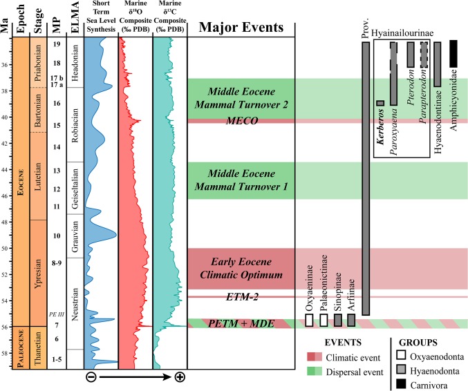
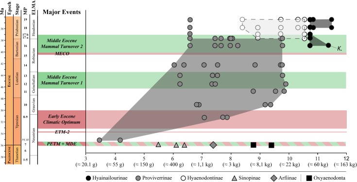
Hyaenodonts are first recorded in Europe during the earliest Eocene (Fig 1). Three subfamilies are known in Europe at this time. Sinopinae and Arfiinae were diverse in Northern Europe and found in Dormaal (Belgium; MP7) [12], Le Quesnoy (France; MP7) [13], and Abbey Wood (England; MP8+9) [14], while the Proviverrinae were restricted to Southern European Province [15–17] (as defined by Marandat [18] and Marandat et al. [19]). During the main part of the Ypresian, Lutetian, and Bartonian the hyaenodont proviverrines were the dominant specialized carnivorous mammals in European ecosystems [17,20–22] (Fig 1). The diversification of proviverrines in Europe, which were small-bodied and restricted to southern Europe during the earliest Eocene, was spurred by the disappearance of the subfamily’s presumed ecological competitors–Oxyaenodonta, and hyaenodonts from the clades Sinopinae and Arfiinae–during the Ypresian (between Dormaal (reference-level MP7) and Avenay (reference-level MP8+9) [13,17,23]).
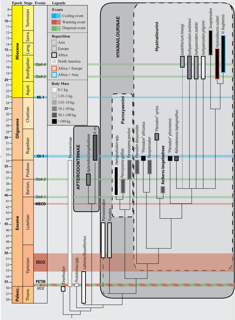
Fig 1. Stratigraphic repartition of the different subfamilies of specialized carnivorous mammals in Europe during the Paleocene and Eocene, with special attention to hyainailourines.
The new taxon described here, Kerberos langebadreae, is in bold. Note that currently i. the stratigraphic extension of Paroxyaena pavlovi during Priabonian is unknown [8] and ii. the age of Parapterodon is unknown but is estimated to be late Priabonian [9]. Stratigraphic scale, eustatic curve, and isotopic curves produced with TSCreator [10] from the data compiled by Gradstein et al. [11]. Abbreviations: ELMA, European Land Mammal Ages; MP, Mammal Palaeogene.
The decline of the proviverrines occurred during the Priabonian and coincides with the arrival in Europe during Bartonian and Priabonian of new competitors from Asia (Carnivora and Hyaenodontinae) and Africa (Hyainailourinae) [9] (Fig 1). The last occurrence of Proviverrinae (Allopterodon minor) is close to the MP19 reference-level in Obergösgen (Priabonian; Germany) [24]. While the Bartonian is characterized by the presence of Proviverrinae and Hyainailourinae, the Priabonian of Europe is characterized by the presence of Proviverrinae, Hyainailourinae and Hyaenodontinae (Fig 1).
One of the co-authors, D.V., discovered in 1981 the specimens described here in the French site of Montespieu–a locality that is very close to the city of Lautrec (Tarn) (Fig 2). As part of the “Castrais” faunas, the locality is considered to be Bartonian in age and close to the MP16 level (Robiac) [26–29]. The fossils belong to a large hyaenodont referred to a new hyainailourine taxon: Kerberos langebadreae gen. & sp. nov. The new taxon described here represents one of the two oldest hyainailourines recorded in Europe. The other, the hyainailourine Paroxyaena, is known in Robiac’s fauna (reference locality of MP16 reference-level) [30]. The study of these hyainailourines is important for understanding their ecological role in the Bartonian of Europe just as proviverrines begin to decline (Fig 1). Moreover, Kerberos langebadreae expands our knowledge of carnivorous mammalian niches in European environments.
Fig 2. Geographic map with indications of the location of the fossiliferous locality of Montespieu (MP16), where material of Kerberos langebadreae was found.
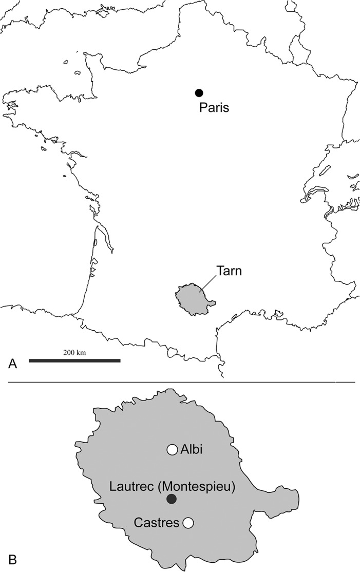
A, Localisation of the department of Tarn (France). B, Localisation of Montespieu in Tarn. B: redrawn after Laurent et al. ([25], Fig 1).
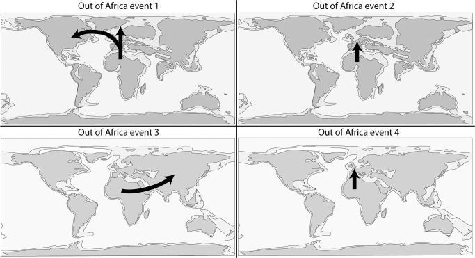
The discovery of this new species also has important implications for the evolution of Hyainailourinae. Solé et al. [6] recently described the earliest hyainailourines from the Gour Lazib Formation (late Early or early Middle Eocene; Algeria). Through subsequent dispersals, this subfamily is known from Africa, Asia, Europe, and North America, but the phylogenetic hypothesis presented in Solé et al. [6] supports an African origin for Hyainailourinae. This new taxon allows i. the study of the evolution of this subfamily, and ii. the examination of hyainailourine dispersals into Laurasia during the Eocene.
Additionally, the cranial, mandibular and dental remains associated with postcranial material are the first known for an early hyainailourine. Because the new taxon is one of the oldest hyainailourines ever recorded, the postcranial material allows us to study the skeletal morphology and locomotion of an early representative of the subfamily.
Material and Methods
Material
The fossils of Kerberos langebadreae were found by D.V. in the French fossiliferous locality of Montespieu, which is situated close to Lautrec (Tarn, France) (Fig 2). The Montespieu locality was discovered during 19th century. The mammals that were found in this locality were notably described and illustrated by Stehlin [26]. Most of the first fossils collected from the locality are housed in the Muséum d'Histoire naturelle de Toulouse (Collection Noulet). The mammal fauna of Montespieu is considered close to that of Robiac in age–the latter locality is the reference-locality for reference-level MP16 [29,30].
The fieldwork undertaken by D.V. in 1970s and ‘80s previously provided the type and only specimen of Cynohyaenodon lautricensis [31].
The material of the new hyainailourine was prepared by D.V. and C. Bouillet (MNHN). The specimens are housed at the Muséum national d’Histoire naturelle (Paris, France).
Specimen numbers: MNHN.F.EBA 517; MNHN.F.EBA 518a; MNHN.F.EBA 518b; MNHN.F.EBA 520; MNHN.F.EBA 521; MNHN.F.EBA 522; MNHN.F.EBA 523; MNHN.F.EBA 524; MNHN.F.EBA 525; MNHN.F.EBA 526; MNHN.F.EBA 527; MNHN.F.EBA 528.
Repository information: Muséum national d’Histoire Naturelle, Collection of fossils, Collection of fossils from the Eocene of Aquitaine Basin (EBA); 57 rue Cuvier, CP38, F-75005, Paris, France.
No permits were required for the described study, which complied with all relevant regulations.
Nomenclatural Acts
The electronic edition of this article conforms to the requirements of the amended International Code of Zoological Nomenclature, and hence the new names contained herein are available under that Code from the electronic edition of this article. This published work and the nomenclatural acts it contains have been registered in ZooBank, the online registration system for the ICZN. The ZooBank LSIDs (Life Science Identifiers) can be resolved and the associated information viewed through any standard web browser by appending the LSID to the prefix “http://zoobank.org/”. The LSID for this publication is: urn:lsid:zoobank.org:pub:8C2E4D2E-4890-4625-ABA9-B3137A6DB276. The electronic edition of this work was published in a journal with an ISSN, and has been archived and is available from the following digital repositories: PubMed Central, LOCKSS.
Terminology
We follow the dental terminology of Van Valen [32] for the molars, and of Ginsburg [33] for the premolars. The dental measurements (Length x Width in mm.) follow Gingerich and Deutsch [34]. The statistical parameters are the observed range (OR) and mean (M).
The osteological terminology follows that of Miller et al. [35]; while the measurements of the postcranial elements follow Argot [36].
Phylogenetic analysis
The taxa that have been included are listed in S1 Text. The data matrix is based on Holroyd [37] with new character definitions. It consists of 49 dental characters (32 binary characters and 17 multistate characters) and one character related to body mass (S2 Text). It includes 18 hyainailourine taxa.
The polarization of the characters was based on outgroup comparison criteria. All the multistate characters were treated as unordered–Holroyd [37] treated characters 34 and 44 as ordered.
We used the basal hyaenodonts Tinerhodon, Eoproviverra eisenmanni, and Prototomus as outgroups to Hyainailouridae. Because the dentition of E. eisenmanni is not entirely known (only several molars have been discovered), we used its contemporaneous Parvagula in order to code the premolars and the proviverrine Proviverra typica for coding the remaining characters. P. typica is considered one of the most primitive hyaenodonts–and thus proviverrines–despite its Eocene age [38]. Prototomus was coded based on the two European species: P. minimus and P. girardoti and the oldest North American species: P. deimos and P. phobos.
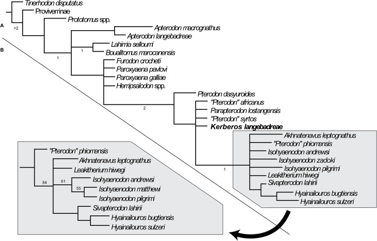
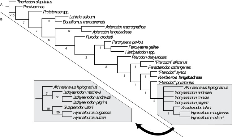
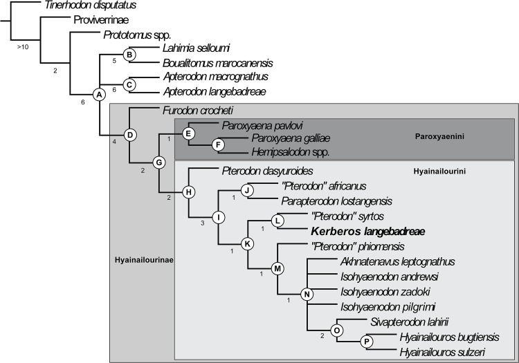
The data matrix (S1 File) was assembled with WinClada [39] and the parsimony analyses were performed with TNT [40] using implicit enumeration. The consensus trees have been assembled and analyzed with WinClada. We performed three analyses: the first one includes all the taxa and characters, while Leakitherium is excluded in the second and third analyses because it is very poorly known.
The genus Leakitherium is only known from two maxillary fragments that display P4, M1 and M2 [3]. Morales et al. [41] referred a fragment of an M2 to the genus. The taxonomic status of Leakitherium among hyainailourines is unresolved [41]. They hypothesized that Leakitherium is a synonym for Isohyaenodon andrewsi. In this analysis Leakitherium is closely related to Miocene hyainailourines such as Megistotherium and Isohyaenodon, but the hypothesis of Morales et al. [41] is not supported here.
Finally, the first character, which concerns body mass (S2 Text), is excluded in the third analysis because it could correspond to ecologic convergence rather than reflecting a phylogenetic signal.
After each analysis, each node of the strict consensus tree was assigned a Bremer support calculated with TNT for ten supplementary steps.
Results
Systematic Paleontology
Placentalia Owen, 1837 [42]
Ferae Linnaeus, 1758 [43]
Hyaenodonta Van Valen, 1967 [44]
Diagnosis (emended after Gunnell [45])
Elongate, narrow skull with narrow basicranium and high, narrow occiput; transversally constricted interorbital region; tritubercular to sectorial molars with carnassial blades in P4, M1, M2, and M1, M2 and M3 (except in Limnocyoninae and Machaeroidinae which have lost the posterior-most molars); M3 present in most taxa; M3 always present (except in Limnocyoninae and Machaeroidinae); manus and pes mesaxonic, plantigrade to digitigrade posture; fibula articulated with calcaneum; astragalar-cuboid articulation reduced or absent; terminal phalanges compressed and fissured at tip in most taxa; central, scaphoid, and lunar not fused (except perhaps in the hyainailourines Hyainailouros and Pterodon).
Distribution
Africa, Asia, Europe, and North America; Selandian (Paleocene) to Serravallian (Miocene).
Included families
Hyaenodontidae Leidy, 1869 [46], emended in this paper; Hyainailouridae Pilgrim, 1932 [47], emended in this paper;? Koholiinae Crochet, 1988 [48].
Notes
As in other specialized carnivorous mammals, hyaenodonts possess secant teeth. These meat-slicing teeth, also called carnassial teeth, are located in M1-2 and M2-3 position in Hyaenodonta [45]. Hyaenodonta was traditionally placed in the extinct order Creodonta together with the Oxyaenidae. Some workers have suggested a diphyletic origin for Creodonta [1,38,49–51]. While the separate origins of Oxyaenidae and Hyaenodontidae have not been established in a cladistic framework, Solé [51] proposed to raise these two families to the ordinal level–Oxyaenodonta and Hyaenodonta, and not Hyaenodontida as mistakenly used by several authors [6,51–53]–to highlight the possible separate evolutionary origins of these two extinct carnivorous mammalian lineages. It is worth mentioning that Spaulding et al. [54] recovered a monophyletic Creodonta, but their phylogenetic analyses, which addressed the relationships of Cetacea among mammals, only included four creodonts (three hyaenodonts and only one oxyaenid).
The systematic position of Koholiinae among Hyaenodonta is presently uncertain because cranial material is unknown (see below).
Discussion of the relationships within Hyaenodonta
Polly [38] established that hypercarnivory (characterized by reduction of the metaconids, simplification of the talonid, extension of the metastyle, reduction of the protocone, among other features) arose at least twice within hyaenodonts: among Hyainailourinae (Pterodontinae sensu Polly) and Hyaenodontinae. The distinction between the two subfamilies includes several cranial features. Polly noted that hyainailourines are characterized by a nuchal crest that does not extend laterally toward the mastoid processes and a circular subarcuate fossa on the petrosal. Hyaenodontines are characterized by a nuchal crest that extends toward the mastoid process, a robust bridge over the foramen stylomastoid primitivum, the absence of a bony ridge dividing the posterior petrosal sinus from the foramen stylomastoid primitivum, the presence of an inflated posterior petrosal sinus, and the presence of a horse-shoe shaped subarcuate fossa.
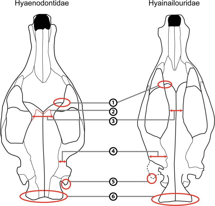
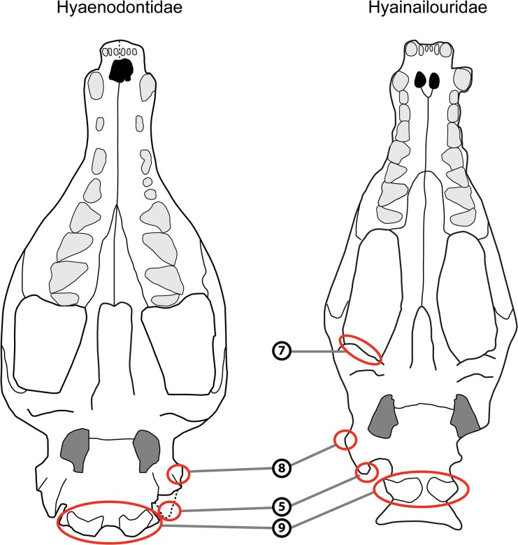
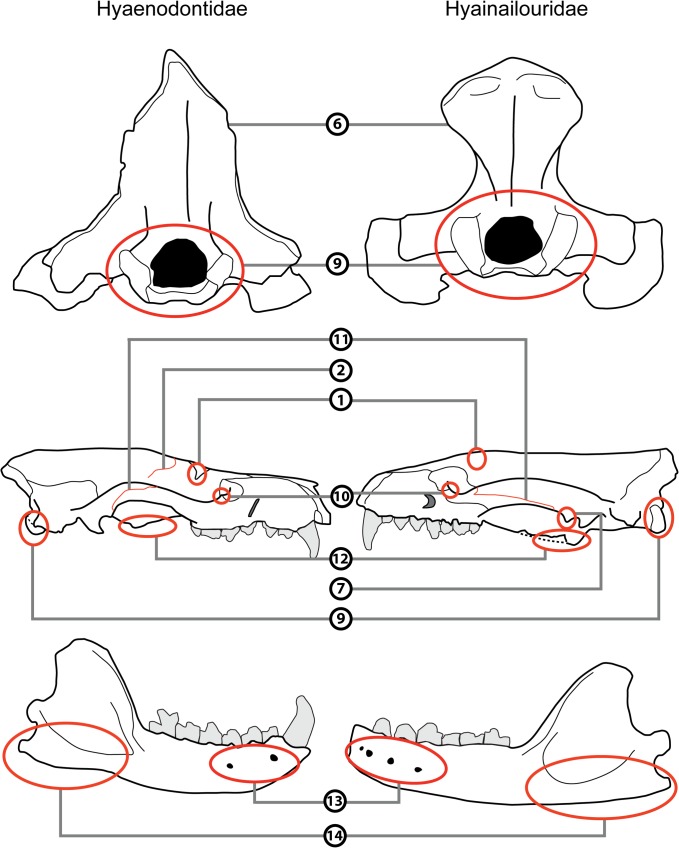
The cranial morphology of the hyainailourines is similar to that of the European and African hyaenodont subfamily Apterodontinae, notably in the short lateral extension of the nuchal crest (Fig 3, Feature 6; Table 1). Moreover, based on the present study, we can add the following features that seem to unite Hyainailouridae (Hyainailourinae+Apterodontinae): presence of a preglenoid crest (Figs 4 and 5, Feature 7; Table 1), early obliteration of the suture between the frontal and parietal bones (Figs 3 and 5, Feature 2; Table 1), anteroposteriorly extended suture between the jugal and squamosal (Fig 5, Feature 11; Table 1), a lateral expansion of the squamosal posterior to the zygomatic arch (Fig 3, Feature 4; Table 1), and large occipital condyles (Figs 4 and 5, Feature 9; Table 1). We can also note that apterodontines and hyainailourines often display more than two mental foramina (Fig 5, Feature 13; Table 1), while hyaenodontids very rarely display more than two, and have a weak ventral concavity anterior to the angular process (Fig 5, Feature 14; Table 1). The younger representatives of the clade (e.g., Hyainailouros) are characterized by a transverse elongation of the mastoid process (Fig 4, Feature 8; Table 1), rather than an anteroposterior elongation (Figs 3 and 4, Feature 5; Table 1) as observed in the earliest species in the order Hyaenodonta. Finally, the narrowest part of the braincase is immediately behind the postorbital processes in Hyaenodontidae, while this constriction is more posterior in Hyainailouridae (at least at the middle of the parietals) (Fig 3, Feature 3; Table 1).
Fig 3. Comparison of the skull in dorsal view of Hyaenodontidae (left) and Hyainailouridae (right).
See Table 1 for description of the numbered features. Left: skull of Sinopa grangeri redrawn after Matthew ([55], Fig 4); right: skull of Apterodon macrognathus redrawn after Osborn ([56]; Fig 1A).
Table 1. Comparison of the cranial features of Hyaenodontidae and Hyainailouridae.
| Feature | Hyainailouridae | Hyaenodontidae |
|---|---|---|
| 1: Postorbital process | Poorly developed; except in Miocene Hemipsalodon | Well developed |
| 2: Suture frontal parietal | Early obliteration of the suture (except in Hemipsalodon) | Present |
| 3: Constriction before the braincase | Absent | Present |
| 4: Squamosal | Laterally expanded posterior to the zygomatic arch | Poorly laterally expanded |
| 5: Expansion of the mastoido-paroccipital apophysis | Weak posterior extension in later forms | Always posteriorly extended |
| 6: Expansion of the nuchal crest to mastoid process | Nuchal crest does not reach the mastoid process | Nuchal crest reaches the mastoid process |
| 7: Preglenoid crest | Present | Absent |
| 8: Lateral expansion of the mastoid process | Mastoid process laterally expanded in later forms | Mastoid process poorly laterally expanded in later forms |
| 9: Occipital condyle | Large | Small |
| 10: Process at the junction between the maxillary and jugal | Rarely present | Often present |
| 11: Suture between the jugal and squamosal | Anteroposteriorly extended | Poorly anteroposteriorly extended |
| 12: Pterygoid | Extended anteroposteriorly and ventrally | Poorly extended anteroposteriorly and ventrally |
| 13: Mental foramina | Often more than two | Very rarely more than two |
| 14: Angular process | Poorly developed; absence of anterior concavity | Robust with a anteriorly located concavity |
Fig 4. Comparison of the skull in ventral view of Hyaenodontidae (left) and Hyainailouridae (right).
See Table 1 for description of the numbered features. Left: skull of Sinopa grangeri redrawn after Matthew ([55], Fig 5); right: skull of Apterodon macrognathus redrawn after Szalay ([57]; Fig 7).
Fig 5. Comparison of the skull in posterior (upper) and lateral (middle) views and of the mandible in labial view (lower) of Hyaenodontidae (left) and Hyainailouridae (right).
See Table 1 for description of the numbered features. Upper: skulls of Pterodon dasyuroides (right) and Hyaenodon compressus (left) redrawn after Polly ([38]; Fig 11A and 11B); middle: skulls of Limnocyon verus (left) and Apterodon macrognathus (right) redrawn respectively after Matthew ([58]; Fig 53) and Szalay ([57], Fig 6); lower: mandibles of Limnocyon verus (left) and Apterodon macrognathus (right) redrawn respectively after Matthew ([58]; Fig 53) and Szalay ([57], Fig 9).
Some representatives of hyainailourines have lost the P1 [9]. This feature is one of the main characteristics of Koholiinae [1]. Koholiines and hyainailourines share the latter feature, as well as the presence of pronounced secant teeth and extensive radiations in Africa. However, the koholiines differ from hyainailourines in featuring a robust angular process that is preceded anteriorly by a concavity (Fig 5, Feature 14; Table 1). It is thus presently difficult to refer the koholiines to either Hyainailouridae or Hyaenodontidae.
The cranial morphology of Hyaenodontidae is on the whole similar to Hyaenodontinae in Polly [38]. The clade is characterized by the morphology of the nuchal crest, which extends toward the mastoid process (Figs 3 and 5, Feature 6; Table 1). We can add several features not described previously to distinguish Hyaenodontidae from Hyainailouridae. Hyaenodontids generally possess: a well-developed postorbital process (Figs 3 and 5, Feature 1; Table 1), a short extension of the jugal/squamosal suture anteroposteriorly (Fig 5, Feature 11; Table 1), absence of a preglenoid crest (Figs 4 and 5, feature 7; Table 1), a process at the junction between the maxillary and the jugal (Fig 5, Feature 10; Table 1), and a small occipital condyle (Figs 4 and 5, Feature 9; Table 1). Unlike Hyainailouridae, Hyaenodontidae exhibit a clear suture between the parietal and frontal (Figs 3 and 5, Feature 2; Table 1) and the suture is located close to the anterior constriction of the braincase (Fig 3, Feature 3; Table 1). Hyaenodontids all possess a single- or double-rooted p1 and very rarely display more than two mental foramina (Fig 5, Feature 13; Table 1). Hyaenodontid pterygoids are generally less developed ventrally and posteriorly than hyainailourid pterygoids (Fig 5, Feature 12; Table 1). In almost all hyaenodontids the mandible possesses a concavity on the ventral margin anterior to the angular process and the latter is generally robust and projects more ventrally in Hyaenodontidae (Fig 5, Feature 14; Table 1).
We tentatively refer the Teratodontinae as recently defined by Solé et al. [6] to Hyaenodontidae based on the skull of Dissopsalis described by Colbert [59] and mandibles available for the teratodontine genera. The fragmentary skull of Dissopsalis seems to display developed postorbital process (Figs 3 and 5, Feature 1; Table 1), constriction before the braincase (Fig 3, Feature 3; Table 1), and a distally elongated mastoido-paroccipital apophysis (Figs 3 and 4, Feature 5; Table 1). Colbert [59] noted that Dissopsalis possesses a developed preglenoid crest (Figs 4 and 5, Feature 7; Table 1), a feature of Hyainailouridae; however, based on his drawings, we assess that the morphology of the mandibular fossa is similar to that of Hyaenodontidae rather than to that of Hyainailouridae. Finally, teratodontines display a concavity anterior to a robust angular process (Fig 5, Feature 14; Table 1) as in other Hyaenodontidae.
The Indohyaenodontidae seem to be endemic in South (India, Pakistan) and Southeast (Myanmar) of Asia [6]. Unfortunately, only few cranial remains are known for this subfamily [60–61]. Cranial and dental specimens show typical features of Hyaenodontidae such as the presence of a concavity anterior to the angular process (Fig 5, Feature 14; Table 1), the presence of only two mental foramina (Fig 5, Feature 13; Table 1), the absence of a preglenoid crest (Figs 4 and 5, Feature 7; Table 1), a process at the junction between the maxillary and the jugal (Fig 5, Feature 10; Table 1), a well-developed postorbital process (Figs 3 and 5, Feature 1; Table 1), and a clear suture between the parietal and frontal (Figs 3 and 5, Feature 2; Table 1). We therefore include this subfamily in Hyaenodontidae.
Machaeroidinae has been often considered to be closely related to Limnocyoninae [62]. Machaeroidinae and Limnocyoninae notably exhibit an extension of the nuchal crest towards the mastoid, as seen in other species in Hyaenodontidae. Species in both subfamilies have moreover lost M3 and M3 (except in their oldest representative Prolimnocyon), supporting a close relationship between these families and the inclusion of both Machaeroidinae and Limnocyoninae in Hyaenodonta. Several authors (e.g. Muizon & Lange-Badré [63]) however referred the Machaeroidinae to Oxyaenodonta; Zack [64] recently proposed arguments based on postcranial elements to support this hypothesis.
The two cranial morphotypes described and discussed here may be useful for clarifying the relationships within Hyaenodonta, though these features await rigorous evaluation in a cladistic analysis to establish plesiomorphic and apomorphic states of characters. Because the two morphotypes imply distinct recruitment of the cranial muscles and both morphotypes seem to be a combination of primitive and derived features, we hypothesize that these two cranial morphotypes appeared early in the radiation of Hyaenodonta. Based on the presence of hyaenodontids in the Paleocene of Asia, we place the radiation of Hyaenodonta and divergence of Hyaenodontidae and Hyainailouridae during the Selandian, if not earlier [2].
Whether or not the order Hyaenodonta is rooted in Africa (Hyainailourindae + Hyaenodontidae) as proposed by Gheerbrant [5], Gheerbrant et al. [50] and Solé et al. [1,6] needs further clarification.
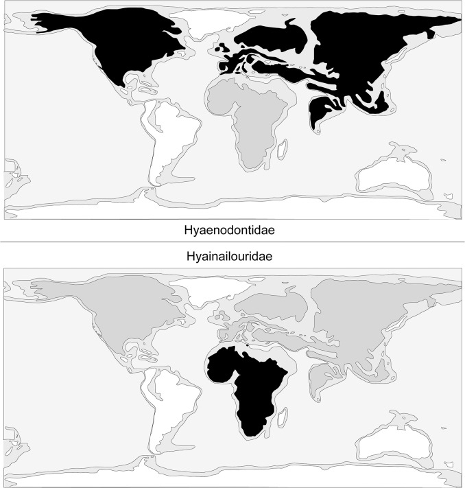
One can note that Hyaenodontidae mainly radiated in Laurasia (Fig 6) where six clades are known (Hyaenodontinae, Limnocyoninae, Sinopinae, Arfiinae, Proviverrinae, and probably Machaeroidinae), while Hyainailouridae principally radiated in Africa (Fig 6), where koholiines, apterodontines and hyainailourines find their earliest record [1,6,52].
Fig 6. Comparison of the geographic distribution of Hyaenodontidae (top) and Hyainailouridae (bottom).
White: no fossils recorded; Grey: limited diversity; Black: main radiation.
Concerning the dispersals of these two families, it is worth mentioning that apterodontines dispersed into Europe and hyainailourines dispersed into North America, Europe and Asia [9,65–66]. Among Hyaenodontidae, only Teratodontinae and Indohyaenodontinae are known exclusively from Gondwanan landmasses (Africa and South/South-East of Asia). If Hyaenodontidae appeared in Laurasia, these two subfamilies may have dispersed into Africa during the earliest Eocene.
Hyaenodontidae Leidy, 1869 emend. [46]
Diagnosis
The members of this family share a cranial pattern characterized by the presence of a visible suture between the parietal and frontal and a well-developed postorbital process, a constriction anterior to the braincase, pterygoid generally shorter than in Hyainailouridae, a short extension of the jugal/squamosal suture anteroposteriorly, the absence of preglenoid crest, a distally elongated mastoido-paroccipital apophysis, a nuchal crest extending to the mastoid process, small occipital condyles, the presence of only two mental foramina, and a concavity anterior to the angular process (less pronounced in hyaenodontines). The P1 (single- or double-rooted) is usually present.
Distribution
Africa, Asia, Europe, and North America; Thanetian (Paleocene) to Serravallian (Miocene).
Included subfamilies
Arfiinae Solé, 2013 [51]; Hyaenodontinae Leidy, 1869 [46]; Indohyaenodontinae Solé et al., 2013 [6]; Limnocyoninae Wortman, 1902 [67];? Machaeroidinae Matthew, 1909 [58]; Proviverrinae Schlosser, 1886 [68]; Sinopinae Solé, 2013 [51]; Teratodontinae Savage, 1965 [3].
Note
The systematic position of the saber-toothed Machaeroidinae among Hyaenodonta or Oxyaenodonta is presently uncertain (see Gunnell [45]).
Hyainailouridae Pilgrim, 1832 emend. [47]
Diagnosis
The members of this family share a cranial pattern characterized by a massive skull, the absence of visible suture between the parietal and frontal, a weak postorbital process, a pterygoid generally extended ventrally and distally, an anteroposteriorly extended suture of the jugal/squamosal, the presence of a preglenoid crest, a lateral expansion of the squamosal posterior to the zygomatic arch, a transversally expanded mastoid process (in later forms), a nuchal crest that does not extend laterally to mastoid process, large occipital condyles, and the presence two or more mental foramina. The P1, if present, is small (single-rooted).
Distribution
Africa, Asia, and Europe; late Ypresian or early Lutetian (Paleocene) to Serravallian (Miocene).
Included subfamilies
Apterodontinae Szalay, 1967 [57]; Hyainailourinae Pilgrim, 1932 [47].
Relationships between the Hyainailourinae and Apterodontinae
Hyainailourinae and Apterodontinae are diverse during the Paleogene in Africa [3,52,69–70]. The two groups are also recorded in Europe: the Hyainailourinae are recorded in the Late Eocene and Early to Middle Miocene, while the Apterodotinae are known in the Early Oligocene [9,33,66,71]. Hyainailourinae is also known in Early to Middle Miocene of Asia [47].
Recent discoveries in North Africa place the youngest possible origin of Hyainailourinae in the late Early or early Middle Eocene [6] and the youngest possible origin of Apterodontinae in the late Middle Eocene [52], implying an Afro-Arabian origin for these subfamilies. The presence of Hyainailourinae and Apterodontinae in Eurasia is likely a consequence of Northern immigrations (see below).
Apterodontinae is a peculiar group among Hyaenodonta [37,38,52,57]. Apterodontines are characterized by the loss of the metaconid, the reduction of the paraconid, and retention of a prominent talonid on the lower molars where most hyaenodonts with reduced metaconids have also reduced the talonid and increased the relative size of the paraconid. Based on postcranial material, Grohé et al. [52] proposed semi-aquatic habits for Apterodon. They suggested that the unusual dentition of Apterodon may have been related to a semi-aquatic lifestyle with simplified and tall teeth, employed for catching aquatic prey such as ‘fish’ and/or ‘invertebrates’ [52].
In contrast to apterodontines, large hyainailourines were likely hunters and scavengers and were probably ecologically analogous to the extant hyenas and the extinct borophagines; the latter however differ from large hyainailourines in exhibiting cursorial adaptations (see [72] and [73]).
The possibility of a close relationship between apterodontines and hyainailourines based on dental and cranial characters has been suggested before [6,38,52,61]. Here we explicitly identify numerous cranial features (e.g., absence of visible suture between the parietal and frontal, weak postorbital process, presence of a preglenoid crest, large occipital condyles) that group Apterodontinae and Hyainailourinae within the family Hyainailouridae (see above). The strongest similarities between apterodontines and hyainailourines lie in the posterior region of the skull related to the characteristic rearrangement of cervical musculature first described by Polly [38] in Hyainailourinae (Pterodontinae in Polly [38]).
The skull of Apterodon is anteroposteriorly elongate. The oldest hyainailourines such as Pterodon and Kerberos also exhibit anteroposteriorly elongate skulls, while others, such as the later hyainailourines Paroxyaena, Hemipsalodon, and Hyainailouros, differ from Apterodon and early hyainailoruines in featuring anteroposteriorly short rostral and basicranial regions. This suggests that Kerberos and Apterodon possess a primitive overall cranial morphology for Hyainailouridae.
Apterodon differs from hyainailourines in several features, which include a more robust zygomatic arch. This feature provides greater surface area for the origin of larger masseter muscles in apterodontines. On the mandible, the masseteric fossa is shallow but anteroposteriorly elongate, providing a long insertion for the masseter relative to the masseteric fossa in Kerberos and Pterodon, which further supports the important role of the masseter muscles in apterodontines.
Apterodon also differs from hyainailourines in the arrangement of the upper tooth rows that are close to parallel along the palate while nearly all other hyaenodonts have tooth rows that are distinctively diverging posteriorly. This parallel alignment may be related to semi-aquatic feeding as well. The facial morphology of apterodontines also differs from hyainailourines that have a strong narrowing of the facial morphology posterior to the canines [8,9]. Besides the differences in the masseteric fossa morphology, the mandibles of apterodontines and hyainailourines are generally similar. It is interesting to note that Apterodon macrognathus and Kerberos share the presence of a dorsally inflected, thin and pointed angular process, and an extended coronoid crest.
Concerning the postcranium, the only elements that can be compared between Apterontinae and Hyainailourinae are the distal part of the fibula, the astragalus, and the calcaneum. The distal part of the fibula is very similar in these two groups. However, there are differences in the tarsal bones. The tuber calcaneus of Kerberos and Hyainailouros is shorter and wider than that of Apterodon and the ectal facet of the hyainailourines, though poorly preserved on the specimen referred to Kerberos, seems to be shorter than the ectal facet of Apterodon. The neck of the astragalus of Kerberos and Hyainailouros is also shorter and narrower than that of Apterodon, though this difference in morphology may be related to the larger body size of the hyainailourine species. Apterodoninae and Hyainailourinae share the shallow groove of the tibial facet. This feature suggests that both groups maintained a plantigrade foot posture, though this morphology seems to be primitive in Hyaenodonta. The differences suggest a more weight-bearing mode of life in Kerberos, while the adaptations of Apterodon may suggest adaptations towards semi-aquatic mode of life.
In conclusion, apterodontines and primitive hyainailourines share a characteristic cranial construction. Broad attachments for nuchal musculature could have been adaptive for scavenging/hunting in terrestrial and aquatic environments, but this hypothesis needs to be tested in the future. However, numerous differences are present in the dentition and postcranium, and we advocate retaining separate subfamily designations for these lineages.
Hyainailourinae Pilgrim, 1932 [47]
Emended diagnosis (emended after Holroyd [69])
Hyainailouridae with high and secant paraconid, connate metacone and paracone on M1-M2, weak to absent P3 lingual cingulum, P4 lacking continuous lingual cingulum, relatively large anterior keels on lower molars, M3 talonid reduced relative to that of M1–M2, lower molar protoconids and paraconids subequal in length, facial region constricted lateromedially at P2 and abruptly expanded caudal to P4, infraorbital process poorly developed, massive zygomatic arch, anterior margin of choanae located at the level of M3, and circular subarcuate fossa present on petrosal.
Note
Hyainailourinae (sensu Solé et al., 2013 [6]) includes 12 genera: Akhnatenavus Holroyd, 1999 [69], Furodon Solé et al., 2013 [6], Hemipsalodon Cope, 1885 [74], Hyainailouros Biedermann, 1863 [75], Isohyaenodon Savage, 1965 [3], Kerberos gen. nov. (present work), Leakitherium Savage, 1965 [3], Parapterodon Lange-Badré, 1979 [9], Paroxyaena Martin, 1906 [76], Parvavorodon Solé et al., 2013 [6], Pterodon Blainville, 1839 [77] and Sivapterodon Ginsburg, 1980 [4].
However, agreement on which species should be assigned to different hyainailourine genera is not firmly established. We follow Morlo et al. [78] by assigning only two species to Hyainailouros: Hyainailouros bugtiensis Pilgrim, 1912 [79] (= Megistotherium osteothlastes Savage, 1973 [80]) and Hyainailouros sulzeri Biedermann, 1863 [75] (= Hyainailouros fourtaui Koenigswald, 1947 [81] = Hyainailouros napakensis Ginsburg, 1980 [4] = Hyainailouros nyanzae Savage, 1965 [3]). Concerning Isohyaenodon, we partially follow Morales et al. [41], by including three species: Isohyaenodon andrewsi Savage, 1965 [3], Isohyaenodon zadoki Savage, 1965 [3] (= Isohyaenodon matthewi Savage, 1965 [3]) and Isohyaenodon pilgrimi Savage, 1965 [3]; however Leakitherium is not synonymized with Isohyaenodon andrewsi herein. Moreover, our analysis shows that ‘Pterodon’ might not be a natural group but represents a polyphyletic genus.
Paroxyaenini Lavrov, 2007 [8]
Diagnosis (emended after Lavrov [8])
Medium-sized Hyainailourinae with short, wide facial region. Dental formula: I3/3, C1/1, P4/4, M3/3. P1 single-rooted, P2 and P3 double-rooted. Protocone well developed on P3. Amphicones of M1 and M2 incompletely fused; apices of paracone and metacone distinctly separate; cingulum well developed on molars and premolars; M1, M2, and DP4 showing well-developed precingula with many cuspules. Metastyle of M1 and M2 approximately half of the tooth length. Postprotocrista absent from M1 and M2; QM1 approximately 120°–130°, QM2 approximately 125°; surface of enamel of P3 –M2 rugose. Fissura orbitalis and foramen rotundum fused into one foramen. Medial part of glenoid fossa of jaw joint very deep. Large maxillo-nasalis fossa located dorsal to alveoli of P4–M2. Tentorium well developed.
Included genera
Hemipsalodon Cope, 1885 [74] and Paroxyaena Martin, 1906 [76].
Distribution
Europe and North America; Bartonian (Eocene) to Priabonian (Eocene).
Hyainailourini Ginsburg, 1980 [4]
Diagnosis
Small to large-sized Hyainailourines: facial region elongated in oldest representatives (e.g., Kerberos), but shorter in youngest ones (e.g., Hyainailouros). Dental formula: I1-3/2-3, C1/1, P4/3-4, M2-3/3. P1 single (e.g., Hyainailouros) or double-rooted (e.g., Kerberos), Amphicones of M1 and M2 nearly to completely fused; protocone on molars very reduced; cingula weakly developed on molars and premolars. Fissura orbitalis and foramen rotundum separated.
Included genera
Akhnatenavus Holroyd, 1999 [69], Hyainailouros Biedermann, 1863 [75], Isohyaenodon Savage, 1965 [3], Kerberos gen. nov., Leakitherium Savage, 1965 [3], Parapterodon Lange-Badré, 1979 [9], Pterodon Blainville, 1839 [77] and Sivapterodon Ginsburg, 1980 [4].
Distribution
Africa and Europe; Bartonian (Eocene) to Serravallian (Miocene).
Kerberos gen. nov.
urn:lsid:zoobank.org:act:CF40CC5A-025F-464B-8769-54471DD888B0
Diagnosis
Same as for the type and only species.
Type and only species
Kerberos langebadreae nov. sp.
Type locality
Montespieu (locality close to Lautrec), Tarn, France; MP16, Bartonian, Eocene.
Type horizon and age
Formation des Molasses de Saix et de Lautrec, Grès de Puech Auriol et de Venès, Bartonian, Eocene.
Etymology
Cerberus (Kerberos in Greek) is an impressive mythological multi-headed dog–Hellhound–that guards the entrance of the Underworld.
Kerberos langebadreae sp. nov.
urn:lsid:zoobank.org:act:8645FC88-4DAF-4084-9E14-64AE1D707EC7 (Figs 7–12)
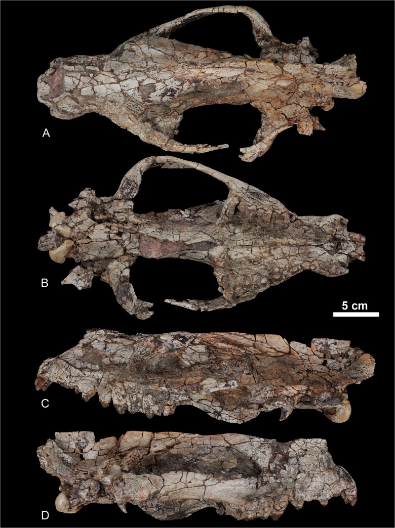
Fig 7. Skull of Kerberos langebadreae gen. & sp. nov (holotype, MNHN.F.EBA 517).
A, dorsal view; B, ventral view; C, left lateral view; D, right lateral view.
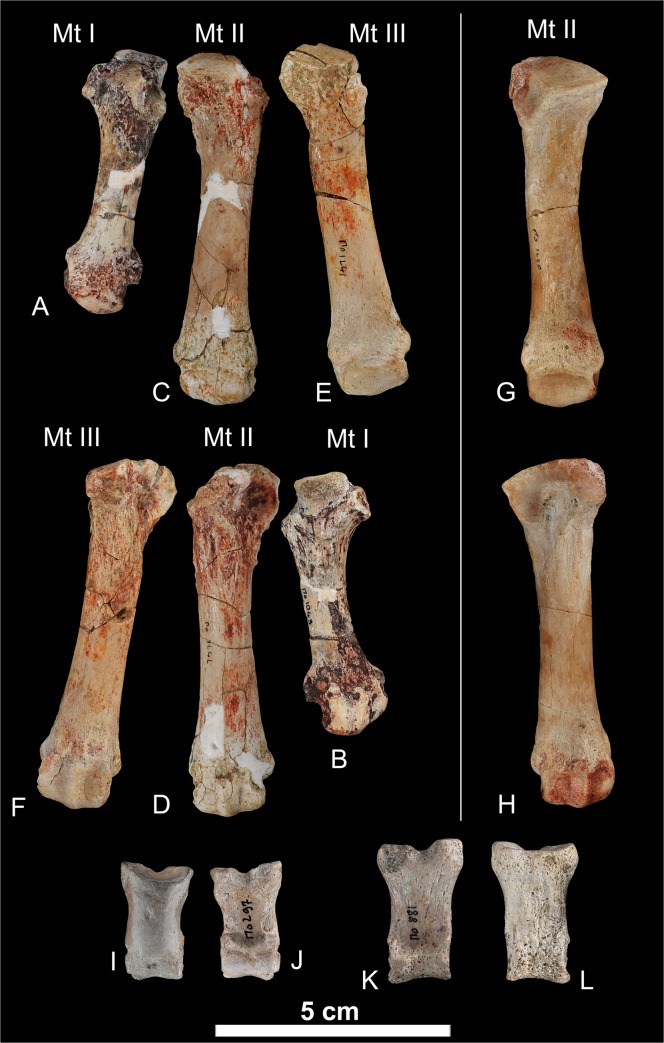
Fig 12. Postcranial elements of Kerberos langebadreae gen. & sp. nov.
A-B, metatarsal I (MNHN.F.EBA 523); A, dorsal view; B, plantar view. C-D, metatarsal II (right MNHN.F.EBA 524); C, dorsal view; D, plantar view. E-F, metatarsal III (MNHN.F.EBA 525); E, dorsal view; F, plantar view. G-H, metatarsal II (left, MNHN.F.EBA 526); G, dorsal view; H, plantar view. I-J, middle phalanx (MNHN.F.EBA 528); I, dorsal view; J, plantar view. K-L, middle phalanx (MNHN.F.EBA 527); K, dorsal view; L, plantar view.
Diagnosis
This large-sized hyainailourine displays a particular combination of primitive features, namely an anteriorly narrow mandible and a small diastema between P1 and P2, and derived features, namely a reduced parastylar area on upper molars, a large protocone on P3, and premaxilla narrow throughout its whole length. It differs from Paroxyaena by having a more closely appressed paracone and metacone, and more reduced protocone on upper molars, and a shorter and narrower talonid on the lower molars. Kerberos differs from Pterodon dasyuroides by possessing larger P1 and P1, and from Parapterodon by retaining the P1, exhibiting a more reduced parastylar area, and a more reduced protocone on upper molars.
Etymology
Dedicated to Dr. Lange-Badré, who greatly improved our knowledge of Eocene carnivorous mammals.
Type locality
Montespieu (locality close to Lautrec), Tarn, France; MP16, Bartonian, Eocene.
Type horizon and age
Formation des Molasses de Saix et de Lautrec, Grès de Puech Auriol et de Venès, Bartonian, Eocene.
Holotype
MNHN.F.EBA 517, nearly complete skull bearing on right side I2-I3 and P2-M3, and on left side I2-I3, P1, P4-M3.
Referred material
MNHN.F.EBA 518a, left hemimandible bearing C and M2-M3; MNHN.F.EBA 518b, right hemimandible bearing P2-P4 and M2-M3; MNHN.F.EBA 520, right fibula; MNHN.F.EBA 521, right astragalus; MNHN.F.EBA 522, right calcaneus; MNHN.F.EBA 523, right metatarsal I; MNHN.F.EBA 524, right metatarsal II; MNHN.F.EBA 525, right metatarsal III; MNHN.F.EBA 526, left metatarsal II; MNHN.F.EBA 527, right middle phalanx; MNHN.F.EBA 528, right middle phalanx.
Measurements
Table 2. Dental measurements (in mm.) of Kerberos langebadreae gen. & sp. nov.
| Locus | n | OR | Mean | Locus | n | OR | Mean | ||
|---|---|---|---|---|---|---|---|---|---|
| I1 | L | 2 | 0.68–0.92 | 0.8 | |||||
| W | 2 | 1.50–1.55 | 1.53 | ||||||
| I2 | L | 2 | 0.44–0.59 | 0.52 | |||||
| W | 2 | 0.91–0.98 | 0.93 | ||||||
| I3 | L | 2 | 0.18–0.28 | 0.23 | |||||
| W | 2 | 0.61–0.67 | 0.64 | ||||||
| C | L | 2 | 2.17–2.19 | 2.18 | C | L | 2 | 1.98–2.12 | 2.05 |
| W | 2 | 1.54–1.63 | 1.59 | W | 2 | 1.76 | 1.76 | ||
| P1 | L | 2 | 1.07*-1.21 | 1.14 | P1 | L | 2 | 1.62*-1.67* | 1.65 |
| W | 2 | 0.65–0.69 | 0.67 | W | 2 | 0.75*-0.77* | 0.76 | ||
| P2 | L | 2 | 1.35–1.64* | 1.5 | P2 | L | 2 | 1.52*-1.68* | 1.60 |
| W | 2 | 0.64–0.65* | 0.65 | W | 2 | 0.68*-0.86* | 0.77 | ||
| P3 | L | 2 | 1.83*-1.86 | 1.85 | P3 | L | 2 | 1.62–1.63* | 1.63 |
| W | 2 | 0.85*-1.18 | 1 | W | 2 | 0.72*-0.91 | 0.82 | ||
| P4 | L | 2 | 2.08–2.13 | 2.11 | P4 | L | 1 | 2.03 | - |
| W | 2 | 1.63–1.76 | 1.7 | W | 1 | 1.01 | - | ||
| M1 | L | 2 | 1.91–1.95 | 1.93 | M1 | L | 1 | 1.57* | - |
| W | 2 | 1.75–1.84 | 1.8 | W | 1 | 1.16* | - | ||
| M2 | L | 2 | 2.30–2.39 | 2.35 | M2 | L | 2 | 1.87–2.09 | 1.98 |
| W | 2 | 1.81–2 | 1.9 | W | 2 | 1.15–1.19 | 1.17 | ||
| M3 | L | 2 | 0.89–0.95 | 0.92 | M3 | L | 2 | 2.50–2.66 | 2.58 |
| W | 2 | 2.44–2.53 | 2.49 | W | 2 | 1.36–1.37 | 1.37 |
*based on roots. Abbreviations: L, mesiodistal length; n, number of teeth measured; OR, Observed range; W, linguolabial width. Observed range.
Table 3. Measurements (in mm.) of postcranial elements of Kerberos langebadreae gen. & sp. nov.
| Measurements of the fibula | ||
| Total length | 193.3 | |
| Anteroposterior depth of the head | 27* | |
| Transverse width of head | 18.3 | |
| Mid-shaft anteroposterior diameter | 12.1 | |
| Mid-shaft transverse diameter | 10.9 | |
| Anteroposterior depth of the distal epiphysis | 26.7 | |
| Transverse width of the distal epiphysis | 19.7 | |
| Measurements of the calcaneum | ||
| Total length | - | |
| Length of the tuber calcanei (up to the ectal facet) | 40.7 | |
| Width of the tuber at mid-length | 21.9 | |
| Transverse width of the ectal facet | 13.5 | |
| Proximodistal length of the ectal facet | 38.6 | |
| Measurements of the astragalus | ||
| Total length | 52.6 | |
| Maximum transverse length | 45.6 | |
| Trochlea length | 35.3 | |
| Trochlea width | 26.5 | |
| Head width | 30.6 | |
| Measurements of the metatarsals and middle phalanges | ||
| Metatarsal I | Length | 57.5 |
| Transverse width at mid-length | 9.6 | |
| Metatarsal II (left) | Length | 76.5 |
| Transverse width at mid-length | 11.4 | |
| Metatarsal II (right) | Length | 76.9 |
| Transverse width at mid-length | 11.9 | |
| Metatarsal III | Length | 78.0 |
| Transverse width at mid-length | 11.6 | |
| Middle phalange MNHN.EBA 528 | Length | 26.7 |
| Transverse width at mid-length | 12.2 | |
| Middle phalange MNHN.EBA 527 | Length | 31.7 |
| Transverse width at mid-length | 14.0 | |
*, estimated.
Description of the skull, mandible and dentition
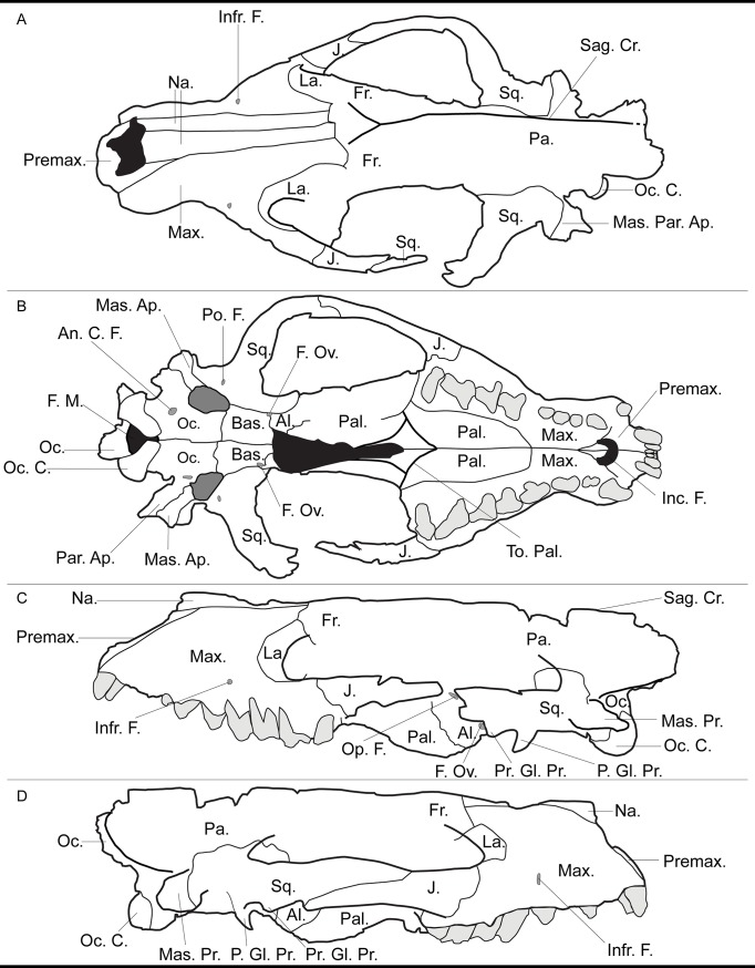
The skull of the holotype is distorted transversally but is almost complete (Fig 7). It is also riddled with postmortem cracks and breaks, which partly obliterate the sutures (see Fig 13 for drawings of the skull with indications of the sutures). The auditory region, which is poorly preserved, will not be described thoroughly. The taxa chosen for comparison assessed from original specimens housed in the MNHN and literature represent the different subfamilies of Hyaenodonta (i.e., the Proviverrinae, Hyainailourinae, Hyaenodontinae, and Limnocyoninae), which are known from the Bartonian, Priabonian and Oligocene.
Fig 13. Drawings of the skull of Kerberos langebadreae gen. & sp. nov. (holotype, MNHN.F.EBA 517).
A, dorsal view; B, ventral view; C, left lateral view; D, right lateral view. Abbreviations: Al., alisphenoid; An. C. F., anterior condyloid foramen; Bas., basisphenoid; F. M., foramen magnum; F. Ov., foramen oval; Fr., frontal; Inc. F., incisive foramen; Infr. F., infraorbital foramen; J., jugal; La., lacrimal; Mas. Ap., mastoid apophysis; Mas. Par. Ap., mastoid-paroccipital apophysis; Mas. Pr., mastoid process; Max., maxilla; Na., nasal; Oc. C., occipital condyle; Oc., occipital; Op. F., optic foramen; P. Gl. Pr., postglenoid process; Pa., parietal; Pal., palatine; Par. Ap., paroccipial apophysis; Po. F., posterior pterygoid foramen; Pr. Gl. Pr., preglenoid process; Premax., premaxilla; Sag. Cr., sagittal crest; Sq., squamosal; To. Pal., torus palatinus.
The most striking feature of the skull is its tremendous size compared to contemporaneous European proviverrines. The condylobasal length (~ 35 cm) approaches that of a female Ursus arctos [82]. The rostrum is very short, even shorter than that of the brevirostrate Hyaenodon brachyrhynchus. The stoutness of the rostrum contrasts with the relatively long ethmoid region. Even though the basicranium is crushed transversally, it seems to have been mediolaterally extensive as in Pterodon dasyuroides.
Premaxilla (Fig 13, Premax.). In lateral view (Fig 7), the opening of the nasal cavity forms an angle of approximately 45° with the anteroposterior lengthening of the skull, which approaches the condition of H. brachyrhynchus. The nasal aperture forms a more opened angle in the proviverrine Cynohyaenodon cayluxi and a slightly sharper angle in P. dasyuroides. The premaxilla differs from that of other hyaenodonts as the ascending ramus is narrow along its entire length. The posterior portion of the premaxilla is not extensive, reaching just posterior to the dorsal edge of the opening of the nasal cavity. The general hyaenodont condition is an extended premaxilla that reaches more posterior to the nasal aperture. The incisive foramina (Fig 13, Inc. F.) are anteroposteriorly shorter than those of P. dasyuroides.
Maxilla (Fig 13, Max.). Dorsal to the canine, the maxilla shows a weak anteroposterior convexity surrounding the long root of the tooth. At the level of P2, the maxillae are conspicuously constricted transversally. The constriction extends to the palate ventrally. Posteriorly the lateral borders of the maxillae flare, reaching more than twice the width of that of the anterior constriction. This morphology is similar to the condition observed in P. dasyuroides. The anterior opening of the infraorbital canal (Fig 13, Infr. F.) is at the level of the mesial root of P4, as in P. dasyuroides, but unlike the more anterior condition at the level of the interradicular space of P3 seen in Hyaenodon [9]. Anterior to the infraorbital canal there is a shallow and round concavity, as described in C. cayluxi [9]. Superior to M1 and M2 is a well-marked fossa for the origin of nasolabialis. The inferior border of the orbit is horizontal, as in C. cayluxi. The posterior margin of the orbit does not slope dorsally as in P. dasyuroides. The orbit is relatively open on its dorsal and ventral margins, as in C. cayluxi, and differing from that of P. dasyuroides and H. brachyrhynchus.
Nasal (Fig 13, Na.). The nasal is a narrow and slightly triangular bone in dorsal view. Its posterior process terminates posterior to the anterior border of the orbit. Similarly long nasals are found in P. dasyuroides, contrasting with the shorter nasals of the proviverrines Quercytherium and C. cayluxi, the hyaenodontine H. brachyrhynchus, and the limnocyonine Thereutherium. There is no lateral expansion of the nasals directed toward the lacrimal as can be found in P. dasyuroides or H. brachyrhynchus. The nasal is mediolaterally convex at its anterior end, and flattens posteriorly.
Lacrimal (Fig 13, La.). The lacrimal is a large bone with a prominent anterior excursion onto the face beyond the medial margin of the orbit. The dorsal suture is on the dorsal border of the orbit, while the ventral suture extends onto the anterior margin of the zygomatic arch at the ventral border of the orbit. Unfortunately, the posterior border of the lacrimal is obscured by the fractures.
Jugal (Fig 13, J.). The fossa for nasolabialis described on the maxilla extends on the anterior portion of the jugal. It is weakly concave ventrally and dorsally. The jugal is almost straight with little lateral flare. Its lateroventral border is a rugose surface. This rugosity indicates a robust origin for the superficial layer of the masseter muscle, and suggests that the taxon was capable of powerful adduction of the mandible [9]. The suture with the squamosal gently slopes posteroventrally and the jugal almost reaches the preglenoid process (Fig 13, Pr. Gl. Pr.).
Frontal-Parietal (Fig 13, Fr.-Pa.). The two bones are well fused and no suture is evident between them. The sagittal crest (Fig 13, Sag. Cr.) is extremely well developed. On the posterior portion of the parietal, the sagittal crest represents more than half of the height of the skull, and forms a large area of origin for the temporal muscle, the other powerful adductor of the mandible.
Squamosal (Fig 13, Sq.). A roughened surface on superior surface of the posterior end of the zygomatic process marks the zygomatic origin of the temporal muscle. The deep masseter and zygomaticomandibularis also originate in this area but anteriorly to the temporal muscle. The squamosal bears both pre- and postglenoid processes (Fig 13, Pr. Gl. Pr. & P. Gl. Pr). The posterior pterygoid foramen (Fig 13, Po. F.) is preserved posterior to the postglenoid process. The otic region is severely crushed and hence hardly describable. The anteroposteriorly short promontorium is preserved and is isolated from the basioccipital by a large carotid foramen. A similar condition is found in P. dasyuroides.
Palatine (Fig 13, Pal.). The anterior border of the choana (posteriormost ventral contact between the palatines) almost reaches the level of the posterior end of the teeth row. Among Hyaenodonta, this feature is shared with the proviverrine Allopterodon and the limnocyonine Thinocyon. The position of the choana contrasts with the fused palatines in P. dasyuroides and Apterodon macrognathus, which finally open to the choana at the mid-point of the orbit. As in P. dasyuroides, there is a well-developed torus palatinus (Fig 13, To. Pal.) at the level of M3. Anterior to M3, the palatine is deeply excavated, for the occlusion with M3. The palatines are as mesiodistally extended as they are in P. dasyuroides.
Alisphenoid (Fig 13, Al.). The morphology of the alisphenoid is difficult to define because of the numerous fractures in the specimen. However, the suture between the alisphenoid and palatine is clearly preserved at the posterior edge of the nasopalatine structure. A single foramen is preserved in the orbital region and we tentatively identify this as the optic foramen (Fig 13, Op. F.).
Basisphenoid (Fig 13, Bas.). The contacts between the alisphenoid, basisphenoid and squamosal are situated close to the foramen oval (Fig 13, F. Ov.). The basisphenoid is almost rectangular in ventral view (Fig 13B). The contact with the pterygoid bones is obscured by matrix.
Mastoid process (Fig 13, Mas. Pr.). The mastoid process is well preserved on the skull of Kerberos. However, the structures that contribute to its composition are difficult to distinguish. We tentatively delimitate its three components: the retrotympanic process of the squamosal, the mastoid apophysis (Mas. Ap.), and the paroccipital apophysis (Par. Ap.). The paroccipital apophysis extends distally.
Occipital region (Fig 13, Oc.). The supraoccipital, exoccipital, and basioccipital bones are all thoroughly fused to each other. The lambdoidal crests are incomplete and do not extend to the mastoid process, a feature characteristic of Hyainailouridae (see above). Instead, the lambdoidal crests converge ventrally, forming a concave, oval posterior surface above the foramen magnum (Fig 8). They are directed ventrally toward the foramen magnum (Fig 13, F. M.), forming a clover-leaf-like shape (Fig 8). This morphology, notably found in P. dasyuroides, is used by Polly [38] to diagnose the Hyainailourinae (“Pterodontinae” sensu Polly [38] = Hyainailouridae). The anterior condyloid foramina are clearly visible and distal to the foramina, the occipital condyles (Fig 13, Oc. C.) are large and positioned around the foramen magnum.
Fig 8. Occipital view of the skull of Kerberos langebadreae gen. & sp. nov. (holotype, MNHN.F.EBA 517).
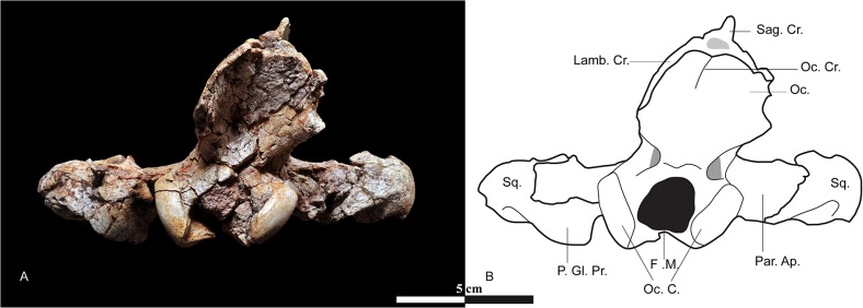
A, occipital view. B, drawing of the occipital view. Abbreviations: F. M., foramen magnum; Lamb. Cr., lambdoid crest; Oc., occipital; Oc. C., occipital condyle; Oc. Cr., occipital crest; P. Gl. Pr., postglenoid process; Par. Ap., paroccipial apophysis; Sag. Cr., sagittal crest; Sq., squamosal.
Upper dentition. The skull (MNHN.F.EBA 517) displays three upper incisors. The three incisors are compressed transversally. The I1 is the smallest. I2 and I3 are simple and conical. I3 is distinctly larger than the two other incisors and is semicaniniform.
Unfortunately, the crowns of the canines are not preserved.
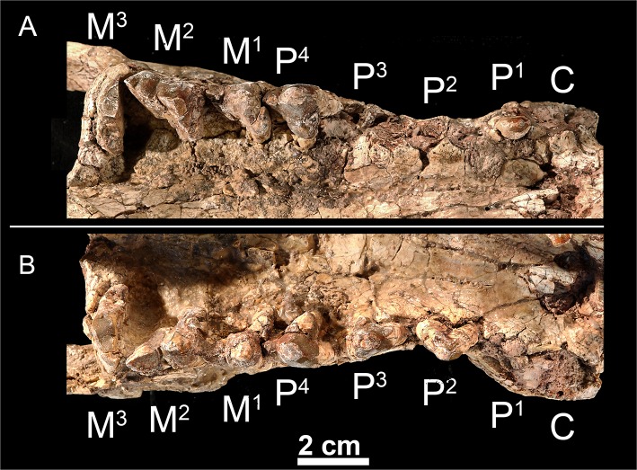
The P1, which is double-rooted, is formed by a single pointed cusp (= paracone) (Fig 9). No parastyle is present. The metastyle is very poorly developed. The crown of the P2 is unknown but the alveoli indicate that it is double-rooted like the P1. The P3 is triple-rooted with a heavily worn parastyle. The metastyle is short and low. A small protocone is present; it is lingually located relative to the paracone. The P4 is the largest premolar. The parastylar region is worn out as on the previous premolars, but it was certainly well developed, which is typical of the hyainailourine P4. The protocone is much more developed than on the P3. The protocone does not bear accessory cusps.
Fig 9. Upper dentition of Kerberos langebadreae gen. & sp. nov. (holotype, MNHN.F.EBA 517).
A, right maxilla bearing P1 and P4-M3 in occlusal view. B, left maxilla bearing P2-M3 in occlusal view.
The three upper molars are known. The M1 is characterized by a short parastyle, a small, mesially located protocone relative to the paracone and metacone, and a long, mesiodistally aligned metastyle. The paracone and metacone are fused into an amphicone [9]. No groove delimitates the paracone from the metacone though the apex of each cusp would have been distinct. The M1 has no cingulum, which is also the case on the M2 and M3. The M2 has a generally similar morphology to the M1. However, the parastylar region and the metastyle are slightly more developed with heavy distal abrasion where the M3 would have shorn against the metastyle. The M3 is transversally elongate. The preparacrista is long. The metacone is absent. The protocone is short and narrow. The P4, M1 and M2 are implanted obliquely. Rather than being positioned parallel to the buccal margin, the cusps of these teeth angle slightly posteriorly and laterally. Such a condition is found in the hyainailourine P. dasuryoides.
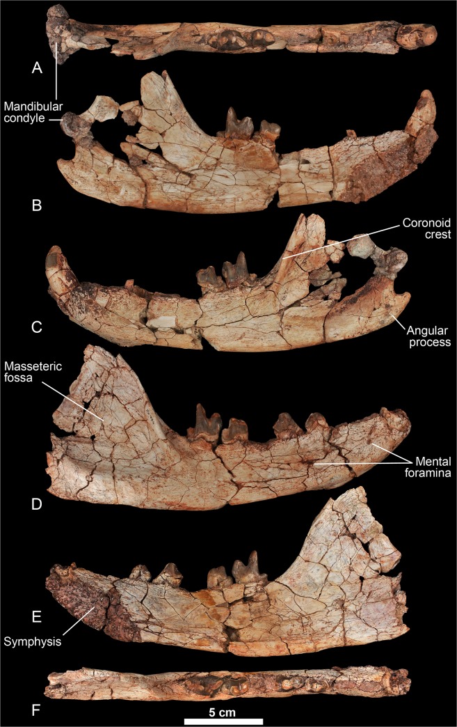
Dentary. One mental foramen is present below the P1 and another is present below the P4 (Fig 10). The symphysis extends posteriorly up to the distal root of the P3. The ventral margin of the mandible is slightly convex, especially posterior to the tooth row. The mandibular condyle is slightly higher than the tooth row. It is cylindrical and mediolaterally elongate. The coronoid crest arises at a 45° angle relative to the horizontal ramus. A deep fossa for insertion of the temporal muscle is present along the anterior margin of the crest. This fossa is particularly prominent at the base and extends below the distal root of the M3. The angular process is curved sharply dorsally and is relatively thin. The masseteric fossa is deep and wide.
Fig 10. Mandible of Kerberos langebadreae gen. & sp. nov. (MNHN.F.EBA 518).
A-C; MNHN.F.EBA 518a, left hemimandible bearing canine, M2-M3; A, occlusal view; B, lingual view; C, labial view. D-F, MNHN.F.EBA 518b, right hemimandible bearing P3-P4 and M2-M3; D, labial view; E, lingual view; F, occlusal view.
Lower dentition. The holotype does not preserve the lower incisors. The crown of P1 is not preserved, but its presence is confirmed by the presence of its single alveolus. The P1 is separated from the P2 by a small diastema. The P2 is double-rooted and is only slightly longer than the P1 (Table 2). The double-rooted P3 is known by one worn tooth. The presence of a slight precingulid indicates the probable presence of a small paraconid. The talonid is large and is constituted by the hypoconid. The P4 is larger than the previous premolars. The relatively small paraconid and the large, hypoconid-bearing talonid recall the morphology of P3.
The M1 is unknown but was clearly smaller than the P4 and the distal molars. The metaconid is absent on the M2 and M3. The talonid on M2 is simple; only the hypoconulid is clearly visible at the distal part of the talonid. A small anterior keel is visible on the buccal aspect of the paraconid. The M3 differs from the M2 by its larger size and shorter talonid. The talonid of M3 is as simple as the talonid of M2.
Description of the postcranial elements
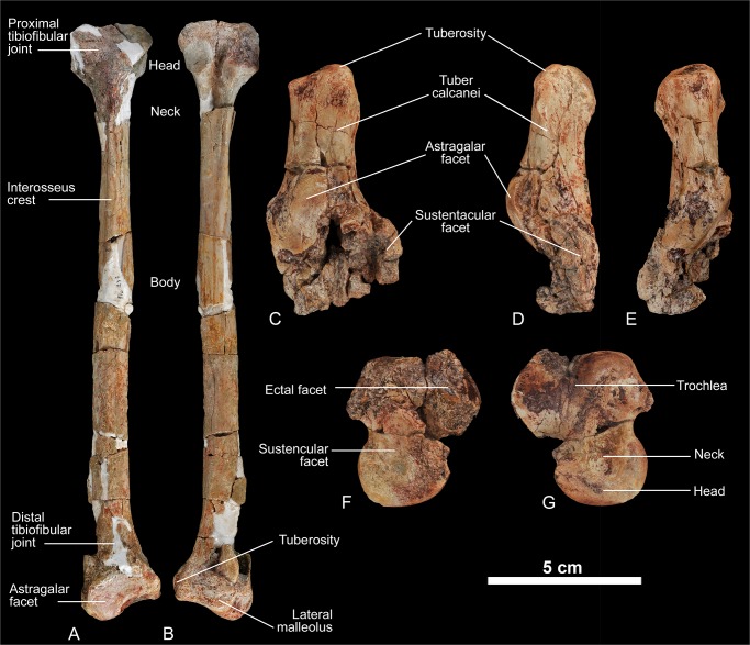
Fibula. The entire fibula is preserved, including both proximal and distal epiphyses. The fibula is partially reconstructed, but this reconstruction does not have any impact on the features described herein. There is no indication that the fibula was fused in any way to the tibia (Fig 11). A tendinal sulcus can be traced on the shaft. The proximal head of the fibula exhibits a semilunar facet for articulation with the tibia. The distal head is broad and possesses two facets that articulate with the calcaneus and the astragalus. The former calcaneo-fibular joint is characteristic of “Creodonta” (e.g., in Hyaenodon and Oxyaena).
Fig 11. Postcranial elements of Kerberos langebadreae gen. & sp. nov.
A-B, fibula (right, MNHN.F.EBA 520); A, dorsal view; B, plantar view. C-E, calcaneus (right, MNHN.F.EBA 522); C, dorsal view; D, medial view; E, lateral view. F-G, astragalus (right, MNHN.F.EBA 521); F, plantar view; G, dorsal view.
Calcaneus. The calcaneus is not well preserved (Fig 11). The distal portion is particularly crushed and difficult to interpret accurately. The tuber calcaneum is long and thickened. The facet for the articulation with the cuboid seems to have been laterally sloping (as in Hyaenodon), because its distal portion is diagonal in proximal view. The sustentacular facet is situated plantar to the astragalar facet. The shape of the sustentacular facet cannot be described, because the bone is damaged in this region. The astragalar facet is elongate and ovoid with a slight proximal inflection that extends onto the tuber calcanei. The astragalar facet is not kidney-shaped as in Hyaenodon.
Astragalus. The astragalus is crushed, especially on its plantar aspect (Fig 11). The trochlea–as well as the head of the astragalus–are asymmetrical. The trochlea is grooved, but rather shallow compared to that of Hyaenodon. The head of the astragalus is widened and transversally flat. In proportion to the trochlea, the head appears unusually large even when compared with the astragalus of the large bodied hyainailourine Hyainailouros. Compared to Hyaenodon the caput astragali is not rounded, but transversally flattened, a condition also found in Hyainailouros. The neck is robust and short. The astragalar neck is relatively shorter and the trochlea shallower than in Hyaenodon. The plantar articular facets for the calcaneus are too poorly preserved to allow any comment.
Metatarsal I. The robust first metatarsal is reduced in length relative to the other metatarsals (Fig 12).
Metatarsal II. The second metatarsal, which is known for both the right and left feet, is subequal in length to the third metatarsal and is short and robust. The proximal articulation facet is proximo-distally elongated. It is asymmetrical with a medial indentation of its shaft. The trochlea is slightly curved. This bone is shorter and wider than the second metatarsal of Hyaenodon. The trochlea is not globular as in Hyaenodon and all recent carnivorans.
Metatarsal III. The third metatarsal is broadly comparable to the second metatarsal except for the morphology of the proximal articulation. The plantar portion of the proximal articulation facet is laterally compressed while the dorsal portion expands dorsally with lateral and medial facets that would have overlapped the metatarsals lateral and medial to the fourth metatarsal.
In general, the columnar shafts of the metatarsals and globular proximal articulations are similar to the metatarsals of Hyainailouros [4] and are completely unlike the more gracile metatarsals present in the digitigrade foot of Hyaenodon [83]. When articulated, the three metatarsals are slightly divergent distally.
Middle phalanx. Two middle phalanges have been recovered. They do not have the same size, indicating that they correspond to distinct digits. This element is short (Fig 12) and wide. The proximal articulation facet is heart-shaped and concave with a plantar indentation. The distal trochlea is asymmetrical. Its shaft is symmetrical and preserves distinct flexor tubercles. Compared to Hyaenodon the middle phalanx is more robust, comparable to the middle phalanx of recent ursids. The middle phalanx of Hyainailouros is more asymmetrical with a unilateral indentation of the shaft [4].
Comparison and discussion
This specimen is especially noteworthy for its tremendous size. Indeed, no hyaenodontid of such a size is presently known for the Bartonian, making Kerberos one of the largest carnivores known from the Eocene of Europe–only younger Parapterodon is slightly larger. In addition to considerations of body size, the material facilitates an examination of the locomotion of the earliest hyainailourines.
The dental formula of the new taxon is reminiscent of the earliest proviverrines, sinopines, arfiines, and indohyaenodontines–as well as the earliest placentals–in the presence of three upper incisors, four premolars and three molars. However, the reduced molar talonids of the new taxon clearly distinguishes it from the contemporaneous proviverrines, which retain three-cusped, basined talonids. The reduction of the talonid is shared with the hyaenodontines. The specimen differs from the hyaenodontines in the morphology of its nuchal crest, which does not extend to the mastoid process, and differs from Hyaenodon in the subequal length of the paraconid and protoconid. These features are characteristic of the Hyainailouridae [38]. The new taxon also displays several dental features of the Hyainailourinae such as are more reduced talonid on M3 relative to that of M2, the absence of metaconids on the molars, and the connate paracone and metacone [38,70].
The African genus Metapterodon, which is morphologically close to hyainailourines, was referred to Koholiinae by Solé et al. [6]; it was previously referred to Hyainailourinae [69–70]. Kerberos notably differs from Metapterodon by the presence of a P1, a short and low metastyle on P4, presence of a small protocone on P3, and absence of an ectoflexus on each upper molar. These features are shared with the hyainailourines.
The Eocene-Oligocene hyainailourines are represented in Africa by four distinct genera: Furodon and Parvavorodon from late Early or early Middle Eocene of Algeria, and Akhnatenavus and Pterodon from the Fayum Depression of Egypt [6,37,69]. Three hyainailourine genera are known for the same period in Europe: Paroxyaena, Parapterodon, and Pterodon. Only one hyainailourine, Hemipsalodon, is known in the Eocene of North America (see below for discussion referring it to Hyainailourinae).
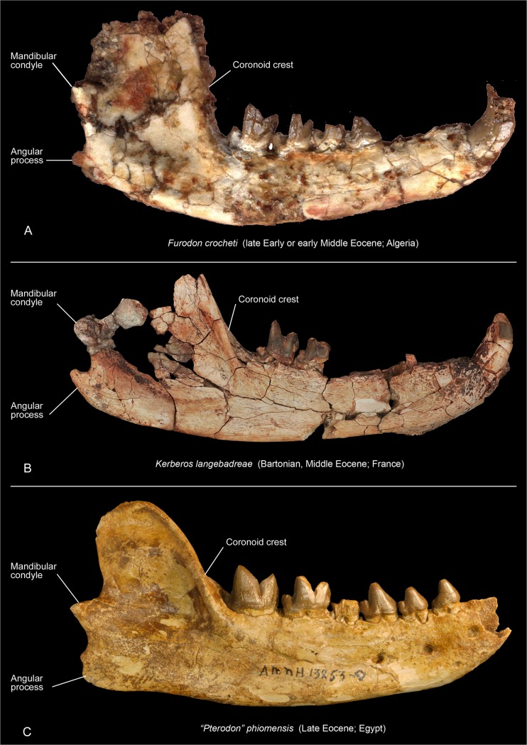
Kerberos shares with Furodon (late Early or early Middle Eocene; Algeria), the oldest hyainailourine, the presence of a single-rooted P1; this tooth is larger than P1 in the younger Pterodon and Akhnatenavus. However, Kerberos differs from Parvavorodon and Furodon by the loss of the metaconid and a more reduced talonid on its lower molars and more connate paracone and metacone on its upper molars. These features are shared with the Late Eocene and Oligocene hyainailourines (e.g., Akhnatenavus Paroxyaena, Parapterodon, and Pterodon). Kerberos and Furodon share a similar mandibular morphology with the mandibular condyle located superior to the tooth row, a relatively superior placement of the angular process, and a steeply inclined ascending ramus and deepened fossa for the insertion of the temporal muscle on the anterior portion of coronoid crest (Fig 14).
Fig 14. Comparison of the hemimandibles in labial view of hyainailourines.
Furodon crocheti (A, left inverted, HGL 50bis-56; HGL, Hammada Gour Lazib, Algeria, Université Montpellier 2, France), Kerberos langebadreae gen. & sp. nov. (B, holotype, left inverted, MNHN.F.EBA 518b) and “Pterodon” phiomensis (C, right, AMNH 13253; AMNH, American Museum of Natural History, New York, USA). Not to scale
The African genus Akhnatenavus is presently represented by only one species: the Oligocene Akhnatenavus leptognathus [69]. Kerberos differs from Akhnatenavus by exhibiting less pronounced diastemata between the premolars than Akhnatenavus, and more simplified molars (e.g., reduced talonid). All these features are derived in Hyainailourinae. Based on dental measurements, Kerberos is 40% larger than Akhnatenavus. Kerberos thus appears more derived than Akhnatenavus. However, Kerberos also differs from Akhnatenavus in the retention of an important primitive feature: a more developed P1.
Kerberos and Pterodon dasyuroides are the only Eocene hyainailourines currently represented by skulls. Both have anteroposteriorly elongate skulls with relatively long rostra and neurocrania. Pterodon includes four European and African species: Pterodon africanus Andrews, 1903 [84] (Oligocene), Pterodon phiomensis Osborn, 1909 [56] (Oligocene), Pterodon syrtos Holroyd, 1999 [69] (Oligocene), and the European Pterodon dasyuroides Blainville, 1839 [77] (Late Eocene), which is the type species. In the analysis presented here, the genus is paraphyletic (see below). The teeth of Kerberos are generally similar to those of “Pterodon”, but Kerberos differs in its larger P1, P1, and M3. The P1 is often absent in P. dasyuroides [9] and small in “P.” africanus and “P.” phiomensis. The talonids on the molars of Kerberos are relatively longer, and the diastema between P1 and P2 is longer than in “Pterodon” species. The mandible of Kerberos is more primitive than that of “Pterodon” species in having a more dorsal mandibular condyle, a more dorsal angular process, a more steeply inclined and taller coronoid crest, and deeper insertions of the temporal muscle on the anterior margin of the coronoid process (Fig 14). The mandibular features are reminiscent of those of Furodon, which is–with Parvavorodon–the oldest hyainailourine presently known (Fig 14). Finally, the metacone and paracone on the molars are more fused, a more derived feature than the less complete fusion present in P. dasyuroides.
Kerberos further differs from the European Pterodon dasyuroides in the presence of large P1 and P1, and an unreduced number of upper incisors (only two upper incisors are present in P. dasyuroides); these features are primitive among hyainailourines. Kerberos also differs from P. dasyuroides in its larger protocone on P3, and reduced parastylar area; these are derived features in hypercarnivores. Finally, based on dental measurements and skull measurements, Kerberos differs from P. dasyuroides by its larger size (10–14%).
In her study of the European hyaenodontids from the Late Eocene to Late Oligocene, Lange-Badré [9] described three genera that she considered to be close to Pterodon: Parapterodon, Paroxyaena, and Schizophagus. Since this study, Schizophagus has been synonymized with Paroxyaena, which is considered to be a peculiar hyainailourine [8,85]. Paroxyaena is known in Robiac’s fauna (reference locality of MP16 reference-level) [27]. Consequently, Kerberos and Paroxyaena are nearly coeval. Sudre [28] noted that the “Castrais” fauna could be slightly older than the Robiac fauna, contra Astruc et al. [29].
Kerberos shares with Parapterodon and P. dasyuroides but not with Paroxyaena the presence of a high, sharp P4, and a more fused paracone and metacone on the upper molars. Kerberos differs from Parapterodon in the primitive presence of a large P1 –this tooth is even vestigial in Parapterodon–and a primitively large M3. Kerberos further differs from Parapterodon by having a more reduced parastylar region, and a more reduced protocone on upper molars, generally derived features among lineages that acquire hypercarnivorous dentition [63,86]. Kerberos shares with Paroxyaena the presence of three upper incisors, but Kerberos differs from Paroxyaena by the presence of P1, a less reduced P1, a less developed protocone on P3, larger M3 (all considered primitive features), more reduced talonids on lower molars, fusion of the paracone and metacone, and reduced protocones on the upper molars (generally derived features).
Kerberos differs from the North American Hemipsalodon in its smaller size (the skull of Hemipsalodon is 45 cm long), its mesiodistally elongate skull (notably in the rostrum and the neurocranium), the distal elongation of the mastoid process (primitive features), and a more closely appressed paracone and metacone on the molars (derived feature).
In summary, the taxon described here departs from other members of the hyainailourine subfamily in featuring a distinctive mixture of primitive and derived features. Thus, we refer this taxon to the new genus, Kerberos. Its discovery increases the taxonomic and morphological diversity of the hyainailourines, especially in Europe.
Discussion
Ecology of Kerberos
The body size of an animal is one of the most important factors in determining its role in its ecosystem. Body size influences dietary preference, predatory behavior, and niche partitioning [87–88]. It is directly related to an organism’s biomechanical and physiological demands. Several methodologies have been proposed for determining the body mass in fossil mammals. Several of them have been applied to hyaenodonts. Depending on the methodology, they correspond to regressions based on dental, cranial or postcranial dimensions [21,89–92]. Most of these studies use extant taxa to correlate known body size to osteological measurements that can be collected from related extinct taxa. The reconstruction of body sizes for Hyaenodonta has been problematic because the entire group is extinct, making direct regressions difficult to apply to the group. Moreover, as mentioned by Van Valkenburgh [89], dentally or cranially derived body size estimates based on extant carnivorans produce unreasonably large values in hyaenodonts because they have relatively large crania compared to carnivorans.
With these caveats in mind, we estimate the body mass of Kerberos langebadreae using three methodologies. (1) The methodology of Morlo [21], which is based on dental dimensions (length of the molars), provides an estimation of 85–90 kg (Table 4). (2) The methodology of Van Valkenburgh [89], based on the skull length, estimates the body mass of K. langebadreae to be about 277 kg. (3) The methodology of Tsubamoto [92], which is based on the dimensions of the astragalus results in ranges from 60 kg to 269 kg (length Li1) and from 49 to 199 kg (length Ar1). The median body mass of these estimates is 140 kg.
Table 4. Mean tooth measurements and body mass estimations.
The data concerning the Oxyaenodonta, Sinopinae and Arfiinae and Proviverrinae are from Solé et al. (2014) [17]. Abbreviations: Hyae, Hyaenodontinae; Hyai, Hyainailourinae; (M1-M3)L, M1 to M3 length. This stratigraphic repartition of H. rossignoli was chosen because the locality of Memerlin (France) where it is recorded is either MP18 or MP19 [93].
| MP | Species | Taxon | (M1-M3)L (mm) | Mass (kg) | LnMass | References |
|---|---|---|---|---|---|---|
| 16 | Kerberos langebreae | Hyai | 60.9 | 87.69 | 11.38 | Present paper |
| Paroxyaena galliae | Hyai | 50.9 | 46.72 | 10.75 | [9] | |
| 17a | Paroxyaena galliae | Hyai | 50.9 | 46.72 | 10.75 | [9] |
| Hyaenodon brachyrhynchus | Hyae | 38.2 | 17.06 | 9.74 | [9] | |
| Hyaenodon minor | Hyae | 30.4 | 7.65 | 8.94 | [9] | |
| Hyaenodon requieni | Hyae | 48.1 | 38.30 | 10.55 | [9] | |
| 17b | Paroxyaena galliae | Hyai | 50.9 | 46.72 | 10.75 | [9] |
| Hyaenodon brachyrhynchus | Hyae | 38.2 | 17.06 | 9.74 | [9] | |
| Hyaenodon minor | Hyae | 30.4 | 7.65 | 8.94 | [9] | |
| Hyaenodon requieni | Hyae | 48.1 | 38.30 | 10.55 | [9] | |
| 18 | Pterodon dasyuroides | Hyai | 52.35 | 51.56 | 10.85 | [9] |
| Parapterodon lostangensis | Hyai | 62.82 | 97.78 | 11.49 | [9] | |
| Paroxyaena pavlovi* | Hyai | 50.9 | 46.72 | 10.75 | [9] | |
| Hyaenodon brachyrhynchus | Hyae | 38.2 | 17.06 | 9.74 | [9] | |
| Hyaenodon heberti | Hyae | 45.09 | 30.53 | 10.33 | [9] | |
| Hyaenodon minor | Hyae | 30.4 | 7.65 | 8.94 | [9] | |
| Hyaenodon requieni | Hyae | 48.1 | 38.30 | 10.55 | [9] | |
| Hyaenodon rossignoli | Hyae | 26.1 | 4.48 | 8.41 | [9] | |
| 19 | Pterodon dasyuroides | Hyai | 52.35 | 51.56 | 10.85 | [9] |
| Parapterodon lostangensis | Hyai | 62.82 | 97.78 | 11.49 | [9] | |
| Paroxyaena pavlovi* | Hyai | 50.9 | 46.72 | 10.75 | [9] | |
| Hyaenodon brachyrhynchus | Hyae | 38.2 | 17.06 | 9.74 | [9] | |
| Hyaenodon heberti | Hyae | 45.09 | 30.53 | 10.33 | [9] | |
| Hyaenodon requieni | Hyae | 48.1 | 38.30 | 10.55 | [9] | |
| Hyaenodon rossignoli | Hyae | 26.1 | 4.48 | 8.41 | [9] |
*Paroxyaena pavlovi is here considered to be equivalent in size to Paroxyaena galliae because the M1 of P. pavlovi, the only molar presently known for the taxon, is similar in size to the M1 of P. galliae.
We present a comparison of the estimated body mass of Kerberos with those of proviverrines from MP7 to MP19 and contemporaneous hyainailourines and hyaenodontines (Fig 15; Table 4). We only use the methodology of Morlo [21] for this comparison because it is based on dental measurements, and cranial and astragalar dimensions are not available for the entire sample. During their evolution, proviverrines never have exceeded 20 kg (see Solé et al. [17]). The two oldest hyainailourines recorded in Europe, Kerberos and Paroxyaena, are clearly distinguished from endemic proviverrines by their much larger size. Thus, neither Kerberos nor Paroxyaena was likely in direct competition with proviverrines. Conceivably, the smaller proviverrines may have even been their prey.
Fig 15. Values of the ln of body mass based on molar dimensions of European oxyaenodontidans and European hyaenodonts from MP7 to MP19.
Values given in Table 4. Abbreviation: K., Kerberos.
It is noteworthy that the body mass range of contemporaneous European hyaenodontines (4–39 kg) partially overlaps with that of proviverrines (0.03–20 kg) (Fig 15). Morlo [21] reconstructed the large proviverrines (e.g., Oxyaenoides) as cursorial hypercarnivorous predators; because this corresponds to the locomotion of Hyaenodon, they thus may have belonged to the same ecological niche. Additionally, one can note that the appearance of Hyaenodontinae in Europe is roughly coeval with the disappearance of the large and hypercarnivorous proviverrines. The body mass range of the hyainailourines (46–98 kg), as noted above, sets them in a very different size class than either hyaenodontines and proviverrines (Fig 15). Consequently, hyaenodontines may have had a greater impact on the evolution of proviverrines than hyainailourines, which did not move into a previously occupied niche space (based on body mass).
The association of the mandible and skull allows detailed discussion of the masticatory musculature in Kerberos and a reconstruction of its feeding habits (see [94–98] for a description of the role and position of each masticatory muscle in carnivorous mammals). It should be noted that the temporal group musculature is more developed than the masseter group as usually observed in carnivorous mammals [9,94]. The anterior base of the coronoid process has a remarkably deep fossa with a significant anterior extension, producing a large insertion for the anterior fibers of the temporal muscle (pars orbitalis). Because this fossa is deeper than in “Pterodon”, it would have allowed Kerberos a more powerful clenching action. As in “Pterodon”, the squamosal and temporal areas, in which the deep masseter, zygomaticomandibularis and temporal muscle (pars temporalis) originate, are important. These three muscles are extensively developed; this observation correlates with the large masseteric fossa observed on the two dentaries. As indicated above, the anterior border of the choana is more anterior than the choana in Pterodon dasyuroides, providing a reduced surface area for the origin of the medial pterygoid muscle. The delicate angular process (relative to other hyainailourines) further supports the feeble contribution of the medial pterygoid to mandibular adduction. The enlargement of the pterygoid muscle in “Pterodon” was noted by Lange-Badré [9]. The important mastoid process, which is larger than in “Pterodon”, suggests that the digastric muscle, an abductor and protruder of the mandible, was also powerful. However, the mastoid is also the site of origin for much of the ventral cervical musculature and the separate sites of origin for these muscles are difficult to interpret (see [99–100] for a description of the muscle attachment sites in the mastoid region in mammals).
Although the cranial morphology of Kerberos is similar to that of “Pterodon”, it has several more primitive features. The younger hyainailourine species expanded the role of the pterygoid muscles and of the masseter muscles, a shift evinced in the more developed angular process and reduced insertion for the temporal in “Pterodon” and younger species. Finally, the emphasis on the temporal musculature and digastric muscles in Kerberos indicates that this taxon would have been capable of powerful slicing and crushing near the molars, but the dentary was only capable of minimal lateral movements.
Savage [80] described a complete skull of Megistotherium osteothlastes (= Hyainailouros bugtiensis) and noticed that the skull is “moulded for maximum efficiency in feeding” (Savage, [80]: p.503). He particularly emphasized the importance of the temporal musculature that originated from the long, tall sagittal crest, a feature shared with Kerberos. Hyainailouros bugtiensis differs from Kerberos in having a shorter rostrum. Savage [80] interpreted the shortened face as important for stabilizing the roots of the canines during food capture. With a more extensive rostrum than M. ostethlastes, the canines may not have been used as actively in Kerberos.
The cervical vertebrae are unknown for Kerberos. However, several of these elements are known for Hyainailourinae and because the basicranial portion of the skull is similar for all known hyainailourines, the cervical vertebrae were likely similar across the subfamily. Savage [80] described the atlas of Megistotherium (= Hyainailouros bugtiensis) and Ginsburg [4] described two cervical vertebrae of Hyainailouros sulzeri. Savage [80] noted that the large size and broad transverse processes of the atlas imply significant extension of the head. The lateral cervical musculature associated with the large, transverse cervical processes indicates the animal was capable of strong lateral flexion of the head, which is particularly useful for restraining struggling prey. Concerning the cervical vertebrae of H. sulzeri, Ginsburg [4] noted the relatively short neural spine on the seventh cervical vertebra. This spine, which is taller and more robust in Hyaenodon, implies that Hyainailouros bore his head low, aligned with the thoracic vertebrae.
The secant molars M2-3 and M1-2 of Hyainailourinae exhibit little morphology that would aid in crushing (e.g., protocone and talonid on molars) and puncturing (e.g., metaconid), but the premolars are robust and display horizontal wear, which indicates they served a separate function from the slicing molars–the addition of a well-developed P3 protocone in hyainailourines illustrates the crushing function hypothesized for the premolars. Based on this dental morphology Ginsburg [4,33] argued that “Pterodon” and Hyainailouros were bone crushers comparable to recent hyaenids, which also possess secant dentition. Stefen [101] noted that Pterodon dasyuroides and Hyainailouros possess only zigzag Hunter-Schreger-Bands (HSB), while limnocyonines Prolimnocyon (probably with the exception of P. elisabethae) and Thinocyon, and sinopines Prototomus and Sinopa display undulating HSB. A transitional state is found in Apterodon and some Hyaenodon and “Pterodon” species: they actually display a transition from undulating HSB at the enamel base through acute angled HSB to zigzag HSB at the tips of the teeth. She concluded that the development of zigzag HSB in the hyainailourines correlates with ossiphagy. The transition from undulating to zigzag HSB is notably found in most European Hyaenodon species–which also exhibits a pattern of wear–suggesting that bone crushing was also part of Hyaenodon´s diet [102].
Consequently, based on the taxon’s robust premolars, the powerful cranial musculature and the phylogenetically bracketed presence of zigzag HSB, we hypothesize that Kerberos was capable of scavenging like its closest relatives. Extant mammal scavengers are also capable hunters, a mode of food acquisition likely partially utilized by Kerberos and hyainailourines more generally. Werdelin [103] distinguishes two morphotypes among ossiphageous carnivores: the bone-crackers (e.g., spotted hyaenas), which break bones with their premolars, and bone-crushers (e.g., wolves) that break bones with their post-carnassial molars. Because Kerberos has extensive premolar wear, it may be considered a bone-cracker with a diet analogous to that of the extant hyenas (except the aardwolf).
The postcranial bones allow a partial reconstruction of the locomotion of Kerberos. The fibula exhibits interesting features related to locomotion. The fibula was not fused to the tibia. Furthermore, the fibula has a large distal head and large facet for contact with the calcaneus and a large proximal head in contact with the tibia. These features indicate a fibula capable of significant rotation for progression over rocky or uneven surfaces [104]. Among extant carnivores, such a rotatory fibula is known in bears and cats for instance and, among Hyaenodontidae, a rotary fibula is known in Hyaenodon, although Hyaenodon shows clear adaptations to cursoriality as well [83,105]. Other postcranial features indicate a plantigrade posture for Kerberos including: a transversally flattened caput astragali, a short astragalar neck, short and robust metatarsals and a short and robust calcaneus [106–107]. The sustentaculum tali has a plantar position, which is an osteological correlate for terrestrial locomotion [104]. The grooving of the astragalar trochlea is a well-known indicator for locomotion with cursors tending to have more deeply grooved astragali, while ambulators have more shallowly grooved astragali [108–109]. The relative depth of the astragalar trochlea also contains a strong phylogenetic signal [89]. The astragalar trochlea of Kerberos is shallow, but grooved. Its development suggests terrestrial locomotion, but clearly not in the cursorial posture indicated by the astragalar trochlea of Hyaenodon [105]. Because O’Leary et al. [110] recently proposed that scansorial features are basal for placental mammals, one must keep in mind that some of the features of Kerberos are potentially primitive, as they are found in many early Tertiary mammals (for instance see [111]).
To conclude, its postcranial anatomy suggests that Kerberos was a plantigrade, terrestrial mammal; we use the term “terrestrial” in the sense of Egi [91], referring to the following particular locomotion: “climbs rarely/never, scarcely running”. This feature is important because it ecologically distinguishes Kerberos from two carnivoran groups that occupied the ossiphageous niches, the Borophaginae and Hyaenidae: these carnivorans are reconstructed to be cursorial [72,73]. In this regard, Kerberos appears more similar to the oxyaenid Palaeonictis because the latter displays dental ossiphageous adaptations [62] and a terrestrial locomotion [23,112].
The locomotor styles of hyaenodonts are poorly known: the postcranial morphology of only a few taxa has been studied, and most of these taxa are from North America (Table 5). Interestingly, a wide range of locomotion has been hypothesized: the limnocyonine Thinocyon is a probable semi-fossorial form, Hyaenodon is cursorial, while Apterodon shows semi-aquatic adaptations [52,83,115]. This lack of knowledge and the apparent diversity of locomotor adaptations among hyaenodonts prevent a rigorous reconstruction of the ancestral locomotor state for this group. However, based on the postcranial elements found in North America we are able to posit a possible primitive locomotor pattern for hyaenodonts. The oldest North American hyaenodonts have adaptations that would have allowed them to move in both arboreal and terrestrial environments. Rose ([113]: p.166) even noted that the “Wasatchian hyaenodontid[an]s, probably [were] reasonably facile in the trees as well as on the ground.” Among North American hyaenodonts, the Sinopinae and Arfiinae differ from the Limnocyoninae in being more terrestrial. For instance, Arfia displays a mosaic combination of cursorial forelimb adaptations and scansorial hindlimb adaptations [34]. The ability to exploit both the trees and the ground was probably the ancestral condition of the hyaenodonts. Finally, the presence of plantigrady in numerous, primitive hyaenodonts allows us to hypothesize a plantigrade foot at the origin of hyaenodonts.
Table 5. Reconstructed locomotor styles and the position of the pes in several hyaenodonts.
| Family | Species | Age | Locomotion | Pes | References |
|---|---|---|---|---|---|
| Sinopinae | Prototomus martis | Early Eocene | Terrestrial/scansorial | - | [34,113] |
| Prototomus secundarius | Early Eocene | Scansorial | - | [113] | |
| Gazinocyon vulpeculus | Early Eocene | Terrestrial (incipient cursorial) | Digitigrade | [38] | |
| Sinopa major | Middle Eocene | Cursorial | Digitigrade | [55] | |
| Tritemnodon agilis | Middle Eocene | Terrestrial | Semi-digitigrade | [58] | |
| Limnocyoninae | Prolimnocyon atavus | Early Eocene | Scansorial | Semi-digitigrade | [114] |
| Limnocyon verus | Middle Eocene | Generalized | Plantigrade | [58,114] | |
| Thinocyon velox and T. medius | Middle Eocene | Semi-fossorial | Plantigrade | [115] | |
| Hyainailourinae | Kerberos langebadreae | Middle Eocene | Terrestrial | Plantigrade | Present paper |
| Hyainailouris sulzeri | Early Miocene | Terrestrial | Digitigrade | [4,33] | |
| Arfiinae | Arfia shoshoniensis | Early Eocene | Terrestrial (incipient cursorial) | Plantigrade | [34,113] |
| Proviverrinae | Lesmesodon * | Middle Eocene | Generalized | Plantigrade | [24] |
| Cynohyaenodon cayluxi | Late Eocene | Generalized | Plantigrade | [21,24] | |
| Hyaenodontinae | Hyaenodon | Oligocene | Cursorial | Digitigrade | [83] |
| Apterodontinae | Apterodon langebadreae and Apterodon indet. | Middle Eocene | Semi-aquatic | Plantigrade | [52] |
*Lesmesodon is only represented by juveniles [24].
The sole hyainailourine for which the locomotion has been studied is Hyainailouros sulzeri. Ginsburg [4,33] reconstructed Hyainailouros not as a fast runner, but as a capable jumper. He also provided arguments for a digitigrade stance. According to our analysis, Hyainailouros is more derived than Kerberos and the postcranial morphology of the new taxon is consistent with the retention of an archaic locomotion.
In conclusion, Kerberos was a heavily built hyaenodont, with a plantigrade posture that predominantly moved across terrestrial substrates. Because of its size and the absence of obvious adaptations to cursoriality, Kerberos was likely not a fast runner but rather complemented active predation with scavenging. Carbone et al. [88,116] demonstrated that extant carnivorous mammals that weigh above 21.5–25 kg generally hunt prey as large or larger than themselves. Because K. langebadreae exceeds this size bracket, it probably focused on large prey such as large artiodactyls (e.g., Choeropotamus) and perissodactyls (e.g., Lophiodon and Palaeotherium). Due to its large body mass, Kerberos langebadreae was probably an important, apex predator among the European faunas of the Bartonian. However, as noted by several authors [117–118], the reconstruction of the ecomorphology of extinct carnivorous mammals is difficult especially when a taxon like Kerberos displays unique character combination, which are not any analogous to those found in extant carnivorans: Kerberos with features similar both to Palaeonictis and several living hyenas, clearly illustrates this issue.
Phylogeny of the Hyainailourinae
As indicated above, Hyainailourinae is characterized by a secant dentition as Hyaenodontinae. Polly [38] demonstrated that the two groups are not closely related and convergently acquired similar shearing dentitions. He provided cranial and dental characters for differentiating the two hypercarnivorous groups. He included Pterodon, Hyainailouros, Hemipsalodon, Sivapterodon, and Megistotherium among hyainailourines. Holroyd [37] added the genus Akhnatenavus and Metapterodon. Peigné et al. [119] removed Dissopsalis and Anasinopa from the “Afro-Asian proviverrines” of Egi et al. [60], and referred them to Hyainailourinae as well. Dissopsalis and Anasinopa differ from Pterodon and its closely affiliated taxa by the presence of a wide talonid and the retention of the metaconid. Lewis & Morlo [70] used this work as a basis for the establishment of a list of African hyainailourines. Hyainailourinae in Morlo et al. [53] further added African “Sinopa” and Metasinopa to the group.
The recent study of Solé et al. [6], based on an analysis of the oldest hyainailourines Furodon and Parvavorodon, restricted Hyainailourinae to its original definition [38]. The authors conserved the following African genera: Pterodon, Hyainailouros, Isohyaenodon, Leakitherium, Sivapterodon, Akhnatenavus, Furodon, and Parvavorodon. The African “Sinopa”, Metasinopa, Dissopsalis and Anasinopa were referred to Teratodontinae. Moreover, Solé et al. [6] found a close relationship between Hyainailourinae and Koholiinae. Our present study of hyaenodont crania allows Apterodoninae and Hyainailourinae to be grouped into Hyainailouridae and the separation of Hyainailouridae from Teratodontinae.
In an effort to better understand the relationships within Hyainailourinae, we performed phylogenetic analyses of the group including closely related taxa. A study of hyainailourines was partially undertaken by Holroyd in her PhD thesis [37]. However, she included several taxa that have been excluded from Hyainailourinae in subsequent work (e.g., Matthodon, Paenoxyaenoides, and Oxyaenoides, which are now referred to Proviverrinae, and Metapterodon, which is referred to Koholiinae), and she restricted her study to the Eocene and Early Oligocene hyainailourines.
Our study includes almost all hyainailourines that are known by a complete lower or upper dentition. We did not include either Parvavorodon or Pterodon sp. [69], because the former is only known by two isolated teeth, and because the maxillary fragment of the latter is much abraded. We also included the apterodontines Apterodon langebadreae and A. macrognathus, as well as the koholiines Lahimia and Boualitomus, because the two subfamilies share many characters with Hyainailourinae, which can potentially help polarizing character transformations within the in-group. The purpose of this study is not to test all hyaenodont relationships. Several taxa, notably species of Hyaenodontidae, were not included and will be considered in future evaluations of the phylogenetic relationships within Hyaenodonta.
Three distinct analyses have been performed (see Material and methods section). The first analysis (all the taxa and characters included) yielded 201 equally parsimonious trees, with a tree length of 117 steps, consistency index (CI) of 0.60 and retention index (RI) of 0.77. The strict consensus tree is 135 steps long (CI = 0.52 and RI = 0.68) (Fig 16A). The majority rule consensus is 120 steps long (CI = 0.59 and = RI 0.75; Fig 16B). The Hyainailourinae includes the genera Paroxyaena and Hemipsalodon. In this analysis the genus Pterodon is not monophyletic and will be closely examined in a future work. The second analysis (Leakitherium deleted) yielded 7 equally parsimonious trees, with a tree length of 116 steps, consistency index (CI) of 0.61 and retention index (RI) of 0.77. The strict consensus tree is 120 steps long (CI = 0.59 and RI = 0.75) (Fig 17A). The majority rule consensus is 117 steps long (CI = 0.60 and RI = 0.76; Fig 17B). The third analysis (Leakitherium deleted and character related to body mass excluded) resulted in 8 equally parsimonious trees (L = 106; CI = 0.63; RI = 0.78; the strict consensus has a length of 111 steps for a CI of 0.60 and a RI of 0.76) (Fig 18). Fig 19 depicts the consensus tree of the third analysis with stratigraphical and geographical information.
Fig 16. Consensuses of the first phylogenetic analysis.
A: strict consensus (135 steps long; CI 0.52; RI 0.68) of the first phylogenetic analysis of the hyainailourines which includes body size character with indications of the Bremer support values. B: relationships among several hyainailourines in the majority rule consensus (120 steps long; CI 0.59; RI 0.75) with percentages of presence of the clades among equally parsimonious trees. In bold: Kerberos langebadreae gen. & sp. nov.
Fig 17. Consensuses of the second phylogenetic analysis of the hyainailourines with Leakitherium excluded.
A: strict consensus (120 steps long; CI 0.59; RI 0.75) of the second phylogenetic analysis of the hyainailourines which includes body size character with indications of the Bremer support values. B: relationships among several hyainailourines in the majority rule consensus (117 steps long; CI 0.60; RI 0.76) with percentages of presence of the clades among equally parsimonious trees. In bold: Kerberos langebadreae gen. & sp. nov.
Fig 18. Strict consensus of the third phylogenetic analysis of the hyainailourines with Leakitherium and body mass character excluded.
111 steps long; CI 0.60; RI 0.76; with indications of the Bremer support values. In bold: Kerberos langebadreae gen. & sp. nov.
Fig 19. Phylogeny of the hyainailourines with stratigraphic and climatic indications based on third phylogenetic analysis with Leakitherium deleted and body mass character excluded.
Abbreviations: Paleoc., Paleocene; Thane., Thanetian; Barton., Bartonian; Priabon., Priabonian; Aquit., Aquitanian; Lang., Langhian; Serra., Serravallian; PETM, Paleocene-Eocene Thermal Maximum; MDE, Mammal Dispersal Event; EECO, Early Eocene Climatic Optimum; MECO, Middle Eocene Climatic Optimum; Oi-1, Oligocene Oi-1 event; Mi-1, Miocene Mi-1 event; OoA, “Out of Africa” event; A., Apterodon; A. m., Apterodon macrognathus; H., Hyainailouros.
The proposed topology does not show temporal incoherence. However, the topology implies a significant ghost lineage for Pterodon dasyuroides. As demonstrated by Holroyd [69], the hyainailourines experienced a significant diversification during the Late Eocene. This diversification should be interpreted cautiously as there are significant gaps in the fossil record of hyainailourines, particularly in the early Paleogene record of Africa. Interestingly, the two Neogene groups–the small hyainailourines represented by Isohyaenodon and the very large Hyainailouros and Sivapterodon–are rooted in Late Eocene taxa.
The consensus of the third analysis differs from the consensus trees of the second analysis by the absence of close relationship between the Apterodontinae and Hyainailourinae although some relationships remain unresolved. Moreover, the Bremer support values are slightly higher. In the second analysis (Fig 17), the clade formed by the Apterodontinae and the Hyainailourinae is supported by the presence of large premolar diastemata [35(2)], an increase of size [1(3)], reduction of the metaconid [37(2) Fast Optimization (= FO) & Slow Optimization (= SO)], a weak paraconule crista [16(1) (SO)] and a paracone greater than the metacone [20(1) SO]. The close relationships between these two subfamilies was hypothesized by Grohé et al. [52] and Rana et al. [61]–together with African Teratodontinae in the latter study–but not by Solé et al. [6]. Our expanded taxonomic sampling allows more complete examination of this relationship. Moreover, as discussed above, the two subfamilies share numerous postcranial and cranial features that would likely lend support to the Apterodontinae+Hyainailourinae clade defined as Hyainailouridae.
We describe in the following paragraphs the consensus tree of the third analysis because the analysis was not disturbed by Leakitherium and not influenced by the body masses (Fig 18). The node A is supported by a weak and short paraconule crista on the molar [16(1)], the reduction of the P2 and P3 relative to P4 [29(1)] and the reduction of the metaconid [37(1)]. The Koholiinae (node B) are supported by the loss of the P1 [28(2)] and a molar series longer than the premolar series [36(1)]. The Apterodontinae (node C) are grouped based on the following features: reduction of the metastyle on P4 [9(1)], completely separated paracone and metacone of equal height [19(0), 20(1)], reduced parastyle on molars [22(1)], absence of ectoflexus [25(0)], reduced distal cuspulid on P3 [31(1)], talonid as wide as the trigonid [44(0) FO], and P3 metastyle present [6(1) SO]. Apterodon macrognathus differs from the earlier A. langebadreae by a single-rooted P1 [3(1)], the reduction of the metaconule [17(1)], the absence of metaconid and entoconid on molars [37(2), 40(2)], and a postprotocristid directed distally [48(1)]. Grohé et al. [52] hypothesized that Apterodon could be a semi-aquatic hyaenodont based on postcranial features. The peculiar morphology of apterodontine dentitions–homogenized premolars and simplified molars–could be an adaptation for a piscivorous diet. This homogenized, simplified dentition is reminiscent of pinniped dentition.
The Hyainailourinae (node D) are supported by a small, mesially located protocone on the molars [24(1), 26(1)], P4 tall and long [33(2)], a protoconid as long as the paraconid [47(0)], a P3 with large lingual cingulum [5(1) FO], P4 mesial root larger than the distal one [14(1) FO], a long metastyle on molars [18(1) FO], large anterior keels [42(1) FO] and higher paraconid on molars [46(1) FO]. The oldest hyainailourine presently known–Furodon from late Early or early Middle Eocene of Algeria–is the sister-group of all other hyainailourines. Parvavorodon has not been included in the phylogenetic analysis (see above) and has been placed at the node C (Fig 19) with an unresolved relationship with other hyainailourines. This position for Furodon contradicts the hypothesis of Solé et al. [6] that Furodon could be closely related to Akhnatenavus.
Numerous features support node E: the absence of the metaconule on the molars [17(1)], the length of the molars equivalent to that of the premolars [36(2)], the loss of the metaconid [37(2)], the reduced talonid on M3 [49(1)], the elongation of the metastyle on molars [18(2) FO], the distally directed postprotocristid [48(1) FO], the reduction of the parastyle on P4 [10(2) SO], and the reduction of the M3 [27(1) SO]. Some of these features are found in hyaenodontines. However, as shown by Polly [38], these features have been acquired convergently in hyaenodontines and hyainailourines.
Node F (= Paroxyaenini) groups the two species of Paroxyaena and the North American genus Hemipsalodon. The synapomorphies at this node are the paracone and metacone of equal height [20(1)], the strong protocone on P3 and P4 [5(2) FO, 7(0) FO, 13(0) FO], the presence of a metastyle on P3 [6(0) FO], the reduced shearing surface of the postparacrista on P4 [12(1) FO], the small anterior keel on the paraconid [42(0) FO], the talonid as wide as the trigonid [44(0) FO], and a paraconid lower than in other hyainailourines [46(0) FO]. It is interesting to note that these two genera have a basal position among hyainailourines in our topology. This early origination agrees with the pleisiomorphic features retained in the group such as the separated paracone and metacone on molars. Surprisingly, Paroxyaena pavlovi is closer to Hemipsalodon than to Paroxyaena galliae (node G); however, it should be noted that Paroxyaena galliae is currently only represented by a juvenile specimen [8], and its permanent dentition is not completely known.
The relationship between Paroxyaena and Hemipsalodon allows the origin of the North American genus to be reconstructed. Contrary to Mellett [65], Gustafson [120] considered a close relationship between Pterodon and Hemipsalodon possible because of similarities in the basicranial regions of both genera. We follow the opinion of Gustafson [120] in considering Hemipsalodon as a hyainailourine. As shown by the phylogenetic analysis, several dental features such as the separation of the paracone and metacone of equal height and the development of a large protocone on P3 support a sister-relationship with Paroxyaena. Moreover, we can add the following features: the presence of three upper incisors, presence of a lingual cingulum on the premolars and molars, the non-reduction of the M3 (compared to other hyainailourines, except Kerberos), the shortening of the rostral part of the skull, and the presence of a depression on the lateral part of the maxilla.
To emphasize the peculiar morphology of Paroxyaena among hyainailourines, Lavrov [8] recognized the tribe Paroxyaenini. The Paroxyaenini are characterized by the fusion of the orbital fissure and the foramen rotundum. Due to this fusion, the Paroxyaenini have only three foramina in the posterior part of the orbital region. Gustafson [120] noted the presence of three foramina instead of four in Hemipsalodon, but he misidentified them; thus, from anterior to posterior, Hemipsalodon has an ethmoid foramen, an optic foramen, and a fused fissura orbitalis and foramen rotundum. As no feature excludes Hemipsalodon from Paroxyaenini, we include this genus in the tribe. The presence of Hemipsalodon among Paroxyaenini has important biogeographic implications as this genus is the only hyainailourine recorded in North America (see below).
Node H is supported by a weak lingual cingulum on the upper premolars [4(0)], weak mesiolabial cingulum on the upper molars [15(1)], fused metacone and paracone [19(2)], small talonid on lower molars [43(1), 45(1)], loss of the paraconule [21(1) FO], and reduction of the talonid cusps [40(2) FO, 41(1) FO]. Pterodon dasyuroides is characterized by the reduction of the number of upper incisors [2(1)] and of the fusion of the symphyseal region [50(1)]–while the latter feature is typical of this species, the first character is also known in Hyainailouros.
Node I is notably supported by the presence of three roots on P3 [8(0)], the reduction of the M1 talonid [39(1)], and the reduction of the P1 [3(1) FO]. Parapterodon and “Pterodon” africanus (node J) are grouped based on the loss of M3 [27(2)]. Node K is supported by the absence of the parastyle on the molars [22(1)], and the increase of the length of the premolars relative to the molars [36(0) FO].
Node L is supported by a mesial root larger than the distal root on P4 [14(0)] and a double-rooted P1 [3(0) FO]. It groups “Pterodon” syrtos and the new genus Kerberos. The former differs from Kerberos by the completely fused paracone and metacone [19(3)SO]. The notable features that support node M are the absence of a talonid on M3 [49(2)], a distally directed postprotocristid [48(1) FO & SO], the loss of the mesiolabial cingulum on the upper premolars and molars [15(2) FO], and reduction of the talonid cusp [40(2) SO, 41(1)]. Node N, which comprises Eocene Akhnatenavus and the Miocene hyainailourines Isohyaenodon, Sivapterodon and Hyainailouros, is notably supported by a tall and short P4 [33(1)], the strong reduction of the talonid on M2 [45(2)], and the presence of very short diastemata between the premolars [35(1) FO]. The relationships within this clade however are poorly resolved. The evolution of the hyainailourines during the later Oligocene is presently poorly known (Fig 19); only several fragmentary fossils of hyainailourines have been reported from this period (see for instance Rasmussen & Gutierrez [121]). Consequently, this lack of fossils could explain the irresolution of the relationships within this clade. This node is comprised of two distinct size groups: node O corresponds to the small Neogene hyainailourines (Isohyaenodon), while node P includes the largest hyainailourines ever recorded (Sivapterodon and Hyainailouros). Consequently, the separation between these two ecological groups occurred during the Oligocene or earlier.
Akhnatenavus is distinguished by the absence of a distal cingulid on P3 [31(1)], reduction of the talonid on P4 [34(1)], presence of large diastemata between the premolars [35(0)], and a less reduced talonid on M2 [45(1)]. Isohyaenodon is not monophyletic in the strict consensus tree of the third analysis, but it was monophyletic in the majority rule consensuses of the first and second analyses. However, the relationships within the Isohyaenodon genus do differ between the first and second analyses. Isohyaenodon is a good example of the trend among hyainailourines towards a simplification of the molars. Isohyaenodon andrewsi–the largest species of the genus–is characterized by the presence of a talonid on M1 [39(0)].
Node O is supported by a distally elongated postprotocrista [48(2)], the loss of the distal cuspulid on P3 [31(1) FO], a low and broad P4 [33(0) FO], molar series longer than the premolar series [36(1) FO], and a very reduced M1 [38(1) FO]. The two species of Hyainailouros form a clade (node P), which supports the monophyly of the genus and the distinctive diagnosis of Sivapterodon (see Pilgrim [47] and Ginsburg [4]); this group is however only supported by a low and broad P4 [33(0) SO] and a very reduced M1 [38(1) SO].
Evolution of size among hyainailourines
The large size of Kerberos was unexpected: it is larger than any other contemporaneous European hyaenodont (Fig 15). With the inclusion of North American Hemipsalodon in Hyainailourinae, there were at least two large-bodied Hyainailourines in Laurasia during the Bartonian. The large body size, exemplified by Bartonian Kerberos and Hemipsalodon, is a common evolutionary tendency in hyainailourines (Fig 19). Hyainailourinae includes one of the largest carnivorous mammals ever known: Megistotherium (= Hyainailourus bugtiensis). The skull of Megistotherium is about 66 cm long—nearly twice as long as that of Kerberos—and the animal may have weighed as much as 800 kg [80].
In his description of Megistotherium, Savage [80] observed that the face of hyainailourines is relatively long when compared to that of carnivorans and Hyaenodontidae. He proposed two hypotheses for the conservation of a long mandible: (1) it is non-adaptive and corresponds to a conserved genetic trait, or (2) it is selective and related to the size of preferred prey (e.g., anthracotheres, pigs, rhinoceroses, mastodonts and deinotheres). The second hypothesis is supported by the abundance of proboscideans in Megistotherium’s fauna. Savage [80] estimated that Megistotherium–due to the length of its jaw–may have had 30 cm of clearance between the tips of the canines, which would allow the animal encircle and bite a proboscidean limb. Rasmussen et al. [122] hypothesized that large hyainailourines, which mainly radiated in Africa, evolved originally as specialized predators or scavengers of the massive herbivores endemic to Africa, such as embrithopods and proboscideans. Ginsburg [4] supported this hypothesis and noted that Hyainailouros appeared in Europe together with the proboscideans (Proboscidean datum; see [123]). Large body size is an ancient feature among proboscideans with the oldest representatives of group already relatively large [124]. Numidotherium, which is known from the latest Early Eocene of Algeria, was one of the largest terrestrial mammals of its time, and initiated an impressive size increase among proboscideans [125]. Following Rasmussen et al. [122], we therefore hypothesize that the large size of hyainailourines may result from co-evolution with endemic African herbivores such as proboscideans, though this hypothesis must be more rigorously tested. However, as demonstrated at a broader mammalian scale, large mammals tend to have longer faces than closely related but smaller ones [126]. Therefore, another hypothesis would be that the long face of Megistotherium is related to the constraints of body size.
The large body size of the hyainailourines, exemplified by Kerberos and by Hemipsalodon, appears early in the history of the subfamily. The large size of the Miocene Hyainailouros and Sivapterodon could result from either conservation or convergence on this trait. Because Hyainailouros and Sivapterodon are close to the small Isohyaenodon in the phylogenetic analyses we performed, it is possible that gigantism evolved at least twice in hyainailourines.
In the same faunas occupied by the gigantic Hyainailouros species, there were also smaller hyainailourines. The smaller hyainailourines are known from the Late Eocene (Akhnatenavus) to Middle Miocene (Isohyaenodon) (Fig 19). The largest one (I. andrewsi) only weighed around 14 kg. Holroyd [69] hypothesized that these differences in sized among hyainailourines are suggestive of the exploitation of different ecological niches. Based on Carbone et al. [88,116], these smaller taxa are below the threshold for hunting prey equal to or larger than their body size. Conversely, they likely exploited prey smaller than them. Unfortunately, the skull and postcranium are unknown for these small hyainailourines, which prevents a clear evaluation of their ecology (i.e. locomotion, choice of prey). The presence of these small-bodied hyainailourines suggests that the hyainailourine bauplan was not specifically adapted for large-bodied predation.
The second cladistic analysis allows discussing the evolution of the body mass among Hyainailourinae in a phylogenetic context. The acquisition of a large body mass (between 31 and 100 kg) [1(3)] is reconstructed for the clade that comprises the hyainailourines and apterodontines. There are convergent secondary return to a smaller body size in Apterodon langebadreae, Akhnatenavus, Isohyaenodon andrewsi [1(2)] (between 11 and 31 kg), Furodon crocheti, Isohyaenodon zadoki [1(1)] (between 1 and 11 kg) and Isohyaenodon pilgrim [1(0)] (below 1 kg). In contrast, independent acquisitions of a large body size state are reconstructed in Hemipsalodon, “Pterodon” phiomensis, “Pterodon” africanus and in the clade formed by Sivapterodon and Hyainailouros [1(4)] (> 100 kg).
The hypothesis of several reductions of the body mass among hyainailourines (and apterodontines) is poorly supported by the analyses of body size evolution among carnivorous mammals. Van Valkenburgh et al. [127] tested the Cope’s rule in extinct canids (borophagines and hesperocyonines) and noticed a clear evolutionary trend toward larger body size in these groups. They argued that energetic constraints and pervasive selection for larger size in carnivorous mammals (e.g., killing and feeding on large preys) also lead to dietary specialization (hypercarnivory). These factors increase vulnerability to extinction, explaining why large hypercarnivorous mammals appear to have been limited in their temporal appearance relative to smaller and more conservative species: a greater vulnerability to extinction ultimately results from evolution towards a larger body size. Based on this remarkable study, the possibility that large sizes evolved at least twice in hyainailourines is more reasonable than several cases of convergent reductions of the body mass. Finally, it is worth reminding that the oldest and most primitive hyainailourine and apterodontines are only of small sizes [6,52].
Paleobiogeographic implications
Based on the discovery of Parvavorodon and Furodon, Solé et al. [6] hypothesized that hyainailourines originated in Africa during the Early Eocene (contra Egi et al. [128]). The presence of a new hyainailourine in the Bartonian of Europe has implications for the terrestrial connection between Europe and Africa during the Eocene. The earliest appearance of the Hyainailourinae in Europe was Paroxyaena galliae, which is known from the MP16, MP17a and MP17b reference-levels [9,27,85] (Fig 1). The presence of Kerberos is consistent with the appearance of Hyainailouridae in Europe at MP 16 (Fig 1). Pterodon dasyuroides is recorded in the MP18 reference-level but is unknown after MP20 reference-level. The stratigraphic position of Parapterodon is also presently uncertain but is probably contemporaneous with Pterodon [9] (Fig 1). As noted by Crochet et al. [85], Paroxyaena and Pterodon were not present in Europe at the same time. The description Paroxyaena pavlovi by Lavrov [8] could contradict this statement, but the stratigraphic position of this species is still uncertain.
These stratigraphic partitions raise the question of the dispersal of Hyainailourinae into Europe and the subfamily’s diversification on the European continent. Did the Hyainailourinae disperse once or several times from Africa to Europe? Did the Hyainailourinae diversify in Europe?
The primitive features of Kerberos (e.g., presence of three upper incisors, large P1 and P1) conforms with an older age for Kerberos compared with the more derived features of “Pterodon” and Parapterodon (Fig 1). Paroxyaena, which is also recorded in the Bartonian of Europe, shares with Kerberos an unreduced number of upper incisors. However, the two genera are clearly different taxa (notably in the reduction of the P1 and P1 in Paroxyaena and the separation of the paracone and metacone). Paroxyaena clearly differs from Kerberos, “Pterodon”, and Parapterodon and represents a distinct lineage (see the phylogenetic analysis). This supports a diversification of the Hyainailourinae during the Bartonian or earlier. However, this diversification is poorly understood due to the lack of fossils, and may have occurred either in Africa or in Europe.
As for the relationships among Kerberos, Parapterodon and “Pterodon”, the new genus is distinguished by its larger size, more derived molars (e.g., parastylar reduced) and a more developed protocone on P3. Because of these differences, Kerberos does not seem to be ancestral to Pterodon dasyuroides or Parapterodon. Consequently, it appears that the Hyainailourini did not diversify and radiate in Europe, but rather dispersed at least twice into Europe. The Paroxyaenini seem to have diversified in Laurasia, but this needs further scrutiny.
The hyainailourines are unknown in Asia until the Miocene. The species Orienspterodon dahkoensis described by Egi et al. [128] from late Middle Eocene of Asia is now considered part of Hyaenodontidae and Indohyaenodontinae rather than to Hyainailouridae [70]. This absence implies that the Bartonian hyainailourines may have dispersed directly from Africa to Europe via the Iberian or Apulian route.
The history of trans-Tethyan Paleogene mammal dispersals is characterized at least by four dispersal phases: during the Early Thanetian, and by the Thanetian/Ypresian, Bartonian/Priabonian, and Priabonian/Rupelian transitions. Two other dispersals between Africa and Laurasia (by the Ypresian/Lutetian and by the Lutetian/Bartonian transitions) have less support [129].
Here we refer to the dispersals of hyainailourines between Africa and Europe as the “Out of Africa event” (= OoA).
Gheerbrant & Rage [129] considered the Lutetian/Bartonian Dispersal Phase as doubtful and minor, but it could explain the dispersal of amphipithecids from Africa to Laurasia. The presence of Kerberos and Paroxyaena in the earliest part of Bartonian (Fig 19) further substantiates this dispersal phase hypothesis. The Lutetian/Bartonian Dispersal Phase corresponds to the first migration of hyainailourines to Laurasia (OoA-1 on Figs 19 and 20). The appearance of the hyainailourines in Europe is related to a modification of the European mammal fauna: the second intra-Eocene mammal turnover of Franzen [130]. This turnover seems to be related to dispersals between Central European Island and Iberian Peninsula [130–131]. Based on this exchange, we propose that the Bartonian hyainailourines may have entered into Europe through the Iberian Peninsula.
Fig 20. Illustration of the different dispersal events supported by the hyainailourines’ distributions.
Out of Africa event 1 = Lutetian/Bartonian Dispersal Phase; Out of Africa event 2 = Bartonian/Priabonian Dispersal Phase; Out of Africa event 3 = MN3 Dispersal Phase; Out of Africa event 4 = MN4 Dispersal phase. (Global map for OoA-1 and OoA-2 is adapted from Ron Blakey, Eocene, http://www2.nau.edu/rcb7/050Marect.jpg and global map for OoA-3 and OoA-4 is adapted from Ron Blakey, Miocene http://jan.ucc.nau.edu/rcb7/020Marect.jpg).
The presence of Hemipsalodon in the Duchesnean NALMA is very interesting. It appeared together with other new migrants. Emry [132] noted that the artiodactyls Brachyhyops and Simimeryx, as well as the hyaenodontine Hyaenodon, were probably migrants from Asia during the Duchesnean. Because Hemipsalodon is not present in all Duchesnean localities, it has been used as an index taxon for separating early and late Duchesnean with Hemipsalodon not present in the early Duchesnean. Because Hemipsalodon and Hyaenodon do not appear in North American localities at the same time, and because there is a close relationship between Paroxyaena and Hemipsalodon (see the phylogenetic analysis), the Paroxyaenini may have dispersed from Europe to North America during the late Duchesnean (Fig 20).
Concerning this issue, it should be emphasized that Orienspterodon displays several features that are reminiscent of Paroxyaena such as the presence of cingulids on premolars. However, the Asian species differ from Paroxyaena in double-rooted P3, better fusion of paracone and metacone on M1, small metaconid on M3 and vestigial metaconid on M1-2 [128]. A close relationship between Orienspterodon and Paroxyaenini would support a dispersal of these hyainailourines to Asia. Such a dispersal represents a competing hypothesis to the dispersal of Paroxyaenini in North America (i.e., a dispersal from Asia to North America as reconstructed for the hyaenodontines). Only the discovery of new material for Orienspterodon will allow its systematic position to be precisely tested and further discussion of these paleobiogeographic scenarios.
The presence of the hyainailourine taxa “Pterodon”and Parapterodon in the Late Eocene of Europe has previously been considered support for a Bartonian/Priabonian Dispersal Phase [6] (OoA-2 on Figs 19 and 20). As noted by Gheerbrant & Rage [129], the Bartonian/Priabonian Dispersal Phase is characterized by the immigration of the “baluchimyine” rodent Protophiomys into Africa. Consequently, the hyainailourines could represent the sole case of a northward dispersal during this phase.
The hypothesized existence of two possible dispersal events results from the lack of support for endemic diversification of the hyainailourines in Europe during the Bartonian and Priabonian. It also implies a turnover, which affected the Europe hyainailourines between these stages. This could result from the inability of hyainailourines to cope with some aspects of European environments. Indeed, “Pterodon” also rapidly disappeared from Europe and is only recorded from MP18-MP20. This is especially conspicuous when compared to the extensive diversification and long stratigraphic record of European Hyaenodon and development of a separate lineage within Europe [133].
Unlike Apterodontinae [66], the hyainailourines did not disperse to Laurasia during the Oligocene, but did so during the Miocene. Two distinct phases can be defined. The OoA-3 corresponds to a migration from Africa to Asia. It resulted in the presence of Hyainailouros and Sivapterodon in Asia (Figs 19 and 20). This dispersal occurred at 19.6 Ma (Mammal Neogene level, MN3). The second dispersal of Hyainailouros occurred only slightly later, when it dispersed to Europe at MN4 (Figs 19 and 20).
By the Early-Middle Miocene, the widely dispersed Hyainailouros and African Isohyaenodon were the last representatives of hyainailourines. Their size differences show that this group remained ecologically and morphologically diverse. The only other hyaenodonts known at that time were the teratodontine Dissopsalis, which was present in Africa and Asia [134] and the koholiine Metapterodon, which was possibly present in Asia [135]. These four taxa are the swan song of Hyaenodonta and they witnessed the ultimate disappearance of their once diverse and successful order.
Conclusion
In this contribution we describe a new hyainailourid hyainailourine, Kerberos langebadreae gen. & sp. nov., based on a skull, mandible, and a few elements of the hind limb. Associated dental and cranial or postcranial elements of hyaenodonts is rare, especially among the hyainailourines, making this description of one of the oldest known hyainailourines particularly important for understanding the evolution of Hyaenodonta.
The skull of Kerberos already displays features typical of the hyainailourines–and by extension of the Hyainailouridae. The hind limb of Kerberos exhibits features consisted with a preference for terrestrial substrates and a plantigrade posture. Because of its size and the absence of adaptations to cursoriality, the new taxon was probably not only an active predator, but also a successful opportunistic scavenger.
The powerful masticatory musculature supports the scavenger behavioral hypothesis usually evoked for Miocene hyainailourines. This contrasts with the contemporaneous Hyaenodon. These hyaenodontines, which probably originated in Asia and were represented by numerous species in Europe [9,136], exhibit terrestrial (semi-plantigrady/digitigrady) and cursorial postcranial features [33,74,137]. Kerberos was ecologically more similar to the striped and spotted hyenas than to contemporaneous hyaenodontines except that Kerberos lacked the cursorial adaptations of these extant hyaenids.
The respective endemic evolution of hyaenodontines (in Asia) and hyainailourines (in Africa) explains both similarities and differences between these two clades, which became competitors in Europe and North America, but our knowledge of this history still needs to be improved.
Supporting Information
*Category established by comparisons with relatives of similar sizes.
(DOCX)
(DOCX)
(ZIP)
Acknowledgments
The optical photographs were taken by C. Lemzaouda and P. Loubry (CNRS). We thank C. Argot, P. Tassy, G. Billet, and C. Sagne (MNHN) for granting access to the material of Hyaenodonta under their care. We are also indebted to C. Bouillet (MNHN) for helping preparing the skull of the holotype of K. langebadreae. We thank R. Tabuce (CNRS-Université Montpellier 2) for having permitted the utilization of the picture of Furodon crocheti. We also thank Shawn P. Zack, Lars Werdelin, Gregg Gunnell, Margaret E. Lewis and one anonymous referee and the Academic Editors–Nathan Jeffery and Matt Friedman–for their very constructive comments that improved the manuscript.
Data Availability
All relevant data are within the paper and its Supporting Information files.
Funding Statement
This research was supported by the National Science Foundation Grant DEB-1311354 to MB (PhD Thesis). The Belgian Science Policy Office supports the project Paleurafrica BR/121/A3/PALEURAFRICA (Postdoctoral position of FS). The funders had no role in study design, data collection and analysis, decision to publish, or preparation of the manuscript.
References
- 1. Solé F, Gheerbrant E, Amaghzaz M, Bouya B (2009) Further evidence of the African antiquity of hyaenodontid (“Creodonta”, Mammalia) evolution. Zoological Journal of the Linnean Society 156: 827–846. [Google Scholar]
- 2. Yans J, Amaghzaz M, Bouya B, Cappetta H, Iacumin P, Kocsis L, et al. (2014) First carbon isotope chemostratigraphy of the Ouled Abdoun phosphate Basin, Morocco; implications for dating and evolution of earliest African placental mammals. Gondwana Research 25: 257–269. [Google Scholar]
- 3. Savage RJG (1965) Fossil mammals of Africa: 19 The Miocene Carnivora of East Africa. Bulletin of the British Museum (Natural History), Geology 10: 242–316. [Google Scholar]
- 4. Ginsburg L (1980) Hyainailouros sulzeri, mammifère créodonte du Miocène d’Europe. Annales de Paléontologie (Vertébrés) 66: 19–73. [Google Scholar]
- 5. Gheerbrant E (1995) Les mammiferes paleocenes du Bassin d'Ouarzazate (Maroc) III. Adapisoriculidae et autres mammiferes (Carnivora,? Creodonta, Condylarthra,? Ungulata et incertae sedis). Palaeontographica Abteilung A 237: 39–132. [Google Scholar]
- 6. Solé F, Lhuillier J, Adaci M, Bensalah M, Mahboubi M, Tabuce R (2013b). The hyaenodontids from the Gour Lazib area (?Early Eocene, Algeria): implications concerning the systematics and the origin of the Hyainailourinae. Journal of Systematic Palaeontology 12, 303–322. [Google Scholar]
- 7. Meng J, Zhai R-J, Wyss AR (1998) The late Paleocene Bayan Ulan fauna of Inner Mongolia, China. Bulletin of Carnegie Museum of Natural History 34: 148–185. [Google Scholar]
- 8. Lavrov AV (2007) A new species of Paroxyaena (Hyaenodontidae, Creodonta) from Phosphorites of Quercy, Late Eocene, France. Paleontological Journal 41: 298–311 [Google Scholar]
- 9. Lange-Badré B (1979) Les Créodontes (Mammalia) d´Europe occidentale de l´Éocéne supérieur à l´Oligocéne supérieur. Mémoires du muséum national d´histoire naturelle, Série C, Sciences de la terre 42: 1–249. [Google Scholar]
- 10.Ogg JG, Lugowski A (2013) TS creator–visualization of enhanced Geologic Time Scale 2012 database. Available: http://www.tscreator.org.
- 11. Gradstein FM, Ogg JG, Schmitz M, Ogg G (2012) The geologic time scale 2012, 2-Volume set. Elsevier, Amsterdam. 1144 pp. [Google Scholar]
- 12. Smith T, Smith R (2001) The creodonts (Mammalia, Ferae) from the Paleocene-Eocene transition in Belgium (Tienen Formation, MP7). Belgian Journal of Zoology 131: 117–136. [Google Scholar]
- 13. Solé F, Gheerbrant E, Godinot M (2013a) The Sinopaninae and Arfianinae (Hyaenodontida, Mammalia) from the Early Eocene of Europe and Asia; evidence for dispersals in Laurasia around the P/E boundary and for an unnoticed faunal turnover in Europe. Geobios 46: 313–327. [Google Scholar]
- 14. Hooker JJ (2010) The mammal fauna of the Early Eocene Blackheath Formation of Abbey Wood, London. Monograph of the Palaeontographical Society London 165: 1–162. [Google Scholar]
- 15. Godinot M (1981) Les mammifères de Rians (Eocène inférieur, Provence). Palaeovertebrata 10: 43–126. [Google Scholar]
- 16. Godinot M, Crochet J-Y, Hartenberger J-L, Lange-Badré B, Russell DE, Sigé B (1987) Nouvelles données sur les mammifères de Palette (Eocène inférieur, Provence). Münchner Geowissenschaftliche Abhandlungen A 10: 273–288. [Google Scholar]
- 17. Solé F, Falconet J, Laurent Y (2014) New Proviverrines (Hyaenodontida) from the Early Eocene of Europe; Phylogeny and Ecological Evolution of the Proviverrinae. Zoological Journal of the Linnean Society 171: 878–917. [Google Scholar]
- 18. Marandat B (1997) La disparité des faunes mammaliennes du niveau MP 7 (Eocène inférieur) des domaines péri-mésogéens et nordiques. Investigation d’un provincialisme intra-européen. Newsletters on Stratigraphy 35: 63–82. [Google Scholar]
- 19. Marandat B, Adnet S, Marivaux L, Martinez A, Vianey-Liaud M, Tabuce R (2012) A new mammalian fauna from the earliest Eocene (Ilerdian) of the Corbières (Southern France): palaeobiogeographical implications. Swiss Journal of Geosciences 105: 417–434. [Google Scholar]
- 20. Morlo M, Gunnell GF, Nagel D (2010). Chapter 10 –Ecomorphological analysis of carnivore guilds in the Eocene through Miocene of Laurasia In Goswami A, Friscia A, editors. New contributions to the Natural History of Carnivora. Cambridge University Press, Cambridge, U.K: pp. 269–310. [Google Scholar]
- 21. Morlo M (1999) Niche structure and evolution in creodont (Mammalia) faunas of the European and North American Eocene. Geobios 32: 397–305. [Google Scholar]
- 22. Solé F (2014) New carnivoraforms from the early Eocene of Europe and their bearing on the evolution of the Carnivoraformes. Palaeontology 57: 963–978. [Google Scholar]
- 23. Solé F, Gheerbrant E, Godinot M (2011). New data on the Oxyaenidae from the Early Eocene of Europe; biostratigraphic, paleobiogeographic and paleoecologic implications. Palaeontologia Electronica: 14 Available: http://palaeo-electronica.org/2011_2/258/index.html. Accessed July 2011 [Google Scholar]
- 24. Morlo M, Habersetzer J (1999) The Hyaenodontidae (Creodonta, Mammalia) from the lower Middle Eocene (MP11) of Messel (Germany) with special remarks on new x-ray methods. Courier Forschungsinstitut Senckenberg 216: 31–73. [Google Scholar]
- 25. Laurent Y, Adnet S, Bourdon E, Corbalan D, Danilo L, Duffaud S, et al. (2010) La Borie (Saint- Papoul, Aude): un gisement exceptionnel dans l'Éocène basal du Sud de la France. Bulletin de la Société d'Histoire Naturelle de Toulouse 146: 89–103. [Google Scholar]
- 26. Stehlin HG (1904) Sur les Mammifeh special reference to the appearance of Hunter-Schreger-Bété géologique de France, 4e serie 4: 445–475. [Google Scholar]
- 27. Matthis C (1985) Contribution à la connaissance des mammifères de Robiac (Eocène supérieur): Creodonta et Carnivora. Bulletin du Muséum National d’Histoire Naturelle; Paris, 4e série, 7, 305–326. [Google Scholar]
- 28. Sudre J (1969) Les gisements de Robiac (Eocène supérieur) et leurs faunes de Mammifères. Palaeovertebrata 2: 95–156. [Google Scholar]
- 29. Astruc JG, Hugueney M, Escarguel G, Legendre S, Rage J-C, Simon-Coicon R, et al. (2003) Puycelci, nouveau site à vertébrés de la série molassique d’Aquitaine. Densité et continuité biochronologique dans la zone Quercy et bassins périphériques au Paléogène. Géobios 36: 629–648 [Google Scholar]
- 30.BiochroM’97 (1997). Synthèses et tableaux de correlation. In Aguilar J-P, Legendre S and Michaux J, editors, Actes du Congrès BiochroM’97 Montpellier. Mémoires et Travaux de l’Ecole Pratique des Hautes Etudes, Institut de Montpellier, 21. pp. 769–805
- 31. Lange-Badré B (1978) Cynohyaenodon lautricensis nov. sp. (Creodonta, Mammalia) et les Cynohyaenodon européens. Bulletin de la Société d’Histoire Naturelle de Toulouse 114: 472–483. [Google Scholar]
- 32. Van Valen L (1966) Deltatheridia, a new order of mammals. Bulletin of the American Museum of Natural History 132: 1–126. [Google Scholar]
- 33. Ginsburg L (1999) Order Carnivora In Rössner GE, Heissig K, editors. The Miocene Land mammals of Europe. Friedrich Pfeil, München, Germany: pp. 105–108. [Google Scholar]
- 34. Gingerich PD, Deutsch HA (1989) Systematics and evolution of Early Eocene Hyaenodontidae (Mammalia, Creodonta) in the Clarks Fork Basin, Wyoming. Contributions from the Museum of Paleontology, the University of Michigan 27: 327–391. [Google Scholar]
- 35. Miller ME, Christensen G, Evans H (1965) Anatomy of the Dog Academic Medicine, Philadelphia, Saunders: p. 941 [Google Scholar]
- 36. Argot C (2004) Functional-adaptative features and palaeobiologic implications of the postcranial skeleton of the late Miocene sabretooth borhyaenid Thylacosmilus atrox (Metatheria). Alcheringa 28: 229–266. [Google Scholar]
- 37.Holroyd PA (1994) An examination of dispersal origins of Fayum mammalia. PhD. Thesis. Durham, Duke University. 328 p.
- 38. Polly PD (1996) The skeleton of Gazinocyon vulpeculus gen. et comb. nov. and the cladisitic relationships of Hyaenodontidae (Eutheria, Mammalia). Journal of Vertebrate Paleontology 16: 303–319. [Google Scholar]
- 39.Nixon KC (1999) Winclada (BETA) ver 099. Published by the author, Ithaca, NY.
- 40. Goloboff PA, Farris JS, Nixon KC (2008) TNT, a free program for phylogenetic analysis. Cladistics 24: 774–786. [Google Scholar]
- 41. Morales J, Pickford M, Fraile S, Salesa MJ, Soria D (2007) New carnivoran material (Creodonta, Carnivora and Incertae sedis) from the early Miocene of Napak, Uganda. Paleontological Research 11: 71–84. [Google Scholar]
- 42.Owen R (1837) Teeth. In Todd RB, editor. The Cyclopaedia of Anatomy and Physiology, 4(2). Sherwood, Gilbert, and Piper, London, U.K. pp. 864–935.
- 43. Linnaeus C (1758) Systema Naturae per regna tria naturae, secundum classes, ordines, genera, species, cum characteribus, differentiis, synonymis, locis Editio decima, reformata. Laurentius Salvius: Holmiae: ii, 824 pp. [Google Scholar]
- 44. Van Valen L (1967) New Paleocene insectivores and insectivore classification. Bulletin of the American Museum of Natural History 135: 217–284. [Google Scholar]
- 45. Gunnell GF (1998) Creodonta In: Janis CJ, Scott KM, Jacobs LL, editors. Evolution of tertiary mammals of North America–Volume 1: Terrestrial Carnivores, Ungulates, and ungulatelike mammals. Cambridge University Press; pp. 91–109. [Google Scholar]
- 46. Leidy J (1869) On the extinct Mammalia of Dakota and Nebraska. Journal of the Academy of Natural Sciences Philadelphia 7: 1–472 [Google Scholar]
- 47. Pilgrim GE (1932) The fossil Carnivora of India. Memoirs of the Geological Survey of India, Palaeontologia indica 18: 1–232. [Google Scholar]
- 48. Crochet J-Y (1988) Le plus ancien Créodonte africain: Koholia atlasense nov. gen., nov. sp. (Eocene inferieur d’El Kohol, Atlas saharien, Algerie). Comptes Rendus de l’Academie des Sciences, Paris 307: 1795–1798. [Google Scholar]
- 49. Polly PD (1994) What if anything is a creodont? Journal of Vertebrate Paleontology 14(supplement): 42A. [Google Scholar]
- 50. Gheerbrant E, Iarochene M, Amaghzaz M, Bouya B (2006) Early African hyaenodontid mammals and their bearing on the origin of the Creodonta. Geological Magazine 134: 475–489. [Google Scholar]
- 51. Solé F (2013) New proviverrine genus from the Early Eocene of Europe and the first phylogeny of Late Paleocene-Middle Eocene hyaenodontidans (Mammalia). Journal of Systematic Paleontology 11: 375–398. [Google Scholar]
- 52. Grohé C, Morlo M, Chaimanee Y, Blondel C, Coster P, Valentin X, et al. (2012) New apterodontinae (Hyaenodontida) from the Eocene locality of Dur At-Talah (Libya): systematic, paleoecological and phylogenetical implications. PLOS One 7:e49054 10.1371/journal.pone.0049054 [DOI] [PMC free article] [PubMed] [Google Scholar]
- 53. Morlo M, Bastl K, Wenhao W, Schaal SFK (2014) The first species of Sinopa (Hyaenodontida, Mammalia) from outside of North America: Implications for the history of the genus in the Eocene of Asia and North America. Palaeontology 57: 111–125. [Google Scholar]
- 54. Spaulding M, O’Leary MA, Gatesy J (2009) Relationships of Cetacea (Artiodactyla) Among Mammals: Increased Taxon Sampling Alters Interpretations of Key Fossils and Character Evolution. PLoS ONE 4(9): e7062 10.1371/journal.pone.0007062 [DOI] [PMC free article] [PubMed] [Google Scholar]
- 55. Matthew WD (1906) The osteology of Sinopa, a creodont mammal of the Middle Eocene. Proceedings of the United States National Museum 30: 203–233. [Google Scholar]
- 56. Osborn HF (1909) New carnivorous mammals from the Fayûm Oligocene, Egypt. Bulletin of the American Museum of Natural History 26: 415–424. [Google Scholar]
- 57. Szalay FS (1967) The affinities of Apterodon (Mammalia, Deltatheridia, Hyaenodontidae). American Museum Novitates 2293: 1–17. [Google Scholar]
- 58. Matthew WD (1909) The carnivora and Insectivora of the Bridger Basin, middle Eocene. Memoirs of the Amercian Museum of Natural History 9: 289–567. [Google Scholar]
- 59. Colbert EH (1933) The skull of Dissopsalis carnifex Pilgrim, a Miocene creodont from India. American Museum Novitates 603: 1–8 [Google Scholar]
- 60. Egi N, Holroyd PA, Tsubamoto T, Soe AN, Takai M, Ciochon RL (2005) Proviverrine Hyaenodontids (Creodonta: Mammalia) from the Eocene of Myanmar and a phylogenetic analysis of the Proviverrines from the Para-Tethys Area. Journal of Systematic Paleontology 3: 337–358. [Google Scholar]
- 61. Rana R, Kumar K, Zack S, Solé F, Rose KD, Missiaen P, et al. (2015) Craniodental and postcranial morphology of Indohyaenodon raoi from the early Eocene of India, and its implications for ecology, phylogeny, and biogeography of hyaenodontid mammals. Journal of vertebrate paleontology. 10.1080/02724634.2015.965308 [DOI] [Google Scholar]
- 62. Denison RH (1938) The broad skull Pseudocreodi. Annals of the New York Academy of Sciences 37: 163–257. [Google Scholar]
- 63. Muizon C, Lange-Badré B (2007) Carnivorous dental adaptations in tribosphenic mammals and phylogenetic reconstruction. Lethaia 30: 353–366. [Google Scholar]
- 64. Shawn Z (2014) Saber-tooth origins: a new skeletal association and the affinities of Machaeroidinae (Mammalia, Creodonta). Journal of Vertebrate Paleontology, Program and Abstracts, 2014: 259–260. [Google Scholar]
- 65. Mellett JS (1969) A skull of Hemipsalodon (Mammalia, Deltatheridia) from the Clarno Formation of Oregon. American Museum Novitates 387: 1–19 [Google Scholar]
- 66. Lange-Badré B, Böhme M (2005) Apterodon intermedius, sp. nov., a new European creodont mammal from MP22 of Espenhain (Germany). Annales de Paléontologie 91: 311–328. [Google Scholar]
- 67. Wortman JL (1902) Studies of Eocene Mammalia in the Marsh collection, Peabody Museum. Part I. Carnivora. Paper 7. American Journal of Science 13: 197–206 [Google Scholar]
- 68. Schlosser M (1886) Paläontologische Notizen. Über das Verhältniss der Cope‘schen Creodonta zu den ubrigen Fleischfressem. Morphologische Jahrbüch 12: 287–294. [Google Scholar]
- 69. Holroyd PA (1999) New Pterodontinae (Creodonta:Hyaenodontidae) from the late Eocene-early Oligocene Jebel Qatrani Formation, Fayum province, Egypt. Paleobios 19: 1–18. [Google Scholar]
- 70. Lewis ME, Morlo M (2010) Creodonta In Werdelin L, Sanders WJ, editors. Cenozoic mammals of Africa. University of California Press; pp. 543–560. [Google Scholar]
- 71. Frey E, Munk W, Böhme M, Morlo M, Hensel M (2011) First creodont carnivore from the Rupelian Clays (Oligocene) of the Clay Pit Unterfeld at Rauenberg (Rhein-Neckar-Kreis, Baden-Würtemberg): Apterodon rauenbergensis n.sp. Kaupia 17: 107–113. [Google Scholar]
- 72. Werdelin L, Solounias N (1991) The Hyaenidae: taxonomy, systematics and evolution. Fossils and Strata 30: 1–104. [Google Scholar]
- 73. Wang X, Tedford RH, Taylor BE (1999) Phylogenetic systematics of the Borophaginae (Carnivora, Canidae). Bulletin of the American Museum of Natural History 243: 1–391. [Google Scholar]
- 74. Cope ED (1885) The White River beds of Swift Current River, Northwest Territory. American Naturalist, 19: 163. [Google Scholar]
- 75. Biedermann WGA (1863) Petrefacten aus der Umgegend von Winterthur: II. Die Braunkohlen von Elgg Anhang: Hyainailouros sulzeri. Bleuler-Hausheer, Wintherthur. 23 pp. [Google Scholar]
- 76. Martin R (1906) Revision der obereocaenen und unteroligocaenen Creodonten Europas. Revue Suisse de Zoologie 14: 405–600. [Google Scholar]
- 77. Blainville HMD de. (1839). Sur l’Hyaenodon leptorhynchus (de Laizer) nouveau genre de carnassiers fossiles d’Auvergne. Annales Françaises et Etrangères d’Anatomie et Physiologie, 3: 17–31. [Google Scholar]
- 78. Morlo M, Miller ER, El-Barkooky AN (2007) Creodonta and Carnivora from Wadi Moghra, Egypt. Journal of Vertebrate Paleontology 27: 145–159. [Google Scholar]
- 79. Pilgrim GE (1912) The Vertebrate Fauna of the Gaj Series in the Bugti Hills and the Punjab. Memoirs of the Geological Survey of India. Palaeontologia Indica, New Series 4:1–83. [Google Scholar]
- 80. Savage RJG (1973) Megistotherium, gigantic hyaenodont from Miocene of Gebel Zelten, Libya. Bulletin of the British Museum of Natural History (Geology) 22: 483–511. [Google Scholar]
- 81. Koenigswald GHR von (1947) Ein Hyaenaelurus aus dem Miocaen Nordafrikas. Société Paléontologique de Suisse, Spécial Volume: Contribution à l’Étude des Vertébrés miocène de l’Egypte: 292–294. [Google Scholar]
- 82. Pasitschniak-Arts M (1993) Ursus arctos. Mammalian Species 439: 1–10. [Google Scholar]
- 83. Mellett JS (1977) Paleobiology of North American Hyaenodon (Mammalia, Creodonta). Contributions to Vertebrate Evolution 1: 1–133. [Google Scholar]
- 84. Andrews CW (1903) Notes on expedition to the Fayum, Egypt, with descriptions of some new mammals. Geological Magazine 4: 337–343 [Google Scholar]
- 85. Crochet J-Y, Remy J-A, Sudre J (1983) Données nouvelles sur la faune mammalienne de Fons 6 (Fons outre Gardon, Gard) datée de l’Eocène supérieur. Géologie Méditerranéenne 10: 49–62. [Google Scholar]
- 86. Van Valkenburgh B (1999) Major patterns in the history of carnivorous mammals. Annual Review of Earth and Planetary Science 27: 463–493. [Google Scholar]
- 87. Gittleman JL (1985) Carnivore body size: Ecological and taxonomic correlates. Oecologia 67: 540–554. [DOI] [PubMed] [Google Scholar]
- 88. Carbone C, Teacher A, Rowcliffe JM (2007) The costs of carnivory. PLoS Biol 5(2): e22 10.1371/journal.pbio.0050022 [DOI] [PMC free article] [PubMed] [Google Scholar]
- 89. Van Valkenburgh B (1987) Skeletal indicators of locomotor behaviour in living and extinct carnivores. Journal of Vertebrate Paleontology 7: 162–182. [Google Scholar]
- 90. Gingerich PD (1990) Prediction of body mass in mammalian species from the long bone lengths and diameters. Contributions from the Museum of Paleontology 28: 79–92. [Google Scholar]
- 91. Egi N (2001) Body mass estimates in extinct mammals from limb bone dimensions: the case of North American hyaendontids. Paleontology 44: 497–528. [Google Scholar]
- 92. Tsubamoto T (2014) Estimating body mass from the astragalus in mammals. Acta Palaeontologica Polonica 59: 259–265. [Google Scholar]
- 93. Rémy JA, Crochet J-Y, Sigé B, Sudre J, de Bonis L, Vianey-Liaud M, Godinot M, Hartenberger J-L, Lange-Badré B, Comte B (1987) Biochronologie des phosphorites du Quercy: mise à jour des listes fauniques et nouveaux gisements de mammifères fossiles.Münchner geowissenschaftliche Abhandlungen A 10: 169–188. [Google Scholar]
- 94. Turnbull WD (1970) Mammalian masticatory apparatus. Fieldiana, Geology 18: 149–356. [Google Scholar]
- 95. Ewer RF (1973) The carnivores Cornell University Press, Ithaca, New York: [Google Scholar]
- 96. Smith KK (1993) The form of the feeding apparatus in terrestrial vertebrates: studies of adaptation and constraint In Hanken J, Hall BK, editors. The skull—functional and evolutionarymechanisms, Vol. 3 University of Chicago Press, Chicago, USA: pp. 150–196 [Google Scholar]
- 97. Laison F, Lautrou A, Azérad J, Pollin B, Lévy G (2001) Superficial architecture of the jaw-closing muscles of the cat (Felis catus): the temporo-masseteric complex. Comptes Rendus de l'Académie des Sciences, Séries III 324: 855–862. [DOI] [PubMed] [Google Scholar]
- 98. Greaves WS (1985). The generalized carnivore jaw. Zoological Journal of the Linnean Society 85: 267–274. [Google Scholar]
- 99. Filan SL (1990) Myology of the head and neck of the bandicoot marsupialia peramelemorphia. Australian Journal Of Zoology 38: 617–634. [Google Scholar]
- 100. Antón M, Salesa MJ, Pastor JF, Sánchez IM, Fraile S, Morales J. 2004. Implications of the mastoid anatomy of larger extant felids for the evolution and predatory behaviour of Sabretoothed cats (Mammalia, Carnivora, Felidae). Zoological Journal of the Linnean Society 140: 207–221. [Google Scholar]
- 101. Stefen C (1997) The enamel of Creodonta, Arctocyonidae, and Mesonychidae (Mammalia), with special reference to the appearance of Hunter-Schreger-Bands. Paläontologische Zeitschrift 71: 291–303 [Google Scholar]
- 102. Bastl K, Semprebon G, Nagel D (2012) Low-magnification microwear in Carnivora and dietary diversity in Hyaenodon (Mammalia: Hyaenodontidae) with additional information on its enamel microstructure. Palaeogeography, Palaeoclimatology, Palaeoecology 348–349: 13–20. [Google Scholar]
- 103. Werdelin L (1989) Constraint and adaptation in the bone-cracking canid Osteoborus (Mammalia, Canidae). Paleobiology 15: 387–401. [Google Scholar]
- 104. Barnet CH, Napier JR (1953) The rotary mobility of the fibula in eutherian mammals. Journal of Anatomy 87: 11–21. [PMC free article] [PubMed] [Google Scholar]
- 105.Bastl K (2012) The ecomorphology of the European Hyaenodon. Ph.D. Thesis. University Vienna. 300 pp.
- 106. Ginsburg L (1961) Plantigradie et digitigradie chez les carnivores fissipedes. Mammalia 25: 1–21. [Google Scholar]
- 107. Laborde C (1987) Caractéres d´adaption des membres au mode de vie aboricole chez Cryptoprocta ferox par comparaison d´autres Carnivores Viverridés. Annales des Sciences Naturelles, Zoologie, Série 13: 25–39. [Google Scholar]
- 108. O’Leary MA, Rose KD (1995) Postcranial skeleton of the early eocene mesonychid Pachyaena (Mammalia: Mesonychia). Journal of Vertebrate Paleonto1ogy 15: 401–430. [Google Scholar]
- 109. Heinrich RE, Rose KD (1997) Postcraniam morphology and locomotor behavior of two Early Eocene miacoid carnivorans, Vulpavus and Didymictis . Palaeontology 40: 279–305. [Google Scholar]
- 110. O’Leary MA, Bloch JI, Flynn JJ, Gaudin TJ, Giallombardo A, Giannini NP, et al. (2013) The Placental Mammal Ancestor and the Post–K-Pg Radiation of Placentals. Science 339: 662–667 10.1126/science.1229237 [DOI] [PubMed] [Google Scholar]
- 111. Gould FHD, Rose KD (2014) Gnathic and postcranial skeleton of the largest known arctocyonid "condylarth," Arctocyon mumak, (Mammalia, Procreodi) and ecomorphological diversity in Procreodi. Journal of Vertebrate Paleontology 34: 1180–1202. [Google Scholar]
- 112. Sinclair WJ, Jepsen GL (1929) A mounted skeleton of Palaeonictis . Proceedings of the American Philosophical Society 68: 163–173. [Google Scholar]
- 113. Rose KD (2001) Compendium of Wasatchian mammal postcrania from the Willwood Formation of the Bighorn Basin. University of Michigan Papers on Paleontology 33:157–183. [Google Scholar]
- 114. Gebo DL, Rose KD (1993) Skeletal morphology and locomotor adaptation in Prolimnocyon atavus, an early Eocene hyaenodontid creodont. Journal of Vertebrate Paleontology 13:125–144. [Google Scholar]
- 115. Morlo M, Gunnell GF (2003) Small Limnocyonines (Hyaenodontidae, Mammalia) from the Bridgerian Middle Eocene of Wyoming: Thinocyon, Prolimnocyon, and Iridodon, new genus. Contributions from the Museum of Paleontology, The University of Michigan 31: 43–78. [Google Scholar]
- 116. Carbone C, Mace GM, Roberts SC, Macdonald DW (1999) Energetic constraints on the diet of terrestrial carnivores. Nature 402: 286–288 [DOI] [PubMed] [Google Scholar]
- 117. Van Valkenburgh V (1985) Locomotor diversity in past and present guilds of large predatory mammals. Paleobiology 11: 406–428 [Google Scholar]
- 118. Janis CM, Wilhelm PB (1993) Were there mammalian pursuit predators in the Tertiary? Dances with wolf avatars. Journal of Mammalian Evolution 1: 103–125. [Google Scholar]
- 119. Peigné S, Morlo M, Chaimanee Y, Ducrocq S, Tun ST, Jaeger J-J (2007) New discoveries of hyaenodontids (Creodonta, Mammalia) from the Pondaung Formation, middle Eocene, Myanmar—paleobiogeographic implications. Geodiversitas 29: 441–458. [Google Scholar]
- 120. Gustafson EP (1986) Carnivorous mammals from the late Eocene and early Oligocene of Trans-Pecos, Texas. Texas Memorial Museum Bulletin 33: 1–66. [Google Scholar]
- 121. Rasmussen DT, Gutierrez M (2009) A mammalian fauna from the Late Oligocene of northwestern Kenya. Palaeontographica Abteilung A 288: 1–52. [Google Scholar]
- 122. Rasmussen DT, Tilden CD, Simons EL (1989) New specimens of the gigantic creodont, Megistotherium, from Moghara, Egypt. Journal of Mammalogy 70: 442–447. [Google Scholar]
- 123. Van der Made J, Mazo AV (2003) Proboscidean dispersals from Africa towards Western Europe. Deinsea 9: 437–452. [Google Scholar]
- 124. Gheerbrant E, Tassy P (2009) L'origine et l'évolution des éléphants. Comptes-Rendus Palevol 8: 281–294. [Google Scholar]
- 125. Sander WJ, Gheerbrant E, Harris JH, Saegusa H, Delmer C (2010) Proboscidea In Werdelin L, Sanders WJ, editors. Cenozoic mammals of Africa. University of California Press; pp. 161–251. [Google Scholar]
- 126. Cardini A, Polly PD (2013) Larger mammals have longer faces because of size-related constraints on skull form. Nature Communications 4: 2458 10.1038/ncomms3458 [DOI] [PubMed] [Google Scholar]
- 127. Van Valkenburgh B, Wang X, Damuth J (2004) Cope’s rule, hypercarnivory, and extinction in North American canids. Science 306: 101–104. [DOI] [PubMed] [Google Scholar]
- 128. Egi N, Tsubamoto T, Takai M (2007) Systematic status of Asian “Pterodon” and early evolution of hyaenaelurine hyaenodontid creodonts. Journal of Paleontology 81: 4770–4778. [Google Scholar]
- 129. Gheerbrant E, Rage J-C (2006) Paleobiogeography of Africa: How distinct from Gondwana and Laurasia? Palaeogeography, Palaeoclimatology, Palaeoecology 241: 224–246. [Google Scholar]
- 130. Franzen JL (2003) Mammalian faunal turnover in the Eocene of central Europe. GSA Special Papers 369: 455–461. [Google Scholar]
- 131. Badiola A, Checa L, Cuesta MA, Quer R, Hooker JJ, Astibia H (2009) The role of new Iberian finds in understanding European Eocene mammalian paleobiogeography. Geologica Acta 7: 243–258. [Google Scholar]
- 132. Emry RJ (1981) Additions to the mammalian fauna of the type Duchesnean, with comments on the status of the Duchesnean “Age.” Journal of Paleontology 55: 563–570. [Google Scholar]
- 133. Bastl K, Nagel D, Peigné S (2014) Milk tooth morphology of small-sized Hyaenodon (Hyaenodontidae, Mammalia) from the European Oligocene–evidence of a Hyaenodon lineage in Europe. Palaeontographica A 303: 61–84. [Google Scholar]
- 134. Barry JC (1988) Dissopsalis, a middle and late Miocene proviverrine creodont (Mammalia) from Pakistan and Kenya. Journal of Vertebrate Paleontology 8: 25–45. [Google Scholar]
- 135. Barry JC (1980) Occurrence of a Hyaenodontine Creodont (Mammalia) in the Late Miocene of Pakistan. Journal of Paleontology 54: 1128–1131. [Google Scholar]
- 136. Bastl K, Morlo M, Nagel D, Heizmann E (2011) Differences in the tooth eruption sequence in Hyaenodon (“Creodonta”, Mammalia) and implications for the systematics of the genus. Journal of Vertebrate Paleontology 31: 181–192. [Google Scholar]
- 137. Scott WB, Jepsen G (1936) The Mammalian fauna of the White River Oligocene. Part I–Insectivora and Carnivora. Transactions of the American Philosophical Society 28: 1–153. [Google Scholar]
Associated Data
This section collects any data citations, data availability statements, or supplementary materials included in this article.
Supplementary Materials
*Category established by comparisons with relatives of similar sizes.
(DOCX)
(DOCX)
(ZIP)
Data Availability Statement
All relevant data are within the paper and its Supporting Information files.


