Abstract
Several studies have reported that the number of crossovers increases with maternal age in humans, but others have found the opposite. Resolving the true effect has implications for understanding the maternal age effect on aneuploidies. Here, we revisit this question in the largest sample to date using single nucleotide polymorphism (SNP)-chip data, comprising over 6,000 meioses from nine cohorts. We develop and fit a hierarchical model to allow for differences between cohorts and between mothers. We estimate that over 10 years, the expected number of maternal crossovers increases by 2.1% (95% credible interval (0.98%, 3.3%)). Our results are not consistent with the larger positive and negative effects previously reported in smaller cohorts. We see heterogeneity between cohorts that is likely due to chance effects in smaller samples, or possibly to confounders, emphasizing that care should be taken when interpreting results from any specific cohort about the effect of maternal age on recombination.
 The question of whether recombination rate increases with maternal age is controversial, with conflicting prior evidence. Here, Martin et al. analyse nine cohorts in the largest SNP-based analysis of this question and find a small positive increase with maternal age in the number of crossovers.
Failure of recombination is known to contribute to human aneuploidies by producing nondisjunction of homologous chromosomes during meiosis I (reviewed in refs 1, 2). Aneuploidies are the leading genetic cause of miscarriage and developmental disabilities in humans, and the vast majority are of maternal origin3. The frequency of aneuploidies increases dramatically in older mothers4: only about 2% of clinically-recognized pregnancies in women under 25 are trisomic, compared with 35% in women over 40 (ref. 1).
It has been proposed that changes in recombination rate in normal oocytes are responsible for the maternal age effect on aneuploidies5. To test this directly, one would need to examine recombination levels in oocytes taken from the same women at multiple time points. However, obtaining such samples is difficult, so several groups have instead used pedigree studies to examine the effect of maternal age on the number of crossovers in liveborn offspring6,7,8,9. These studies have yielded conflicting results.
Kong et al.6 analysed about 1,000 microsatellite markers in 14,140 maternal meioses from Iceland, and used data on age rounded to the nearest 5 years. They fitted a linear mixed model that accounted for differences in baseline rates between mothers to estimate the expected change in the number of crossovers per year of maternal age, βage. They reported that βage=0.082 (s.e.=0.012, P<10−8) but noted that the relationship between recombination rate and maternal age was not linear in their data, and that there was a large jump from age 30–35. They hypothesized that the apparent increase in the number of crossovers with age was due to a selection effect, such that eggs with more crossovers were more likely to give rise to a live birth. Thus, they predicted that women with higher recombination rates would have more children, and indeed, they found that the number of crossovers was positively associated with family size (+0.0109 children per crossover, s.e. 0.0041, P=0.0076). It is for this reason that it is important to account for the baseline recombination rate of the mother when analysing the maternal age effect, since the women who have children later might simply have higher rates throughout their lives.
In the first study of recombination using genome-wide SNP data, Coop et al.7 analysed Affymetrix 500K chip data from 725 Hutterite individuals, part of a large 13-generation pedigree. They found an increase of 0.19 crossovers per year using crossover counts and ages adjusted by the mean for the parent (s.e. 0.093, P=0.035 by permutation). Like Kong et al.6, they found a more pronounced increase in older women: mothers over 35 had an average of 3.1 more crossovers than those under 35 (P=0.028 using a within-family permutation test). Coop et al. also found that the mean maternal recombination rate was significantly correlated with family size (Spearman ρ=0.24, P=0.027).
Two more recent pedigree studies8,9 found that the recombination rate decreases with maternal age in humans, as it does in mice10. Hussin et al.8 analysed 195 maternal meioses from a French Canadian cohort. Using a linear mixed model with the mother as a random effect, they estimated that βage=−0.49 crossovers per year (P=0.0017), and they obtained similar results using adjusted counts within families (βage=−0.42, P=0.0047). Bleazard et al.9 analysed 338 meioses from Mongolian and Korean families. They regressed maternal crossover count on maternal age, including mother as a factor, and found βage=−0.29 crossovers per year (P=0.03).
Recently, Campbell et al.11 reported an analysis of recombination in about 4,200 families from the 23andMe data set in which they found an increase of 0.067 maternal crossovers per year. However, as they noted, this analysis could be confounded if mothers with higher recombination rates tend to have more children later in life, since they fitted a simple linear regression that did not account for the baseline rate of the mother. When Campbell et al. restricted the analysis to 2,184 phase known meioses (from families with more than two children) and ran the regression on the adjusted counts, they did not find a significant association with age (P=0.11).
Thus, there is a lack of consensus about the size and direction of effect of maternal age on recombination rate.
There are likely several factors contributing to the conflicting results between different cohorts in the previous studies6,7,8,9. There was considerable heterogeneity in the number of genetic markers used, in genotype data filtering and quality control, in the accuracy of the age data available, and in the methods used to call crossovers and to test the age effect. Here, we revisit this question in nine different cohorts (Table 1; Supplementary Table 1). These include the French Canadians in which Hussin et al. had previously investigated the maternal age effect, six cohorts that O'Connell et al. had used in a study on phasing in populations with high levels of relatedness12, and two cohorts from twin registries not previously studied for this purpose. We attempt to eliminate some of the possible confounders by applying the same methodology, as far as possible, to all cohorts, to see whether applying a standardized approach makes the results more consistent. We find that the maternal age effect on the number of crossovers is small and positive, and that the differences between cohorts are likely due to chance effects in small samples.
Table 1. Cohorts included in the study.
| Cohort | Number of samples | Duos from informative nuclear families | Informative duos | Duos from families with >1 child |
|---|---|---|---|---|
| Carlantino, Italy | 630 | 6 (2) | 29 (11) | 53 (23) |
| French Canadians | 477 | 106 (29) | 158 (50) | 218 (80) |
| Friuli Venezia Giulia, Italy | 1,236 | 13 (4) | 72 (26) | 160 (70) |
| Korcula, Croatia | 897 | 0 (0) | 7 (3) | 34 (17) |
| Netherlands Twin Registry | 2,729 | 298 (96) | 398 (130) | 889 (379) |
| ORCADES, Orkney, Scotland | 2,215 | 48 (15) | 118 (40) | 252 (109) |
| Queensland Twin Registry (QTR370) | 3,754 | 890 (239) | 1,310 (323) | 1,441 (389) |
| Queensland Twin Registry (QTR610) | 7,364 | 1,283 (403) | 1,337 (420) | 2,898 (1202) |
| Queensland Twin Registry (QTRCoreExome) | 4,444 | 234 (75) | 329 (106) | 781 (332) |
| Val Borbera, Italy | 1,664 | 30 (9) | 72 (26) | 266 (123) |
| Vis, Croatia | 960 | 0 (0) | 11 (4) | 37 (17) |
| Total | 3,036 (910) | 4,253 (1268) | 7,688 (3003) | |
| Total analysed | 2,889 (866) | 3,823 (1132) | 6,011 (2305) |
The three rightmost columns show the number of maternal meioses for which we had age data, followed by the number of families in parentheses. The total number analysed excludes the cohorts (or, for the rightmost column, family configurations within a cohort) with fewer than 20 meioses. The SNP chips used, number of SNPs and sample sizes for paternal meioses are given in Supplementary Table 1.
Results
Calling crossovers
We analysed the maternal age effect on recombination in the largest sample to date using SNP chip data (Table 1; Supplementary Table 1). We applied two methods to call crossovers; these use different approaches to phase the parents and to account for genotyping errors that can look like recombination events over short intervals. NFTOOLS8, based on the method by Coop et al.7, phases the parental chromosomes using patterns of transmission to the children, and then identifies positions where a child's chromosome switches from copying one parental haplotype to the other. It requires both parents and at least three children (‘informative nuclear families') to assign crossovers to children. In contrast, the Hidden Markov Model (HMM) method, duoHMM12, phases all samples in the cohort as though they are unrelated, and then incorporates family information to correct phasing, detect genotyping errors and call crossovers. It requires only a duo (parent–child pair) to call crossovers, but as O'Connell et al. showed12, it has greatest power for duos from informative nuclear families or families in which the grandparents are also genotyped. We refer to duos/meioses from such families as ‘fully informative', and all other duos/meioses as ‘partially informative' (termed ‘uninformative' in ref. 12).
The number of crossovers called by NFTOOLS agreed well with expectation (Supplementary Fig. 1), but duoHMM seemed to be over-calling for several of the cohorts, notably those from more outbred populations with low background relatedness (Queensland Twin Registry (QTR), Netherlands Twin Registry (NTR) and French Canadians (FC)) (Supplementary Fig. 2; Supplementary Table 2). We suspected this was due to phasing errors, since we would expect the phasing to be better in cohorts with more low-level relatedness. We found that removing double crossovers called within short intervals (see Methods), which were probably due to switch or genotype errors, brought the number in line with expectation for all cohorts (Supplementary Fig. 2). All duoHMM results described below were based on these filtered calls, but we also obtained very similar results using the raw calls (compare Supplementary Table 3 with Table 2). There was good agreement between the final NFTOOLS and duoHMM callsets on the same meioses (that is, from informative nuclear families; Supplementary Fig. 3), with 96% of crossovers overlapping. We estimated that 70–76% of maternal crossovers occurred in HapMap II recombination hotspots, after correcting for the number expected by chance (Supplementary Table 4). This is similar to previous reports in non-African cohorts8,9,13.
Linear mixed models and meta-analysis
Exploratory analyses of fully informative meioses suggested a small increase in the number of crossovers with maternal age (Fig. 1), with some suggestion of a larger increase in mothers over 35, as described by Coop et al.7 and Kong et al.6, although not in mothers over 39, as described by Campbell et al.11. However, this simple analysis does not account for differences in baseline recombination rates between mothers. Thus, for each cohort separately, we fitted a linear mixed model to the duoHMM counts from fully informative meioses, treating mothers as a random effect (Supplementary Table 5), following refs 6 and 8. Estimates of the effect of maternal age on the number of crossovers, βage, varied between −0.26 for the FC cohort and 0.34 for Val Borbera (VB) (Fig. 2; Supplementary Table 5), but only the three QTR cohorts were significantly different from 0 at the 0.05 level (QTR370: βage=0.09, P=0.03, QTR610: βage=0.15, P=0.01, QTRCoreExome: βage=0.28, P=0.01; P-values are from a t-test in the linear mixed model).
Figure 1. Plot of the number of crossovers as a function of binned parental age.
Ages are grouped into 5-year bins, relative to parents of between 20 and 25 years old. The number of meioses in each age bin is indicated along the top, in red for maternal and blue for paternal. Note that age data were not always available for both parents, hence the total sample size differs for maternal and paternal meioses. Points show means and error bars 95% confidence intervals. This plot is based on data from fully informative duos analysed with duoHMM.
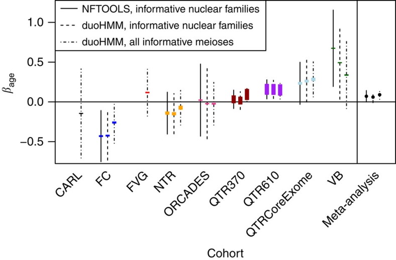
Figure 2. Estimates of the maternal age effect from linear mixed models.
The length of the coloured boxes is proportional to sample size. The black lines show 95% confidence intervals, and the line type indicates which data set was used. On the far right are estimates from the meta-analysis, indicated as points with their confidence intervals. The sample sizes for each cohort are listed in the second last column of Table 1.
Combining the estimates from all cohorts in a fixed-effects meta-analysis, we found that the effect of maternal age was small but significant (βage=0.09, P=0.002; Z-test) (Supplementary Table 6). Similar results were obtained using only informative nuclear families with both NFTOOLS and duoHMM data (Fig. 2; Supplementary Tables 5 and 6), but the effects for FC and VB were more extreme (βage=0.49, P=0.049 for VB; βage=−0.42, P=0.008 for FC, using duoHMM counts). We suspect that these estimates were more affected by outliers, given the smaller sample size imposed by restricting analysis to informative nuclear families (Supplementary Fig. 4), but we discuss this further below. In the meta-analysis, there was significant heterogeneity in the estimated effects between cohorts, as assessed by Cochran's Q test (Supplementary Table 6).
Bayesian analysis of the maternal age effect
Hierarchical models that share information between cohorts, while allowing for differences between them, may add power to discern an effect, and have a more natural interpretation. We fitted a variety of such models in a Bayesian framework (see Methods). We found very similar results for NFTOOLS and duoHMM counts on the same set of meioses from informative nuclear families (Supplementary Table 7; Supplementary Fig. 5). Thus, we focus on the duoHMM results below, because this method also allows us to include meioses from all families, not just informative nuclear families, and hence to substantially increase the number studied.
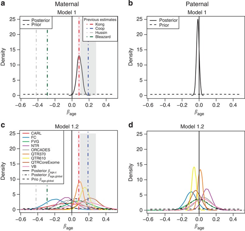
We began by analysing only the fully informative meioses (n=3,823 maternal meioses from 1,132 families; 3,558 paternal meioses from 1,073 families). Figure 3 shows the posterior distribution of the age effect, βage, under a normal model for the number of crossovers in which the mother (or father) effects for parent j in cohort c, αj,c, are drawn from a normal distribution with cohort-specific mean μc and variance  , with the cohort-specific means and variances being independently sampled from a common distribution across cohorts. Posterior distributions for the other parameters are shown in Supplementary Fig. 6. We fitted either a common βage for all cohorts (Model 1; Fig. 3 top), or a different βage,c for each cohort c, drawn from the same normal distribution with mean βage, global and variance
  (Model 1.2; Fig. 3 bottom). Our results for Model 1 fitted to maternal crossovers (Fig. 3a) strongly support a small positive effect (Table 2): the 95% credible interval was (0.0267, 0.1451), which is consistent with the effect found by Kong et al.6 using microsatellite markers, and 99.8% of the posterior distribution for βage was concentrated on positive values. Our results are not consistent with the larger positive effect reported in the Hutterites by Coop et al.7 or the negative effects reported in the original analysis of the French Canadians by Hussin et al.8 or in the Asian cohort by Bleazard et al.9.
Figure 3. Bayesian posteriors for the age effect from normal models fitted to fully informative meioses.
These plots show the priors (dashed lines) and posteriors (solid lines) for βage from a normal model fitted to the number of crossovers called in fully informative duos by duoHMM, with either the same (panels a,b) or different (panels c,d) age effects for each cohort (see Models 1 and 1.2 in Methods). In the bottom plots, the posterior for βage, global is also indicated. The vertical lines show the estimates from previous studies, and the shaded boxes the corresponding standard errors, if reported. Under this model, the expected number of crossovers increases by βage per year.
Table 2. Summary of posteriors for β age for different Bayesian models.
| Parent | Model | 2.5% | 25% | 50% | 75% | 97.5% | Pr(βage >0) |
|---|---|---|---|---|---|---|---|
| Maternal | Model 1 | 0.02672 | 0.06608 | 0.08582 | 0.10706 | 0.14513 | 0.99803 |
| Maternal | Model 2 | 1.00081 | 1.00171 | 1.00217 | 1.00266 | 1.00367 | 0.99963 |
| Maternal | Model 2* | 1.00098 | 1.00172 | 1.00213 | 1.00251 | 1.00333 | 0.99995 |
| Paternal | Model 1 | −0.04766 | −0.02582 | −0.01492 | −0.00410 | 0.01769 | 0.17883 |
| Paternal | Model 2 | 0.99820 | 0.99906 | 0.99951 | 0.99997 | 1.00089 | 0.23465 |
| Paternal | Model 2* | 0.99849 | 0.99925 | 0.99965 | 1.00006 | 1.00082 | 0.28315 |
This table shows several quantiles of the posterior of βage for Model 1 and of  for Models 2 and 2*, as well as the posterior probability that βage is greater than 0. Models 1 and 2 were fitted to fully informative meioses, and Model 2* to fully and partially informative meioses. Note that the interpretation of βage is additive for Model 1 and multiplicative for Models 2 and 2*. These results are for duoHMM counts.
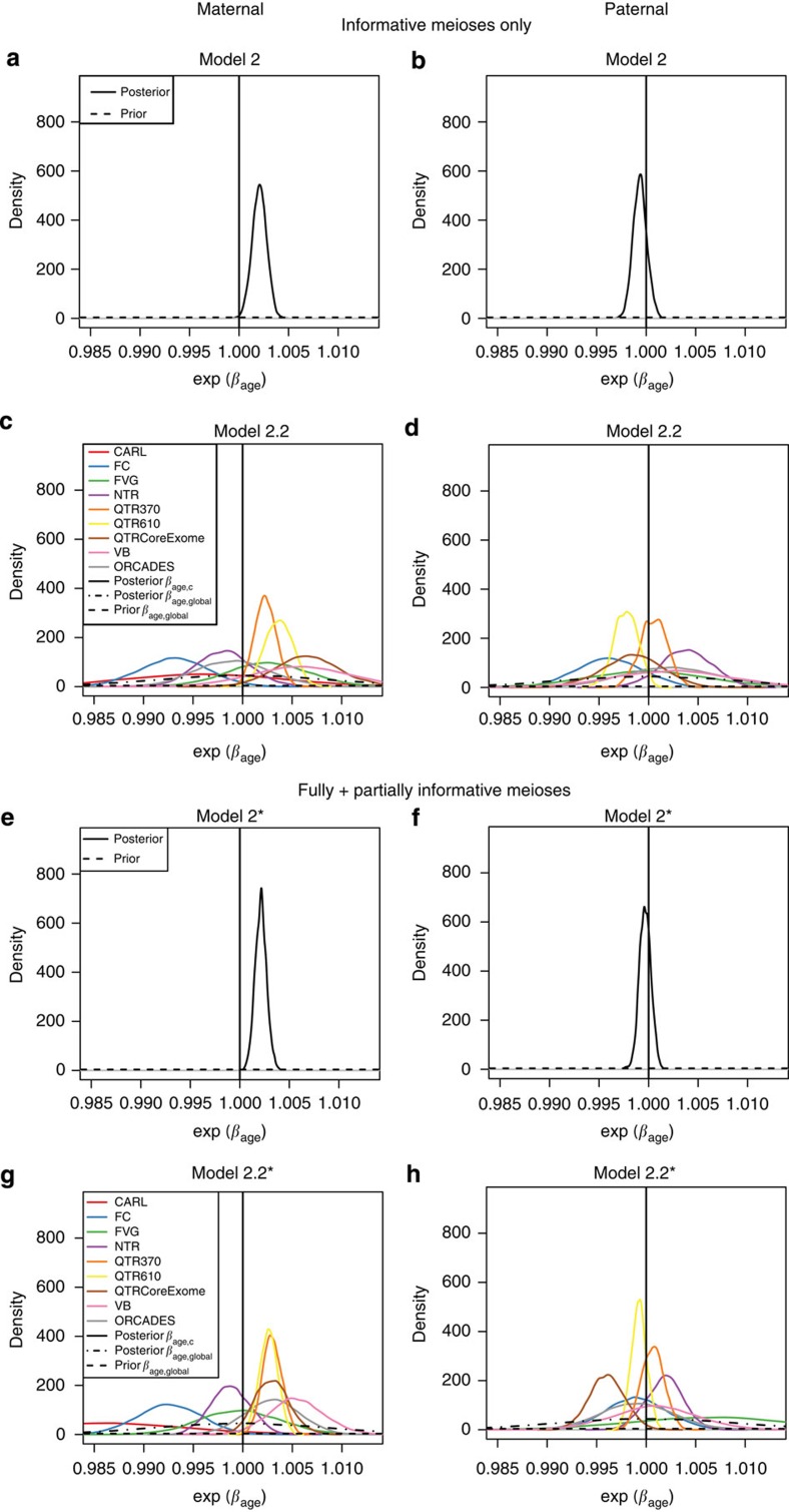
When we fitted a more general normal model with cohort-specific age effects (Model 1.2) to maternal meioses, there was some evidence for heterogeneity of effects, with FC showing the strongest negative effect (Pr(βage,FC<0)=0.95), and QTR610 the strongest positive (Pr(βage, QTR610>0)=0.98; Fig. 3c). Since it seemed more appropriate for count data, we also fitted a negative binomial model with a common age effect, in which the number of crossovers increased multiplicatively by a factor of  per year (Model 2). The evidence in favour of a positive effect was very similar to that under the normal model (Fig. 4a–d; Supplementary Fig. 7): 99.96% of the posterior distribution for βage was concentrated on positive values (Table 2).
Figure 4. Bayesian posteriors for the age effect from negative binomial models.
These plots show the priors and posteriors for  from a negative binomial model fitted to the number of crossovers called by duoHMM in either informative duos only (a–d) or both informative and partially informative duos (e–h), with either the same (Models 2 and 2*) or different (Models 2.2 and 2.2*) age effects for each cohort (see Methods). We have plotted
  since, under the negative binomial model, the expected number of crossovers increases by this factor per year. The axes have been chosen to facilitate comparison with the normal model shown in Figure 3, assuming a baseline of 38, that is, the bounds [0.985,1.013] on
  under the negative binomial model are approximately equivalent to the bounds [38 × log(0.985), 38 × log(1.013)]=[−0.57,0.49] on βage under the normal model.
The results presented thus far have all been based on data from 3,823 fully informative maternal meioses from 1,132 families. However, duoHMM can call crossovers in any duo, since it uses all individuals in the cohort for phasing. Its performance depends on the amount of low-level relatedness in the cohort, which affects phasing accuracy. Supplementary Figure 8 shows that duoHMM is under-calling crossovers in meioses that are only partially informative, but to a different extent in each cohort, consistent with the results of O'Connell et al.12. We adapted the negative binomial model to allow for this effect by instead drawing the mother (or father) effects, αj,c,f, from a normal distribution with mean μc,f and variance  corresponding to the family configuration f in cohort c (see Methods for a description of Model 2* and the family configurations). We fitted this to the 6,011 maternal meioses from families with at least two children, requiring multiple children so that the mother effect could be estimated. With this increased sample size, the results were broadly similar (Fig. 4e–h; Supplementary Fig. 9), though the posterior on βage (or βage,c) was tighter than when using only the fully informative meioses (compare top four with bottom four plots in Fig. 4).
We emphasize that the interpretation of βage is different for the negative binomial models versus the normal model. In the normal model (Model 1), the number of crossovers is expected to increase linearly from the baseline by βage per year, whereas in the negative binomial models (Models 2 and 2*), it increases by a factor of  per year. In practice, because βage is so small, this makes very little difference. For our estimated βage values, the multiplicative
  -fold increase is close to a linear increase by (βage × baseline) per year, at least over a relatively small number of years.
To check that our results were robust to the choice of priors, we fitted Model 2* with an uninformative prior placed in turn on each parameter (see Methods). Our inference about βage was not substantially changed: in each model, at least 99% of the posterior density was concentrated on positive values (Supplementary Fig. 10).
All previous studies had found no significant effect of the father's age at birth on the number of paternal crossovers6,7,8,9,11. Our results are consistent with this (Table 2; Figs 3 and 4; Supplementary Figures 5, 6, 7 and 9), but it is notable that we see some heterogeneity of estimated paternal age effects between cohorts. For example, for Model 1.2 (Fig. 3d), we find Pr(βage>0)=0.94 for NTR and 0.09 for FC. We discuss possible explanations for and implications of this heterogeneity later.
Posterior predictive checks
We conducted posterior predictive checks to assess how well these hierarchical models fitted the data, by comparing the distribution of the observed crossover counts to data simulated under a model with the estimated parameters (Supplementary Figs 11–13). The median number of crossovers was similar between the observed and simulated data for all models. The minimum tended to be too low in data simulated under the normal model (Model 1), which is expected given that it has no lower bound. There was an asymmetry in the distribution of the observed data (Supplementary Fig. 11, red histograms), and, for the fully informative meioses, the negative binomial model (Model 2) captured this better than the normal model (Model 1) (Supplementary Fig. 13).
Discussion
We have investigated the maternal age effect on recombination in data from over 6,000 meioses from nine cohorts. This is by far the largest SNP-based sample analysed for this question. Kong et al.6 studied a much larger sample of over 14,000 meioses, using 1,000 microsatellite markers, and had age data rounded to the nearest 5 years.
To estimate the age effect we first applied the same statistical approach as in refs 6 and 8, namely a linear mixed model (Fig. 2; Supplementary Table 5). We found significant effects in some cohorts (positive in VB and the QTR cohorts, negative in FC), and a meta-analysis of all cohorts combined showed a small and significant positive effect (Supplementary Table 6).
In addition to the approach of earlier authors, we also fitted various hierarchical models that accounted for differences between mothers and cohorts, fitting these in a Bayesian statistical framework. Model 2* allowed us to account for the uncalled crossovers in partially informative meioses, so we could increase our sample size by including these. These hierarchical models had good frequentist properties, including low-type I error, and we had good power to detect a multiplicative effect greater than about 1.0015 crossovers per year with the informative meioses (Supplementary Fig. 14).
Our results on 3,823 fully informative meioses (posterior mean for βage=0.0862 crossovers per year) were very similar to the small positive maternal age effect reported by Kong et al.6 (0.082 crossovers per year). They are also in line with the estimate reported recently by Campbell et al.11 (βage=0.067 crossovers per year), although we note that this effect became nonsignificant when they restricted the analysis to the families in which crossovers could be assigned to children (2,184 meioses). We could reject there being no effect with high probability: 99.96% of the posterior density was concentrated on positive values (Model 2 fitted to informative meioses). When we included partially informative meioses (Model 2*), increasing the sample size to 6,011 meioses, this gave a tighter posterior on the age effect, and we estimated, using the posterior mean of βage, that the expected number of crossovers increases by 2% over 10 years (95% credible interval (1.1%, 3.0%)). Translation between parameters in this multiplicative model and the additive model we and others have used for fully informative meioses is not immediate. Nonetheless, assuming a baseline number of crossovers of 38, as inferred from the data (Supplementary Fig. 6), shows the multiplicative parameter estimate to be in good agreement with the additive change of ∼0.08 crossovers per year that we and Kong et al.6 inferred.
Following previous studies7,8, we removed double crossovers over short intervals since we thought these were likely to be due to genotyping or, for duoHMM, switch errors. This brought the distribution of the number of crossovers closer to expectation for the QTR, NTR and FC cohorts. If, however, some of these double crossover events were real, we would be underestimating the number of crossovers, and if their frequency changed with age, as might be expected given the recent report by Campbell et al.11, this could be a confounding factor in our analysis. We thus fitted our hierarchical models again using the raw duoHMM data. The 95% credible intervals for βage were very similar to those obtained using the filtered data (compare Supplementary Table 3 with Table 2) and we therefore concluded that the filtering did not greatly affect our results.
Although we applied the same methods to all cohorts, we still saw substantial heterogeneity between cohorts in the magnitude and direction of the maternal age effect. There were differences between cohorts in the number of SNPs (Supplementary Table 1), but although this does affect μc, the mean of the mother effects for cohort c (Supplementary Figs 6, 7 and 9), it is unlikely to affect the estimate of βage,c, since, within a cohort, there is no association between maternal age at birth and the number of SNPs. There could also be inter-cohort differences in family size, which has previously been found to be associated with recombination rate6,7. If women with higher baseline recombination rates have more children later in life, the number of recombinations will appear to be higher in older mothers. We could not explicitly include family size as a covariate in our analyses, since we did not have that information, but we should, in theory, be capturing this effect by including the mother effect, α, in our model. Since the α for each mother is drawn from the same distribution as the other mothers in the cohort, we might expect some shrinkage towards the mean mother effect. This could potentially, for example, reduce the estimate of α for high-recombining mothers and thus inflate βage,c. However, it is not clear how such a phenomenon could cause the negative βage,c estimates in some cohorts, notably in the French Canadians. Notably, we still see heterogeneity when fitting the linear mixed model to each cohort separately (Fig. 2), in which the mother effects are independent of one another, so we do not think differences in family size can account for this.
There is information about the levels of between-cohort heterogeneity expected under the null from analyses of paternal meioses. All previous studies (and our analyses) have suggested that there is no paternal age effect on numbers of crossovers. Further, the explanations proposed for the maternal age effect on aneuploidies (the production line model5, or loss of cohesin during the long meiotic arrest14) are not relevant to males. There is clearly still some heterogeneity between cohorts for paternal age effect estimates (Figs 3d, 4d-4h, ), which must just reflect variation under the null hypothesis.
We observed that the number of crossovers was more over-dispersed in females than in males, relative to the mean: the posteriors for ω, the over-dispersion parameter, put more weight on smaller values for males versus females in Supplementary Figs 7 and 9. We should thus expect more variation, and less precise estimation, in females than in males, and indeed this is the case in our data: the posteriors for βage are tighter for fathers than for mothers (for example, compare Fig 3c–d). Taken together, this suggests that the differences in βage we see between cohorts may simply reflect chance effects. We note that the large negative effect reported by Hussin et al.8 in informative nuclear families from the French Canadians was attenuated when the sample was expanded from 106 to 158 to include all fully informative meioses (that is, adding families with only two children, but with a third generation; see Supplementary Fig. 4).
It is possible that the heterogeneity in effects between cohorts is due, at least in part, to biological differences or to confounders. We think that true biological differences are unlikely, given that meiotic recombination is such a fundamental process. However, if the relationship between maternal age and recombination rate were nonlinear, and if the cohorts had different maternal age distributions, this might explain why they show varying directions and magnitudes of effect. The age distributions do differ between cohorts (Supplementary Fig. 15), but we saw no evidence for nonlinearity with age in the residuals (Supplementary Fig. 16), so we do not think this a likely explanation.
We can think of two possible confounders that may be contributing to the heterogeneity. The first is the use of assisted reproductive technologies (ART), which might interfere with normal recombination processes (for example, in vitro fertilization might select for eggs with more crossovers). These are common in mothers of twins, who make up a substantial fraction of our sample (the QTR and NTR cohorts). About 27 and 10% of the mothers of informative duos in the QTR and the NTR reported that they had some form of ART. We found that stratifying QTR and NTR mothers by ART use in the hierarchical models did not reveal any clear differences in the age effect (Supplementary Fig. 17a), although we had limited power for these smaller sample sizes.
A second possible confounder is oral contraception. This suppresses ovulation, and so, if eggs are released in the order they are produced during fetal development, as happens in mice15, the age of eggs released at a certain point in a woman's lifetime will differ depending on whether and for how long she has taken oral contraception. If there is an association between meiotic entry and recombination, as in the production line model suggested by Henderson and Edwards5, oral contraceptive use could confound the measured effect of maternal age on the number of crossovers. We note that a recent study argued that the production line model could not explain the maternal age effect on aneuploidies16. However, there is evidence that mothers over 40 who have taken oral contraception for longer have a significantly lower risk of having a fetus with a common trisomy17, suggesting that suppressing ovulation could somehow affect the recombination levels of the eggs subsequently released. It is notable that the largest positive estimate of βage previously reported was in the Hutterites7, most of whom probably do not use any birth control. We had some data on oral contraceptive use for the NTR and ORCADES cohorts. We saw no clear difference in βage between those who had taken oral contraception before the birth of their children and those who had not (Supplementary Fig. 17b). Nonetheless, we had reduced power for these smaller samples, and thus, the effects of oral contraception on recombination levels and aneuploidy risk merit further investigations in much larger samples for which more detailed data, for example on the length of oral contraceptive use, are available.
In conclusion, we analysed over 6,000 meioses from across nine cohorts, and found that the maternal age effect on the number of crossovers is small and positive. We can rule out the larger positive and negative effects previously reported, and we do not replicate the larger increase in number of crossovers for mothers over 39 described by Campbell et al.11 (Fig. 1). Neither our results nor those of other studies apply directly to either maternal recombination rates nor to the maternal age effect on aneuploidies because all studies count crossovers in live births. Since the chance of fetal survival may increase with the amount of recombination, observed effects on numbers of crossovers may not reflect changes in the recombination rate in oocytes. Nonetheless, our results are still of inherent biological interest, and further insights into the recombination process could shed light on factors affecting aneuploidy rates. The positive effect we observe can probably be explained by the selection hypothesis proposed by Kong et al.6: because the factors that cause aneuploidies increase with maternal age, eggs with more crossovers are more likely to be able to overcome these and give rise to a live birth in older mothers. We see some evidence for heterogeneity between cohorts, and we can exclude differences in crossover-calling methodology or in statistical approaches as explanations for this. It may instead be attributable to hidden confounders, possibly including, but not limited to, assisted reproductive technologies or oral contraception, or simply to chance effects in smaller samples. Thus, our findings also emphasize that care should be taken in interpreting results from any specific cohort.
Methods
Cohorts
The cohorts used are detailed in Table 1 (maternal meioses) and Supplementary Table 1 (paternal meioses and genotyping chips used). The QTR and NTR cohorts were collected for a variety of studies on behavioural and medical traits. In both cohorts, all families include one or more pairs of monozygotic and/or dizygotic twins, as well as one or both parents and, in some cases, additional siblings. For the analyses in this paper, one child of each monozygotic pair was removed. Permission was received to use these data for studying recombination from the Central University Research Ethics Committee of the University of Oxford, from the Ethics Committee of the Queensland Institute of Medical Research, and from the Central Ethics Committee on Research Involving Human Subjects of the VU University Medical Centre, Amsterdam. Informed consent was given by study participants.
The French Canadian cohort consists of 478 individuals from 89 overlapping nuclear families. This is the same cohort studied by Hussin et al.8. The remaining cohorts were studied in O'Connell et al.12, and are composed of both unrelated individuals and pedigrees of various sizes. These are from isolated populations: the Orkney Islands (ORCADES18), the Dalmatian islands of Vis and Korcula off Croatia19 (treated separately), and three Italian populations, VB20, Friuli Venezia Giulia21 and Carlantino21.
Data on the use of ART and oral contraception were available for a subset of families in the QTR, NTR and ORCADES cohorts. For the QTR, some mothers had been surveyed during clinic visits, online and/or by phone or postal questionnaire about whether they had ever had hormone treatment, in vitro fertilization or alternative or natural fertility treatments. For the NTR, the mothers had been asked whether each child was conceived spontaneously or not (for example, using hormones or in vitro fertilization), whether they had ever taken oral contraception, and whether they had taken it before the birth of the twins. For the ORCADES cohort, we had data on whether each mother had ever taken oral contraception. For some analyses, we stratified QTR, NTR and/or ORCADES mothers by ART or oral contraceptive use before the birth of their children.
Preparation of genotype data
We first ran KING22 on each cohort to detect any pedigree errors and find any cryptic first-degree relationships. In addition to the quality control performed on individual cohorts before this project, we also removed SNPs with a minor allele frequency in the founders <5%, a P-value from a test of Hardy–Weinberg equilibrium in the founders <0.01, or >1% Mendelian errors. We set all remaining Mendelian errors to missing, as well as any genotypes flagged as unlikely by Merlin's error detection algorithm23. SNPs with >1% missingness were then removed. For the QTR, we split the sample into three sets of families to maximize the intersection of the SNPs genotyped on all family members: QTR610—families in which all individuals had been genotyped on the Illumina Human610 or Human660W chip; QTR370—families in which at least one individual had been genotyped on the Illumina CNV370 chip; QTRCoreExome—families in which all individuals had been genotyped on the HumanCoreExome chip. For ORCADES, individuals were genotyped on either the Illumina HumanHap300 or the Illumina Omni Express chip. Since most families had individuals genotyped on different chips, for all families we used the intersection of the chips across the whole cohort. The final number of SNPs used for all cohorts is shown in Table 1.
Calling crossovers
There are several different methods6,7,8,9 for calling crossovers using genotype data from pedigrees. Essentially, these rely on inferring the transmission patterns of polymorphic markers from parents to their offspring. A key requirement is that they take into account the possibility of genotype errors which can look like recombination events over short intervals. We used two different methods for calling crossovers. The first, NFTOOLS8, based on the approach of Coop et al.7, is a heuristic method that phases the parental chromosomes using patterns of transmission to the children, and then identifies positions where a child's chromosome switches from copying one parental haplotype to the other. Following Hussin et al.8, we removed double crossovers within five informative markers of each other, since these were likely due to genotyping errors.
The second method, duoHMM12 involves first phasing the samples with SHAPEIT2 (ref. 24) ignoring family information, and then running a HMM on every parent–child duo. This HMM corrects phasing errors inconsistent with the pedigree structure, detects genotyping errors, and calls crossovers, assigning a posterior probability P to each. Following O'Connell et al., we kept only the crossovers with P>0.5. We found the number of crossovers called at this threshold was somewhat higher than expected in some cohorts (relative to established genetic maps). Hence we applied some further filtering, excluding double crossovers within X SNPs of each other, where X was the average number of SNPs per megabase for that cohort, given in Supplementary Table 2. We also fitted the hierarchical models described below on the raw counts, to verify that our results were robust to this filtering (compare Table 2 with Supplementary Table 3).
NFTOOLS requires families with both parents and more than two siblings genotyped to assign crossovers to particular children; we refer to these as ‘informative nuclear families'. In contrast, duoHMM can, in theory, call crossovers in any parent–child duo, because it uses all individuals in the cohort to phase the parents rather than relying on having three offspring. However, it has reduced power if there are insufficient first-degree relatives to phase the parents exactly. We refer to duos/meioses from families with both parents and at least three children, or from three-generation pedigrees, as ‘informative', and all other duos/meioses as ‘partially informative' (these were termed ‘uninformative' in ref. 12).
We applied the hierarchical models presented below to either fully informative meioses (Models 1 and 2), or to fully and partially informative meioses (Model 2*), in both cases from families with at least two children. For Model 2*, we use the following six family configurations: families with at least three children and one or both parents (configurations 1 and 2, respectively), families with two children and one or both parents (configurations 3 and 4), and families with two children, the grandparents (on the side of the relevant parent) and one or both parents (configurations 5 and 6). Configurations 2 and 6 are fully informative, and the rest partially informative.
Exploratory analysis, linear mixed models and meta-analysis
To test the effect of parental age on recombination, we began by examining the mean number of crossovers for parental ages binned into 5-year intervals. The 95% confidence intervals shown in Fig. 2 were calculated assuming the mean number of crossovers followed a normal distribution (as would be expected by the Central Limit Theorem). Next, to test the effect more formally and account for differences in baseline recombination rates between individuals, we followed the approach of Hussin et al.8 and fitted a linear mixed model on the maternal or paternal crossover counts:
 |
where Yi,j is the number of crossovers for child i of parent j, μ is an intercept, βage is the effect of the age of parent j at the birth of child i (in crossovers per year), αj is the normally distributed random effect of parent j, and  is a normally distributed error term. Hussin et al.8 included the number of children as a covariate, but since, for most cohorts, we only knew the number of genotyped children, not the total, we could not do this. This model was fitted to the meioses from informative nuclear families for every cohort separately using the nlme package in R (ref. 25). We then carried out a fixed-effect meta-analysis on the βage estimates using the metafor package26, weighting them by their inverse variances27.
Hierarchical models
We next fitted a variety of hierarchical models to test the maternal age effect. These accounted for differences between families and between cohorts. All models were fitted on the paternal crossovers too, using the priors detailed at the end of this section. The first model was a simple normal model on crossover counts:
 |
where Yi,j,c is the number of maternal crossovers for child i of mother j from cohort c, αj,c is the effect of the mother, μc and  are the mean and variance respectively of the mother effects for cohort c, and τ2 is the residual variance.
We chose conjugate distributions for the priors and hyperpriors, and used an empirical Bayes approach to choose some of the parameters for the prior on the residual variance τ2 and for the hyperpriors for μc and  . We specified the mean value of the prior for μc to match approximately the mean number of crossovers for females (41) reported in Hussin et al.8, and we set the variance to be large (100) since we found that a tight prior on μc influenced the estimates of βage. The
  prior was chosen so that its 95% credible interval encompassed the empirical variances in the average number of crossovers for a parent for each cohort. Given that the previous estimates for βage had ranged between −0.49 and +0.19, we chose a N(0,1) prior for this parameter. We chose the prior for τ2 so that its mean would be approximately equal to the variance in the number of maternal crossovers across all fully informative meioses.
We also fitted a more general model which allowed for heterogeneity in the age effect between cohorts:
 |
where βage,c is the age effect for cohort c, and βage, global and  are the mean and variance respectively for the age effects. We used the same hierarchy for αj,c and the same priors for τ2, μc and
  as in Model 1.
Although the normal model has an easy interpretation and seemed to fit the data quite well in the posterior predictive checks, it is not strictly appropriate for count data. Rather than use a Poisson model, which constrains the variance to be equal to the mean, we considered a negative binomial model, using the parameterization:
 |
for  ,
,  and
and  . This gives:
 |
We fitted the following model:
 |
This gives E(Yi,j)=exp(βage × agei,j,c+αj,c) and Var(Yi,j)=ωE(Yi,j), where ω is the over-dispersion parameter. We put a uniform U(0,1) prior on ω−1 so that ω would be constrained to the range (1, ∞). We used the exponential link function since the shape parameter of the negative binomial distribution, a, needs to be strictly positive. We adjusted the priors to account for the transformation of the parameters, so that they would be similar to the priors for Model 1, for example, E(μc)=log(41)≈3.7.
The models described so far were fitted to data from informative meioses from families with at least two children (with only one child, we cannot estimate αj,c), excluding cohorts with fewer than 20 such meioses. Since including partially informative meioses substantially increased our sample size, but as O'Connell et al. showed12, there was decreased power to call crossovers for these meioses, we adapted Model 2 to account for this by simply drawing the mother effects, α, from a distribution with a mean and variance that were specific to the family configuration f from cohort c:
 |
We fitted Model 2* to all meioses from families with at least two genotyped children; we excluded a particular family configuration for a cohort if it included fewer than 20 meioses. The family configurations used are explained in the section above on calling crossovers.
As for Model 1, we also fitted a more general version of Model 2 with cohort-specific age effects. The following model was fitted to the informative meioses:
 |
with the hyperpriors on αj,c and the other priors the same as for Model 2. For the informative and partially informative meioses combined, we kept the μ and σ2 parameters cohort- and family configuration-specific, but had a common βage,c for all families in cohort c:
 |
with the hyperpriors on αj,c,f and the other priors the same as for Model 2*.
To check our results were robust to the choice of priors, we set each parameter in turn to have an uninformative prior, and fitted Model 2* again. Specifically, we used these priors:
 |
 |
 |
 |
We chose a uniform prior on σc,f following the recommendation of Gelman28.
We fitted these models on the paternal crossovers too, using the same priors as above, with the following exceptions:
 |
 |
We fitted all models using a Markov Chain Monte Carlo (MCMC) method, specifically the No U-Turn Sampler extension to Hamiltonian Monte Carlo (HMC), implemented in RSTAN29. HMC takes a series of steps informed by the first-order gradient of the log-posterior to avoid the random walk behaviour inherent in other MCMC methods such as Metropolis and Gibbs sampling30.
We used four chains with 10,000 draws each, and discarded the first half as burn-in. Convergence was assessed using the  statistic, which measures the between- versus within-chain variance31. This should be close to 1 if convergence has been reached. If
  exceeded 1.05 for any parameter, the chains were run for a further 10,000 draws or until
  was less than 1.05. For some models, we also tried running the MCMC for 100,000 draws, discarded the first half, and thinned the remainder by taking every 100th draw. The 95% credible intervals were almost identical, and so we report results from 10,000 draws here.
Posterior predictive checks
To assess the fit of these hierarchical models, we carried out posterior predictive checks to compare empirical properties of the observed data, Y, to data simulated under a model with our estimated parameters, which we denote Yrep. For all parameters in all models, the draws were concentrated in a single mode, and we took the draw with the highest posterior density as our Bayesian estimator. We denote the parameter estimates from this draw as  ,
,  etc. Using these estimated parameters, we simulated 1,000 data sets of the same size as our actual data set, starting from the highest level of the hierarchy in the model. As an example, for Model 1, we simulated αj,c for each mother from N
 , then simulated
  for each child from
  . Then we compared these simulated data sets to the observed data by examining the empirical cumulative distribution functions and distributions of summary statistics that are ancillary to our inference. We also tried simulating data sets using 1,000 different sets of parameters drawn from the joint posterior distribution and comparing these to the observed data. Results were very similar to those from the simulations using the draw with the highest posterior density, so we show only results for the latter.
Assessing the frequentist properties of the Bayesian method
We wanted to assess the probability that, if βage had some true non-zero value βage, true, our Bayesian procedure would have estimated it well. We therefore estimated the coverage and power of the credible intervals under our Bayesian model. Formally, the 95% credible interval has a 100γ% coverage for a parameter θ if
 |
and 100π % power if
 |
where F−1(n) is the 100nth quantile of the cumulative distribution function of the posterior of θ.
We selected 10 bins of βage such that there were at least 1,000 draws in each bin, and then sampled, with replacement, 100 sets of parameters from among those draws. Note that we did this rather then specifying a particular value of βage that we wanted to test, since we needed all the parameters in the model to be consistent. For each of those sets of parameters, we simulated a data set and fitted the model on it in RSTAN, using four chains with 10,000 draws. We then asked in what proportion of data sets the credible interval, at different levels, overlapped 0 (1−π) or overlapped the value of βage used for simulation (γ).
Additional information
How to cite this article: Martin, H. C. et al. Multicohort analysis of the maternal age effect on recombination. Nat. Commun. 6:7846 doi: 10.1038/ncomms8846 (2015).
Supplementary Material
Supplementary Figures 1-17, Supplementary Tables 1-7 and Supplementary Note 1
Acknowledgments
We thank Chris Holmes, Davis McCarthy, Deepti Gurdasani and Stein Belderok for fruitful discussions, and the members of all cohorts for their participation in the study. Additionally, we thank the team members at the Queensland Institute of Medical Research who contributed to the Queensland twin studies, Lorraine Anderson and the research nurses in Orkney, the administrative team in Edinburgh and the people of Orkney who contributed to the ORCADES project, and Fiammetta Vigan2, Prof. Clara Camaschella, the local administrations, the ASL-Novi Ligure and the San Raffaele Hospital for support of the Val Borbera project. Due to the large number of funding agencies that supported this work, these are listed in a Supplementary Note.
Footnotes
J.O. is an employee of Illumina, Inc., a public company that develops and markets systems for genetic analysis. P.D. is a founder and shareholder of Genomics plc., a company that develops genome analytics. The remaining authors declare no competing financial interests.
Author contributions H.C.M. conceived the study, analysed the data and wrote the paper along with P.D., who supervised the study. R.C. contributed to the development of the Bayesian hierarchical models. J.G.H. helped supervise the study and provided the FC cohort. J.O. helped with running duoHMM and interpreting results. S.G., H.M., J.J.H., G.W.M., K.M. and G.W. helped prepare the data. P.G., N.P., P.N., N.S., D.T., V.V., J.F.W., D.I.B., and N.G.M. contributed data. J.M. advised on methodology and facilitated the inclusion of more cohorts. All authors critiqued the manuscript.
References
- Hassold T. & Hunt P. To err (meiotically) is human: the genesis of human aneuploidy. Nat. Rev. Genet. 2, 280–291 (2001). [DOI] [PubMed] [Google Scholar]
- Hou Y. et al. Genome analyses of single human oocytes. Cell 155, 1492–1506 (2013). [DOI] [PubMed] [Google Scholar]
- Nagaoka S. I., Hassold T. J. & Hunt P. A. Human aneuploidy: mechanisms and new insights into an age-old problem. Nat. Rev. Genet. 13, 493–504 (2012). [DOI] [PMC free article] [PubMed] [Google Scholar]
- Chiang T., Schultz R. M. & Lampson M. A. Meiotic origins of maternal age-related aneuploidy. Biol. Reprod. 86, 1–7 (2012). [DOI] [PMC free article] [PubMed] [Google Scholar]
- Henderson S. A. & Edwards R. G. Chiasma frequency and maternal age in mammals. Nature 218, 22–28 (1968). [DOI] [PubMed] [Google Scholar]
- Kong A. et al. Recombination rate and reproductive success in humans. Nat. Genet. 36, 1203–1206 (2004). [DOI] [PubMed] [Google Scholar]
- Coop G., Wen X., Ober C., Pritchard J. K. & Przeworski M. High-resolution mapping of crossovers reveals extensive variation in fine-scale recombination patterns among humans. Science 319, 1395–1398 (2008). [DOI] [PubMed] [Google Scholar]
- Hussin J., Roy-Gagnon M. H., Gendron R., Andelfinger G. & Awadalla P. Age-dependent recombination rates in human pedigrees. PLoS Genet. 7, e1002251 (2011). [DOI] [PMC free article] [PubMed] [Google Scholar]
- Bleazard T., Ju Y. S., Sung J. & Seo J. S. Fine-scale mapping of meiotic recombination in Asians. BMC Genet. 14, 19 (2013). [DOI] [PMC free article] [PubMed] [Google Scholar]
- Polani P. E. & Jagiello G. M. Chiasmata, meiotic univalents, and age in relation to aneuploid imbalance in mice. Cytogenet. Cell Genet. 16, 505–529 (1976). [DOI] [PubMed] [Google Scholar]
- Campbell C. L., Furlotte N. A., Eriksson N., Hinds D. & Auton A. Escape from crossover interference increases with maternal age. Nat. Commun. 6, 6260 (2015). [DOI] [PMC free article] [PubMed] [Google Scholar]
- O'Connell J. et al. A general approach for haplotype phasing across the full spectrum of relatedness. PLoS Genet. 10, e1004234 (2014). [DOI] [PMC free article] [PubMed] [Google Scholar]
- Fledel-Alon A. et al. Broad-scale recombination patterns underlying proper disjunction in humans. PLoS Genet. 5, e1000658 (2009). [DOI] [PMC free article] [PubMed] [Google Scholar]
- Chiang T., Duncan F. E., Schindler K., Schultz R. M. & Lampson M. A. Evidence that weakened centromere cohesion is a leading cause of age-related aneuploidy in oocytes. Curr. Biol. 20, 1522–1528 (2010). [DOI] [PMC free article] [PubMed] [Google Scholar]
- Polani P. E. & Crolla J. A. A test of the production line hypothesis of mammalian oogenesis. Hum. Genet. 88, 64–70 (1991). [DOI] [PubMed] [Google Scholar]
- Rowsey R., Gruhn J., Broman K. W., Hunt P. A. & Hassold T. Examining variation in recombination levels in the human female: a test of the production-line hypothesis. Am. J. Hum. Genet. 95, 108–112 (2014). [DOI] [PMC free article] [PubMed] [Google Scholar]
- Nagy G. R., Gyrffy B., Nagy B. & Rigó J. Jr. Lower risk for Down syndrome associated with longer oral contraceptive use: a case-control study of women of advanced maternal age presenting for prenatal diagnosis. Contraception 87, 455–458 (2013). [DOI] [PubMed] [Google Scholar]
- McQuillan R. et al. Runs of homozygosity in European populations. Am. J. Hum. Genet. 83, 359–372 (2008). [DOI] [PMC free article] [PubMed] [Google Scholar]
- Vitart V. et al. Heritabilities of ocular biometrical traits in two croatian isolates with extended pedigrees. Invest. Ophthalmol. Vis. Sci. 51, 737–743 (2010). [DOI] [PMC free article] [PubMed] [Google Scholar]
- Colonna V. et al. Small effective population size and genetic homogeneity in the Val Borbera isolate. Eur. J. Hum. Genet. 21, 89–94 (2013). [DOI] [PMC free article] [PubMed] [Google Scholar]
- Esko T. et al. Genetic characterization of northeastern Italian population isolates in the context of broader European genetic diversity. Eur. J. Hum. Genet. 21, 659–665 (2013). [DOI] [PMC free article] [PubMed] [Google Scholar]
- Manichaikul A. et al. Robust relationship inference in genome-wide association studies. Bioinformatics 26, 2867–2873 (2010). [DOI] [PMC free article] [PubMed] [Google Scholar]
- Abecasis G. R., Cherny S. S., Cookson W. O. & Cardon L. R. Merlin—rapid analysis of dense genetic maps using sparse gene flow trees. Nat. Genet. 30, 97–101 (2002). [DOI] [PubMed] [Google Scholar]
- Delaneau O., Zagury J.-F. & Marchini J. Improved whole-chromosome phasing for disease and population genetic studies. Nat. Methods. 10, 5–6 (2013). [DOI] [PubMed] [Google Scholar]
- Pinheiro J., Bates D., DebRoy S. & Sarkar D. R Core Team. nlme: Linear and Nonlinear Mixed Effects Models (2014) http://CRAN.R-project.org/package=nlme R package version 3.1-118 .
- Viechtbauer W. Conducting meta-analyses in R with the metafor package. J. Stat. Softw. 36, 1–48 (2010). [Google Scholar]
- Cooper H., Hedges L. V. & Valentine J. C. The Handbook of Research Synthesis and Meta-analysis Russell Sage Foundation (2009). [Google Scholar]
- Gelman A. et al. Prior distributions for variance parameters in hierarchical models (comment on article by Browne and Draper). Bayesian Anal. 1, 515–534 (2006). [Google Scholar]
- Stan Development Team. RStan: the R interface to Stan, Version 2.5.0 http://mc-stan.org/rstan.html (2014).
- Hoffman M. D. & Gelman A. The no-U-turn sampler: adaptively setting path lengths in Hamiltonian Monte Carlo. Preprint at http://arxiv.org/abs/1111.4246 (2011).
- Gelman A., Carlin J. B., Stern H. S. & Rubin D. B. Bayesian Data Analysis Chapmann & Hall/CRC (2004). [Google Scholar]
Associated Data
This section collects any data citations, data availability statements, or supplementary materials included in this article.
Supplementary Materials
Supplementary Figures 1-17, Supplementary Tables 1-7 and Supplementary Note 1


