Figure 4.

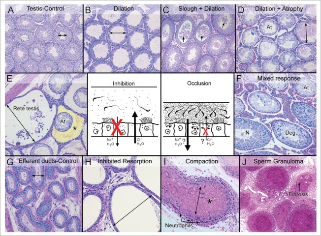
Two mechanisms lead to efferent ductule dysfunction and fluid accumulation in the testis. The central drawing illustrates the two mechanism of efferent ductule dysfunction that will result in the accumulation of luminal fluids and cause backpressure damage to the seminiferous tubules. The ‘inhibition’ mechanism involves the blockage of fluid resorption by inhibiting Na+ and water update and possibly an increase in Cl- and water movement into the lumen, thereby diluting the sperm and exceeding the drainage capacity of the ductules into the epididymis. The ‘occlusion’ mechanism involves excessive resorption and possibly an inhibition of Cl- secretion into the lumen. This mechanism results in a more viscous luminal environment, sperm stasis and eventually the occlusion or blockage of the ductule. (A) Control testis showing normal cross-sectional widths of the seminiferous tubular lumens. (B) Testis showing dilation of the seminiferous tubular lumen caused by the inhibition mechanism. Spermatogenesis appeared normal but there was thinning of the epithelium. (C) Testis showing dilation of the tubules caused by the occlusion mechanism. Sloughing of germ cells into the lumen (arrows) was also involved. (D) Testis showing seminiferous tubular atrophy (At), with some evidence of residual dilation after long-term occlusion of the efferent ductules. (E) Rete testis region showing excessive buildup of fluid and dilation, adjacent to atrophic (At) seminiferous tubules following the inhibition of fluid resorption. The yellow highlighted area (*) illustrates an edematous buildup around the atrophic tubules, which occurs in some cases but not in others. (F) Testis showing a mixed response following long-term occlusion of the efferent ductules. Atrophic tubules (At) are mixed with normal spermatogenesis (N) and degenerative changes (Deg). (G) Control efferent ductules in the conus region showing a normally narrow luminal diameter. (H) Efferent ductules at the proximal/conus junction following the inhibition of fluid resorption show excessive dilation and thinning of the epithelium. (I) Compaction of sperm within the lumen of the efferent ductules leads to dilation of the lumen and occlusion. This response caused the recruitment of polymorphonuclear leukocytes (neutrophils) into the wall lining the epithelium. (J) A long-term consequence of efferent ductule occlusion is the formation of sperm granulomas. The hyaline area shows the beginning of fibrosis.
