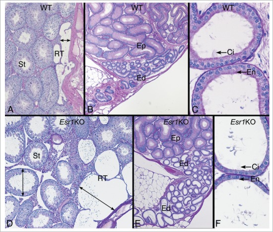
Figure 5.

Testis and efferent ductules in the wild type (WT) and Esr1KO mice. (A) WT testis showing the narrow width of the rete testis and normal seminiferous tubules (St). (B) WT head of the epididymis region showing the coiled common efferent ductule (Ed) adjacent to the initial segment epididymis (Ep). (C) WT proximal region of the efferent ductules have a wider lumen than the common duct and show a PAS+ endocytic and brush border of microvilli (En) on the nonciliated cells and long cilia (Ci) protruding in the lumen from the ciliated cells. (D) Esr1KO testis showing dilated rete testis (RT) filled with fluid and causing dilation of seminiferous tubules (St). (E) Head of the epididymis region in the Esr1KO showing dilated efferent ductules (Ed) adjacent to the initial segment epididymis (Ep). (F) Esr1KO showing the dilated proximal region of the efferent ductules. The epithelium is shorter in height and appears to have lost PAS+ endocytic and brush border lining (En) on the nonciliated cells. Cilia (Ci) are noted but they appear to be thinner in density.
