
Figure 2.
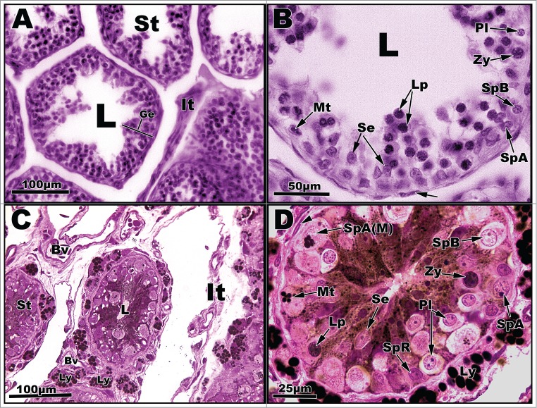
Light micrographs of seminiferous tubules of Graptemys pseudogeographica kohnii from April (A–B) and May (C–D). A. Seminiferous tubules (St) with central one exhibiting seminiferous epithelium (Ge) with an interstitium (It) to its right; L = lumen. B. Segment of seminiferous epithelium reveals several cell types, including Spermatogonia A undergoing mitosis (Mt), type A spermatogonium (SpA), type B spermatogonium (SpB), pre-leptotene spermatocyte (Pl), leptotene spermatocyte (Lp), zygotene spermatocyte (Zy), and Sertoli cells (Se). Arrow points to a fibroblast nucleus; L = lumen. C. Seminiferous tubules (St) surrounded by an interstitium (It), blood vessels (Bv), and individual Leydig cells (Ly) containing lipid droplets. D. Seminiferous tubule circumscribed by Leydig cells (Ly) full of lipid droplets. Germ cell types include a resting spermatogoium (SpR) and spermatogonia A (SpA) undergoing mitosis (Mt) as well as other cells mentioned in B. Paraffin, (A-B); plastic, (C-D).