Abstract
We describe a case of recurrence of chromophobe renal cell carcinoma 8 years after successful surgical treatment of primary localized disease in the left kidney. The primary tumour had exhibited neither gross nor histological evidence of lymphovascular infiltration. The recurrence occurred in the residual left ureteric stump – a finding that, to the best of our knowledge, has not previously been reported.
Introduction
Chromophobe renal cell carcinoma (RCC) is a neoplasm of the kidney with clinicopathological peculiarities; it has a better prognosis than clear cell RCC of similar grade and stage. Chromophobe RCC tends to remain localized even when they have grown to a substantial size. Two histological variants exist: classical and eosinophilic types. It is thought to originate from intercalated cells of the collecting duct. Complete surgical resection results in a good prognosis.
Case report
A 43-year-old male underwent left radical nephrectomy for a 5 × 4-cm renal mass in 2005. On histopathological analysis, the tumour was described as a Furhman grade 2, chromophobe RCC, (tumour size, vascular invasion, necrosis, sarcomatoid features, ureter on histology); it grew into the renal pelvis and was completely excised. Prior to this original surgery, there was uncertainty about the origin of his renal tumour; therefore, a ureterenoscopy was performed to rule out upper tract urothelial cell carcinoma, which revealed a normal urothelium throughout his urinary tract.
Subsequent routine surveillance up to 5 years revealed no evidence of disease recurrence. Following episodes of visible hematuria with clots in April 2010, he was investigated with a flexible cystoscopy and a computed tomography urogram, which were normal. He was consequently discharged from outpatient follow-up in 2011, 6 years after his original surgery. This was in accordance with the guidelines from the European Association of Urology for surveillance after treatment for intermediate-risk RCC.1
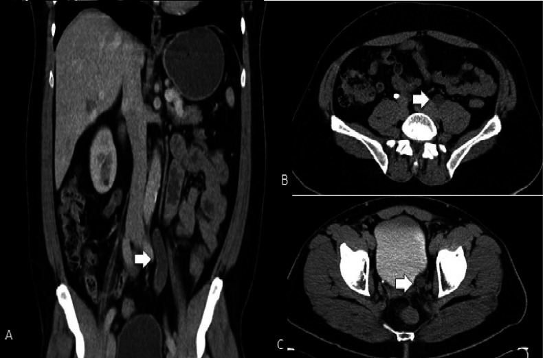
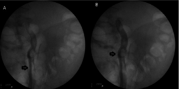
In August 2013, the patient re-presented with further visible hematuria. On this occasion, flexible cystoscopic evaluation failed due to an abundant clot within the bladder, preventing accurate inspection of his bladder urothelium. A subsequent computed tomography urogram, however, revealed a newly dilated left ureter along its full length, with no other significant or suspicious findings (Fig. 1). A retrograde left ureterogram showed multiple filling defects (Fig. 2). This prompted ureteroscopy under general anesthetic, which revealed a long clot in the ureter with multiple polypoid lesions within the left ureteric stump (Fig. 3). These lesions were biopsied and sent for histology, which confirmed that these were deposits of chromophobe RCC.
Fig. 1.
A computed tomography urogram (coronal [a] and axial [b, c]) showing a dilated left ureter.
Fig. 2.
A left ureterogram demonstrating multiple filling defects within the ureter.
Fig. 3.
Ureteroscopic view of the polypoid tumour within the left ureteric stump.
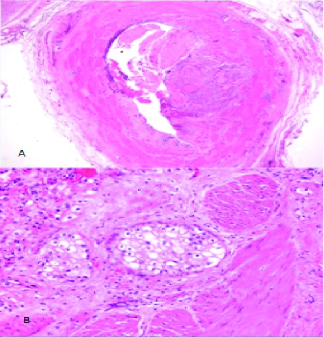
The patient underwent an open left ureterectomy in December 2013. Histology showed islands and nests of tumour confirming a T2 chromophobe RCC with metastatic deposits (Fig. 4) from his previous RCC. The patient made a full recovery. At the 18-month follow-up, he was free of recurrence.
Fig. 4.
A low-power overview of the ureter showing a reduction in the lumen diameter due to the tumour (hemtoxylin and eosin ×1.25 [a] and ×5 [b]).
Discussion
RCC accounts for 86% of all kidney cancers within the United Kingdom.2 The chromophobe subtype represents 5% of cases,3 and confers favourable prognosis in terms of duration of disease-free survival.4 This is the 54th reported case of ureteric metastasis from RCC (43 to the ipsilateral ureter, 10 contralateral).5 Length of time from nephrectomy to detection of metastasis is twice as long compared to that of other disease subtypes, such as clear cell or papillary RCC,6 which may explain the late presentation in this case compared to the other reported cases.
Invasion into the renal pelvis of the tumour at presentation may increase the risk of ureteric metastasis; however. there are reports of similar metastasis in the absence of primary involvement of the renal pelvis. Current evidence supports surgical resection as the only effective treatment option for solitary ureteric metastasis from RCC. The overexpression of KIT (CD117), a type III receptor tyrosine kinase, mTOR signalling pathway, vascular endothelial growth factor receptor and platelet derived growth factor receptor all provide potential targets for chemotherapy.4,7 There is no evidence supporting treatment with radiotherapy.
Conclusion
This case represents a rare finding of metachronous ureteric metastasis from RCC, presenting 8 years after initial diagnosis and treatment. This highlights the possibility that metastatic recurrence can occur at any time and that the possibility of ureteric metastasis should not be overlooked, especially following episodes of visible hematuria. Surgical resection remains the mainstay of treatment in such cases and there is no current evidence to support neoadjuvant chemotherapy or radiotherapy to prevent metastasis from intermediate-risk RCC. Close radiological surveillance with associated cystoscopic and flexible ureteroscopic investigation should be pursued, particularly in cases with visible hematuria.
Footnotes
Competing interests: The authors declare no competing financial or personal interests.
This paper has been peer-reviewed.
References
- 1.Ljungberg B, Bensalah K, Canfield S, et al. EAU guidelines on renal cell carcinoma: 2014 update. Eur Urol. 2015:67913–24. doi: 10.1016/j.eururo.2015.01.005. [DOI] [PubMed] [Google Scholar]
- 2.UK kidney cancer statistics. Cancer Research UK. http://www.cancerresearchuk.org/cancer-info/cancer-stats/types/kidney/. Accessed August 27, 2015. [Google Scholar]
- 3.Eble NJ, Sauter G, Epstein JI, et al. Pathology and Genetics of Tumours of the Urinary System and Male Genital Organs. Lyon, France: IARC Press; 2004. [Google Scholar]
- 4.Stec R, Grala B, Maczewski M, et al. Chromophobe renal cell cancer: Review of the literature and potential methods of treating metastatic disease. J Exp Clin Cancer Res. 2009;28:134–9. doi: 10.1186/1756-9966-28-134. [DOI] [PMC free article] [PubMed] [Google Scholar]
- 5.Cheng KC, Cho CL, Chau LH, et al. Solitary metachronous metastasis of renal cell carcinoma to the ureter. Int J Case Rep Med. 2013;2013 [Google Scholar]
- 6.Beck SD, Patel MI, Snyder ME, et al. Effect of papillary and chromophobe cell type on disease-free survival after nephrectomy for renal cell carcinoma. Ann Surg Oncol. 2004;11:71–7. doi: 10.1007/BF02524349. [DOI] [PubMed] [Google Scholar]
- 7.Vera-Badillo FE, Conde E, Duran I. Chromophobe renal cell carcinoma: A review of an uncommon entity. Int J Urol. 2012;19:894–900. doi: 10.1111/j.1442-2042.2012.03079.x. [DOI] [PubMed] [Google Scholar]


