Abstract
The aim of the work
was to present clinical material referring to rarely occurring abdominal cavity hernias in semilunar line – Spigelian hernias diagnosed with the help of ultrasound.
Material and methods
In the period from 1995 to 2001 785 anterior abdominal wall hernias were diagnosed including 11 Spigelian hernias (1.4%) diagnosed in 10 patients (7 women and 3 men) aged from 38 to 65 years old (average age 48). Eight patients complained of spastic pain in abdomen, in 5 of them it was accompanied by bloating and sometimes loud peristalsis. All the patients had been observing the mentioned symptoms from 2 to 5 years. Each of them had had colonoscopy and abdominal cavity ultrasound examination performed, some of them even three times. In 3 women with uterine fibroid the uterus was removed which did not eliminate the symptoms.
The ultrasound examination of the abdominal integument was performed mainly with the use of linear transducers of the frequency of 7–12 MHz; in obese patients also convex transducers were used (3,5–6 MHz). Each examination of abdominal integument included the assessment of the following areas: linea alba from xiphoid process to pubic symphysis including umbilicus, both semilunar lines from costal margins to pubic bones, and also inguinal areas. Moreover, all types of postoperative scars were examined. Each hernia was assessed in terms of size (the greatest dimension), hernia sac contents, width of the ring and reducibility under the compression of the transducer. Moreover, cough test and Valsalva's maneuver were performed. Generally, the examination was performed in a standing position.
Results
In 9 patients hernias were localized unilaterally, in one patient bilaterally. In 7 cases the hernia sac contained small bowel, in 2 cases the preperitoneal and omental fat, and in 2 cases preperitoneal fat only. Eight patients presenting with clinical symptoms underwent operative repair.
Conclusion
Ultrasound examination is beneficial in confirming the diagnosis of Spigelian hernias especially in terms of proper, therapeutic decision-making.
Keywords: Spigelian hernias, hernia sac contents, abdominal integument, ultrasound examination, treatment
Abstract
Celem pracy
było zaprezentowanie materiału klinicznego dotyczącego rzadko występujących przepuklin jamy brzusznej w kresie półksiężycowatej – przepuklin Spigla rozpoznanych za pomocą ultrasonografii.
Materiał i metoda
W okresie od 1995 do 2011 roku w badaniu ultrasonograficznym rozpoznano 785 przepuklin powłok przedniej ściany brzucha, w tym 11 przepuklin Spigla (1,4%), stwierdzonych u 10 chorych (7 kobiet i 3 mężczyzn) w wieku od 38 do 65 lat (średnia 48 lat). Ośmiu pacjentów skarżyło się na kurczowe bóle w podbrzuszu, przy czym u 5 towarzyszyły temu wzdęcia brzucha i niekiedy głośna perystaltyka. Wszyscy wymienione objawy obserwowali od 2 do 5 lat. Każdy z nich miał wykonane badanie kolonoskopowe i ultrasonograficzne jamy brzusznej, niektórzy nawet trzykrotnie. U 3 kobiet usunięto zmienioną mięśniakowato macicę, co nie wyeliminowało dolegliwości. Badania ultrasonograficzne powłok brzusznych przeprowadzono głównie głowicami liniowymi o częstotliwości 7–12 MHz; u osób otyłych stosowano również głowice konweksowe (3,5–6 MHz). Badanie powłok brzusznych obejmowało ocenę następujących okolic: kresy białej od wyrostka mieczykowatego mostka do spojenia łonowego łącznie z pępkiem, obu kres półksiężycowatych od łuków żebrowych do kości łonowych, a także okolic pachwin. Ponadto badano wszelkie blizny pooperacyjne. Każdą przepuklinę oceniano pod względem wielkości (największy wymiar), zawartości worka przepuklinowego, szerokości wrót i odprowadzalności przy ucisku głowicą. Ponadto stosowano próbę kaszlową i Valsalvy. Zasadniczo badanie przeprowadzano w pozycji stojącej.
Wyniki
U 9 osób przepukliny były zlokalizowane jednostronnie, u 1 osoby obustronnie. W 7 przypadkach worek przepuklinowy zawierał jelito cienkie, w 2 przypadkach – tłuszcz przedotrzewnowy i sieć, a w pozostałych 2 przypadkach – tylko tłuszcz przedotrzewnowy. U 8 chorych przepukliny powodowały objawy kliniczne; osoby te zostały zoperowane.
Wniosek
Badanie ultrasonograficzne spełnia wymogi skutecznej metody diagnostycznej w diagnostyce przepuklin Spigla i pozwala na podjęcie właściwej decyzji terapeutycznej.
Introduction
Anterior abdominal wall hernias located in semilunar line constitute a rare type of pathology and are named, after the name of their discoverer, Spigelian hernias (Flemish anatomist Adriaan van der Spieghel, Spigelius in Latin). Earlier also the caudate lobe of liver and semilunar line used to be named after him(1). Lately in Polish scientific literature there has been a review article with regard to Spigelian hernias, however in imaging documentation no ultrasound (US) images of this pathology were presented(2).
The aim of the work
The aim of the work is the presentation of our own clinical material of Spigelian hernias in ultrasound examination.
Material and methods
In the period from 1995 to 2001 785 anterior abdominal wall hernias were diagnosed, in which in 10 patients (7 women and 3 men aged from 38 to 65) 11 Spigelian hernias (1.4%) were diagnosed by means of ultrasound examination. Eight patients complained of spastic pain in abdomen, in 5 of them it was accompanied by bloating and sometimes loud peristalsis. All the patients had been observing the mentioned symptoms from 2 to 5 years. Each of them had had colonoscopy and abdominal cavity ultrasound examination performed, some of them even three times. In 3 women with uterine fibroid the uterus was removed which did not eliminate the symptoms.
The US examination of the abdominal integument was performed mainly with the use of linear transducers of the frequency of 7–12 MHz; in obese patients also convex transducers were used (3,5–6 MHz). Each examination of abdominal integument included the assessment of the following areas: linea alba from xiphoid process to pubic symphysis including umbilicus, both semilunar lines from costal margins to pubic bones, and also inguinal areas. Moreover all types of postoperative scars were examined. Each hernia was assessed in terms of size (the greatest dimension), hernia sac contents, width of the ring and reducibility under the compression of the transducer. Moreover, cough test and Valsalva's maneuver were performed. Generally, the examination was performed in a standing position.
Results
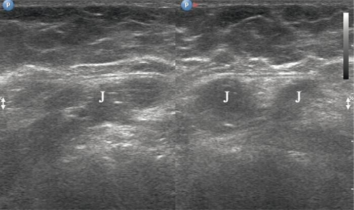
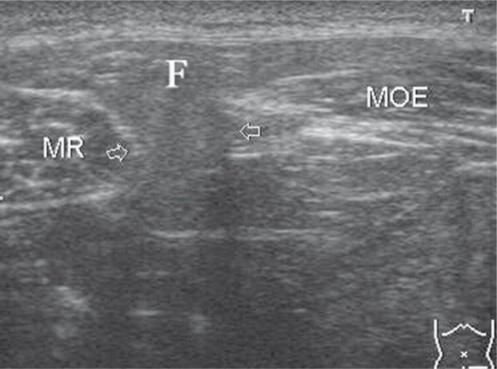
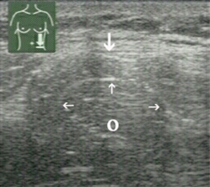
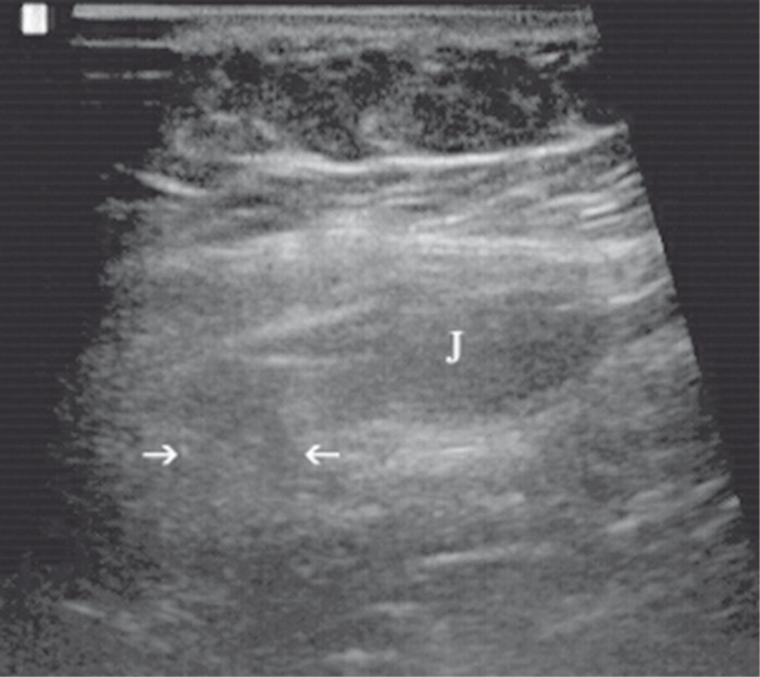
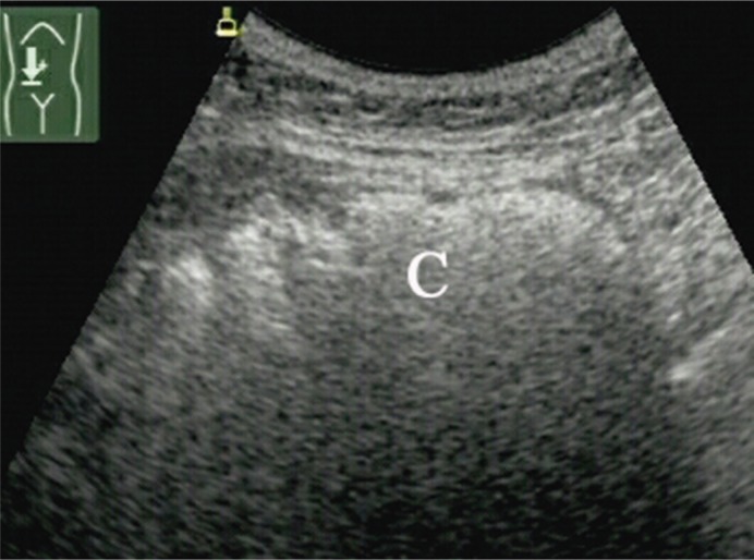
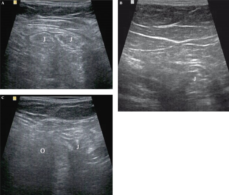
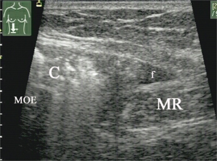
In 10 patients 11 Spigelian hernias were diagnosed, all of them typically over inferior epigastric vessels: 6 on the right side, 5 on the left side. In 56 years old woman 4 years after hysterectomy with oophorectomy a semilunar line hernia was diagnosed on the level of the right end of the postoperative scar going transversally in the abdomen (fig. 1). In the remaining 9 patients the mentioned hernias were of idiopathic character. The size of the hernias was in the range from 3 to 8 cm. In 2 cases the hernia sac contained only preperitoneal fat (fig. 2), similarly in the 2 preperitoneal fat and omentum were stated (fig. 3). Small intestine was found in hernia sac in 6 cases (fig. 4), in 1 right hernia caecum and some fluid were present (fig. 5 A, B). In 62 years old woman bilateral hernias of the semilunar lines containing small intestine were diagnosed, but the pain was related with the greater right hernia (fig. 6 A, B). Only one hernia with preperitoneal fat was totally reducible towards abdomen under the compression of the transducer. Seven hernias decreased in this test, 3 were immobile (fig. 6 C). The rings were usually narrow – average size equaled 15 mm (9–35 mm). Eight hernias increased under the influence of cough or Valsalva's maneuver, 3 did not respond to these tests. Moreover, in 4 patients from this group 5 hernias were diagnosed in a different localization: 3 indirect inguinal hernias and 1 umbilical and 1 femoral.
Fig. 1.
In two scans hernia in right semilunar line in 56 years old woman who underwent hysterectomy with oophorectomy 4 years before. J – small intestine
Fig. 2.
Left idiopathic Spigelian hernia. In the sac there is only preperitoneal fat (F). Arrows indicate the width of the ring. MR – rectus abdominis muscle, MOE – abdominal external oblique muscle
Fig. 3.
Left Spigelian hernia in which the sac contains preperitoneal fat (downward arrow) and greater omentum (O). Small arrows indicate peritoneum
Fig. 4.
Right intramuscular hernia in semilunar line. The hernia sac contains small intestine loop (J). Arrows indicate the width of the ring
Fig. 5 A.
Large (8 cm) right Spigelian hernia containing caecum (C). Sonogram obtained with the use of convex transducer in obese patient
Fig. 6.
In 62 years old woman bilateral Spigelian hernias were visualized: A – greater on the right side contains small intestine loop (J); B – smaller on the left side with small fragment of small intestine (J); C – the same hernia as in fig. 6 A decreased under the compression of the transducer. In this figure small fragment of intestine (J) and greater omentum (O) are visible
Fig. 5 B.
Fragment of the same hernia as in fig. 5 A, but examined with a linear transducer. C – caecum, f – trace of fluid in hernia sac, MR – rectus abdominis muscle, MOE – abdominal external oblique muscle
Discussion
Spigelian hernia, also referred to as lateral ventral hernia, in the majority of cases is of acquired character and is related to hard physical work, overweight, multiple labors, cough and ascites, so the conditions in which there is an increase in intra-abdominal pressure(2–10). Congenital hernia of this type occurs mostly in children and in this age group in the hernia sac except for small intestine, large intestine and omentum also gonads can be found(11).
In our material the most common was small intestine, only in one patient the presence of caecum was stated. Seven patients with hernias containing small intestine loop reported the greatest complains, probably related to the hernia strangulation in its ring. Because of the obstruction risk these patients agreed for operative treatment. In the remaining patients the content of the sac was only preperitoneal fat (in 2 asymptomatic patients) and in 2 others – preperitoneal fat with omentum, which manifested itself with mild local pain. In the group with the sac filled with intestine the hernia ring was narrow and the hernia itself was poorly reducible towards abdominal cavity which threatened with incarceration. The visualization of Spigelian hernia in 3 obese patients required the use of a convex transducer (fig. 5 A, B), in the remaining patients high frequency linear transducer was sufficient.
Basing on literary data(2–5, 7–11), and also on our experience it can be said that ultrasound fulfills the requirements of the first diagnostic method in cases suspected of the presence of such pathology.
This examination has unquestionable advantages, including the possibility of precise visualization of the abdominal wall structure, performing the examination in a standing position and the use of provocative tests in the form of cough, Valsalva's maneuver and transducer compression. This latter test enables the assessment of the degree of reducibility of hernia sac contents towards abdominal cavity. Such a full model of examination enables neither computed tomography nor magnetic resonance – these examinations are expensive and hardly accessible and moreover computed tomography exposes the patient to the negative influence of ionizing radiation.
In the differentiation a series of lesions localized in abdominal integument (hematomas, abscesses, granulomas, primary and secondary neoplasms) and its vicinity (appendicitis, Meckel's diverticulum, colon diverticulitis, omental appendices and many others) should be taken into account.
Summing up, the US examination enables with a great precision the diagnosis of ventral lateral hernias and also facilitates making decision as it comes to the appropriate medical intervention.
References
- 1.Dorland's Illustrated Medical Dictionary. 28. Philadelphia – Tokyo: W.B. Saunders Company; 1994. [Google Scholar]
- 2.Ciesielska-Krosny A, Mazur D, Waszczuk E, Paradowski L. Przepuklina Spigela. Gastroenterol Pol. 2011;18:80–83. [Google Scholar]
- 3.Hodgson TJ, Collins MC. Anterior abdominal wall hernias: diagnosis by ultrasound and tangential radiographs. Clin Radiol. 1991;44:185–188. doi: 10.1016/s0009-9260(05)80867-0. [DOI] [PubMed] [Google Scholar]
- 4.Hiller N, Alberton Y, Shapira Y, Hadas-Halpern I. Richter's hernia strangulated in a Spigelian hernia: ultrasonic diagnosis. J Clin Ultrasound. 1994;22:503–505. doi: 10.1002/jcu.1870220808. [DOI] [PubMed] [Google Scholar]
- 5.Miller PA, Mezwa DG, Feczko PJ, Jafri ZH, Madrazo BL. Imaging of abdominal hernias. Radiographics. 1995;15:333–347. doi: 10.1148/radiographics.15.2.7761639. [DOI] [PubMed] [Google Scholar]
- 6.Ganz E, Tazzioli G, Mosca D, Amorotti C, Speranza M. Ventrolateral hernias of the abdominal wall. The anatomicopathologic, clinical and therapeutic considerations. Minerva Chir. 1997;52:1441–1445. [PubMed] [Google Scholar]
- 7.Kuś H, Mackiewicz Z. Przepukliny brzuszne. Wydawnictwo Lekarskie PZWL, Warszawa. 1997:76. [Google Scholar]
- 8.Ianora AA, Midiri M, Vinci R, Rotondo A, Angelelli G. Abdominal wall hernias: imaging with spiral CT. Eur Radiol. 2000;10:914–919. doi: 10.1007/s003300051036. [DOI] [PubMed] [Google Scholar]
- 9.Rettenbacher T, Hollerweger A, Macheiner P, Gritzmann N, Gotwald T, Frass R, et al. Abdominal wall hernias: cross-sectional imaging signs of incarceration determined with sonography. AJR Am J Roentgenol. 2001;177:1061–1066. doi: 10.2214/ajr.177.5.1771061. [DOI] [PubMed] [Google Scholar]
- 10.Aguirre DA, Casola G, Sirlin C. Abdominal wall hernias: MDCT findings. AJR Am J Roentgenol. 2004;183:681–690. doi: 10.2214/ajr.183.3.1830681. [DOI] [PubMed] [Google Scholar]
- 11.Levy G, Nagar H, Blachar A, Ben-Sira L, Kessler A. Pre-operative sonographic diagnosis of incarcerated neonatal Spigelian hernia containing the testis. Pediatr Radiol. 2003;33:407–409. doi: 10.1007/s00247-003-0879-8. [DOI] [PubMed] [Google Scholar]


