Abstract
Legionella pneumophila (Lp) is the etiological agent responsible for Legionnaires’ disease, a potentially fatal pulmonary infection. Lp lives and multiplies inside protozoa in a variety of natural and man-made water systems prior to human infection. Fraquil, a defined freshwater medium, was used as a highly reproducible medium to study the behaviour of Lp in water. Adopting a reductionist approach, Fraquil was used to study the impact of temperature, pH and trace metal levels on the survival and subsequent intracellular multiplication of Lp in Acanthamoeba castellanii, a freshwater protozoan and a natural host of Legionella. We show that temperature has a significant impact on the short- and long-term survival of Lp, but that the bacterium retains intracellular multiplication potential for over six months in Fraquil. Moreover, incubation in Fraquil at pH 4.0 resulted in a rapid decline in colony forming units, but was not detrimental to intracellular multiplication. In contrast, variations in trace metal concentrations had no impact on either survival or intracellular multiplication in amoeba. Our data show that Lp is a resilient bacterium in the water environment, remaining infectious to host cells after six months under the nutrient-deprived conditions of Fraquil.
Introduction
Identified as the etiological agent of Legionnaires’ disease in the late 1970’s, Legionella pneumophila (Lp) is a Gram-negative, water-borne bacterium [1]. Inhalation of Legionella-contaminated aerosols can lead to Legionellosis, which is comprised of the mild, flu-like Pontiac fever and the more serious pneumonia, Legionnaires’ disease [2, 3]. Legionella species are often found in freshwater bodies as well as an assortment of man-made water distribution systems [4–7] with the exception of Legionella longbeachae whose presence in potting soils is linked to the incidence of Legionnaires’ disease in Australia and New Zealand [8]. In Europe and North America, Lp is responsible for over 90% of reported Legionellosis cases [9]. Public health concerns related to Lp are mainly associated with its contamination of cooling towers and other man-made water distribution systems [10].
In the natural or man-made water environment, Lp can be found in a motile planktonic state, a sessile state within mixed species biofilms, growing intracellularly in amoeba and in a persistent state (viable but non-culturable, VBNC) [11]. In the water niche, Lp is exposed to a changing environment, such as variations in temperature, concentration of dissolved oxygen, minerals, anthropogenic chemicals and organic matter [11]. The most important parameter associated with the presence of Lp in a given water system is the heterotrophic plate count (HPC), which measures the general contamination of a system. The higher the HPC of a system, the higher the odds are of finding Lp within it [12–15]. Elevated total organic content (TOC) in water distribution systems has also been correlated with an increase in the incidence of Legionella [16]. Presumably, amoeba are attracted to contaminated sites in water systems to feed on susceptible bacteria and multiply, providing a source of prey for Lp [11]. In addition, Lp can persist in mixed species biofilms in natural and human-made water systems [17]. Nevertheless, growth of Lp associated with such biofilms requires the presence of amoeba, such as Hartmannella vermiformis [18]. Symbiotic and competitive interactions with other bacterial species seem to have a major impact on the net number of Lp in the water environment [19]. As an example, the persistence of Lp in water is impaired by the presence of Pseudomonas aeruginosa [20, 21]. Physico-chemical factors, such as temperature, pH, and the concentration of dissolved metals also seem to have an impact on the presence of Lp in water systems [10, 13, 14, 22]; however, there have been conflicting results regarding the extent of their effects on Lp persistence in water [23–27].
To date, no standardized system has been presented to study the effect of individual environmental factors on the survival of Lp. Knowledge pertaining to environmental conditions that affect the presence of Lp in such systems has been gained mainly through prospective studies in the field [12–15, 26, 28] or by using tap water models [13, 14, 29, 30]. The impact of different environmental factors such as water temperature, pH, and the presence of trace metals has been recorded mainly as a result of on-site and environmental samplings, or using sterilized tap or distilled water [21, 26, 31–34]. As a result, the interpretation of this data does not take into account variations in water composition depending on the locality or the seasonality. Thus, there is lack of a standardized artificial freshwater medium to study Legionella and environmental factors affecting survival, and their subsequent effect on the virulence potential of the bacterium. A defined water medium will allow elimination of variations in water composition that are dependent on seasonality and geography. It will also provide a foundation to build water microcosm, whose complexity can be increased gradually, thus allowing the evaluation of each additional parameter separately.
We have previously shown that the sigma factor RpoS, and the stringent response is required for Lp to survive in water [35]. Consequently, the RpoS mutant was unable to survive in tap water, and in two defined freshwater media, DFM and Fraquil. DFM composition was derived from the salt and buffer content of the chemically defined media used for Legionella growth [36], and contains 50mg/L NaCl, 20 mg/L KH2PO4 and 50 mg/L KCl. Fraquil is an approximation of freshwater found in North America [37] (see Table 1 for the composition of Fraquil). Our previous results show that Lp survive in Fraquil as well as in tap water for 30 days, but show a slight defect in DFM [35]. Therefore, we decided to further investigate the behaviour of Lp in Fraquil. Here, we use Fraquil to study the impact of individual environmental factors on Lp in a controlled water environment. To further this goal, the consequences of changing temperature, acidity and trace metal content over a short time period were investigated by tracking the capacity of Lp to survive in Fraquil under these conditions, and its subsequent capacity for intracellular multiplication (ICM) in amoeba. Furthermore, we investigated the long-term influence of temperature on the survival of Lp and on its ICM potential.
Table 1. Composition of Fraquil.
| Components | Concentration |
|---|---|
| CaCl2∙2H2O | 0.25uM |
| MgSO4∙7H2O | 0.15uM |
| NaHCO3 | 0.15uM |
| K2HPO4 | 10nM |
| NaNO3 | 0.1uM |
| FeCl3∙6H2O | 10nM |
| CuSO4∙5 H2O | 1nM |
| (NH4)6Mo7O24∙4 H2O | 0.22nM |
| CoCl2∙6 H2O | 2.5nM |
| MnCl2∙4 H2O | 23nM |
| ZnSO4∙7 H2O | 4nM |
Methods
Bacterial Strains and Media
All experiments were conducted using JR32 or its derivatives. JR32 is a salt-sensitive, streptomycin-resistant, restriction negative mutant of Legionella pneumophila (Lp) strain Philadelphia 1 [38]. The dotA - strain, used as a negative control in intracellular multiplication (ICM) assays, is a transposon mutant carrying a mutation in the type IVb secretion system that is essential for intracellular multiplication in Lp [38]. Strains stored at -80°C in 10% glycerol were grown on BCYE (ACES-buffered charcoal yeast extract) agar supplemented with 0.25mg/ml L-cysteine and 0.4mg/ml ferric pyrophosphate. AYE broth (BCYE without agar and charcoal) was used as the liquid medium. The defined water medium used for water exposure experiments, Fraquil was prepared as described by Morel et al. [37] with a final iron concentration of 10nM and was filter-sterilized using a 0.2um filter (Sarstedt). Ultrapure Type 1 water (18.2 MΩ·cm at 25°C), produced with a Synergy Ultrapure Water System (EMD Millipore), was used to prepare Fraquil. The complete composition of Fraquil is presented in Table 1.
Water Exposure Experiments
JR32 cultured on BCYE agar at 37°C for 3 days was washed three times with Fraquil and suspended in fresh Fraquil at an OD600nm of 0.1. Then, 1ml of the bacterial suspension was mixed with 4ml of fresh Fraquil in a 25cm2 cell culture flasks. Three biological replicates were used for each experiment. To test the impact of temperature, the flasks were incubated at six temperatures: 4°C, 10°C, 25°C, 30°C, 37°C, 42°C. For water experiments with varying pH, aliquots of Fraquil were adjusted to pH 4, 5, or 6 with 0.01M HCl and Lp was suspended at each pH as described above. To test the effect of varying trace metal concentrations, Fraquil was prepared without the addition of trace metal (0X), by adding twice the volume of trace metals than standard Fraquil (2X) or by adding 10 times the volume of trace metals (10X). Standard Fraquil (1X metals, please refer to Table 1 for composition) was used as a control. Lp was suspended in each trace metal concentration as described above. For each environmental parameter tested, CFU counts on BCYE agar were used at defined time points to track survival over time.
Intracellular Multiplication Assays
Intracellular multiplication was measured in the amoeba Acanthamoeba castellanii and THP-1-derived human macrophages with a multiplicity of infection (MOI) of 0.1. A. castellanii was cultured in peptone yeast glucose (PYG) broth [39]. PYG contained 20g/L proteose peptone, 1g/L yeast extract, 0.1M glucose, 0.4mM MgSO4, 0.05mM CaCl2, 0.1mM sodium citrate, 0.005mM Fe(NH4)2(SO4)2, 0.25mM Na2HPO4 and 0.25mM KH2PO4, and the pH was adjusted to 6.5 using 1M HCl. Each infection well in a 24-well plate was seeded with 5x10⁵ A. castellanii cells in 1ml of PYG. One hour prior to infecting A. castellanii with Lp, the media in each well was replaced with 1ml of Ac buffer (PYG without proteose peptone, yeast extract and glucose). The plate was incubated for an additional hour at 30°C before introducing approximately 5 x 10⁴ Lp to each well. THP-1 monocytes were cultured in RPMI (GIBCO) supplemented with L-glutamine and 5% FBS. For the THP-1 infection, 5x10⁵ cells treated with 10−7 M phorbol 12-myristate 13-acetate (PMA) were seeded into a 24-well plaque in 1ml of RPMI 3 days prior to infection and left to incubate at 37°C in 5% CO2. One hour prior to infecting the macrophages with L. pneumophila, the media in each well was replaced with fresh RPMI. THP-1 cells were infected with approximately 5 x 10⁴ Lp to each well.
The infection wells were sampled daily to detect the extracellular increase of CFUs relative to time zero. The laboratory wild type JR32 was used as a positive control, while the dotA - mutant, deficient in intracellular replication, served as a negative control. Both control strains were grown on BCYE agar and suspended in AYE broth at an OD600 of 0.1, and were then further diluted 10 fold to obtain an approximate OD600 of 0.01. 2μl of this final solution was used to infect the cells.
For bacterial samples originating from water experiments testing temperature, pH or trace metal content, a CFU count was done 3 days prior to the infection. On the day of the infection assay, this count was used to determine the volume representing 1 x 10⁴ bacterial cells, thus resulting in an MOI of 0.1. When necessary, bacteria were diluted in Fraquil.
Statistical analysis
The graphs show the average of at least three biological replicates and the standard deviation. We used unpaired the one tail Student’s T-test to access statistical significance.
Results
Short-term effect of temperature on the survival of Lp and subsequent ICM potential
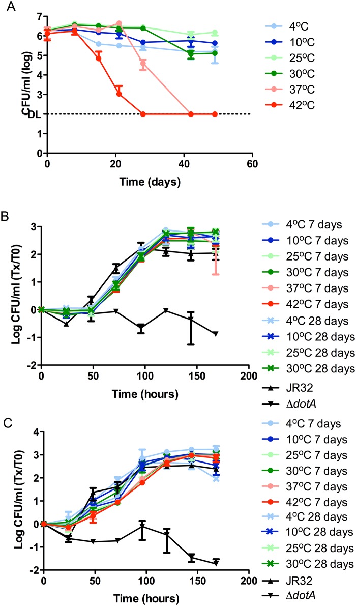
We tested the survival of Lp in Fraquil, hereafter called water, exposed to six different temperatures ranging from refrigeration to the high end of the temperature spectrum recorded as supporting Legionella growth [40]: 4°C, 10°C, 25°C, 30°C, 37°C and 42°C. Using CFU counts, the survival of the JR32 strain was monitored for 49 days (Fig 1A). It was clear that changes in temperature had a definite impact on the survivability of Lp in this water system. The 42°C experimental condition resulted in the fastest CFU decline starting at 15 days, reaching the detection limit of 100 CFU/ml in all three replicates after 28 days. At 37°C, which is in the range of optimal growth temperatures for Legionella, bacterial numbers were stable for approximately 1 month after which the CFU counts decreased dramatically reaching the detection limit at the end of 42 days. A moderate dip in the CFU counts was observed at 30°C at day 42, while Lp incubated at 25°C showed no decrease in bacterial counts during the 49-day tracking period testing survivability in water. At 10°C, Lp seems to maintain relatively stable bacterial counts. The set of samples at 4°C showed a small decrease in CFU counts after approximately two weeks and then stabilized for the remainder of the time tested (49 days). Our results show that even moderate temperatures between 30°C to 42°C significantly impact the survivability of Lp in a minimal water system (Fig 1A).
Fig 1. Impact of temperature on the short-term survival of Lp in Fraquil and intracellular multiplication (ICM) after exposure to different temperatures.
A) The JR32 strain was exposed to Fraquil at six different temperatures. Weekly CFU counts were performed to track survival. DL, detection limit. At 4°C, 10°C, 37°C and 42°C, CFU counts from 8 days were statistically different (P≤0.05) than CFU counts at 25°C. At 30°C, CFU counts from day 42 were statistically different that CFU counts at 25°C. B) A. castellanii was infected with JR32 that had been exposed to the respective temperatures tested for 7 days or 28 days at an MOI of 0.1. Daily CFU counts monitored the ICM inside amoeba and are presented as the ratio over CFU counts at day 0. JR32 from BCYE was used as the positive control and dotA - was used as the negative control. C) Cultured human macrophages (THP-1) were infected with JR32 that had been exposed to the respective temperatures tested for 7 days or 28 days at an MOI of 0.1. Daily CFU counts monitored the ICM and are presented as the ratio over CFU counts at day 0. JR32 from BCYE was used as the positive control and dotA - was used as the negative control.
Following water exposure, we tested the infectivity of Lp incubated at different temperatures towards Acanthamoeba castellanii, a natural host of Legionella found in natural and man-made water systems [41]. The dotA - mutant strain was used as a negative control in the infection assays due to its deficiency in intracellular multiplication (ICM) in A. castellanii as a result of a Type IVb secretion system defect [38]. To detect any changes of virulence potential in response to different incubation times in water, we used bacteria that had been exposed to the respective temperatures for both 7 and 28 days (Fig 1B). Lp exposed to 37°C and 42°C was not used in the 28 day infections since the CFU counts were either too low (37°C) or non-existent (42°C). The volume of the 37°C samples required to infect the cells at an MOI of 0.1 was too large and would have compromised the dynamics of the infection assay, thus making the results unreliable. At the end of the infection cycle (168 hours), there was no significant difference in the CFU increase during infection between the different temperatures. Moreover, no significant differences were observed between 7-day and 28-day exposure times. The ICM potential in cultured human macrophages was also tested in parallel (Fig 1C). As for the infection of amoeba, there were no significant differences between the positive control and the bacterial cultures originating from water. Since no significant difference was observed between temperature-treated samples in macrophages, subsequent ICM experiments were conducted exclusively in A. castellanii, which is more relevant in the context of the water environment.
These infection assay results show that Lp is able to maintain its ICM potential for at least 28 days post-incubation in the nutrient-deficient water environment at temperatures ranging from 4°C to 30°C, and retains ICM at least 7 days after exposure to water at 37°C and 42°C, temperatures that are environmentally relevant (Fig 1B and 1C).
Effect of trace metal concentrations on the survival and subsequent ICM of Lp
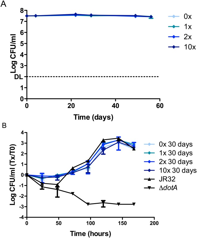
To test whether a water environment with a reduced level of trace metals impairs the ability of Lp to survive, or if an increase in these metal concentrations may help it survive better, we tested four different trace metal concentrations in Fraquil; namely without the addition of trace metals (0X), standard Fraquil (1X), double the concentration of trace metals (2X) and 10 times the concentration of trace metals (10X) used in standard Fraquil (Fig 2A). Over a period of 56 days, there were no observable significant differences in the CFU counts between Lp exposed to different trace metal concentrations.
Fig 2. Survival of Lp at different trace metal concentrations and subsequent intracellular multiplication (ICM).
A) The JR32 strain of Lp was exposed to Fraquil at four different metal concentrations: no addition of trace metals (0X), standard Fraquil (1X), double the quantity of trace metals (2X) and 10 times the quantity of trace metals (10X) than in standard Fraquil. Weekly CFU counts were performed to track survival. DL, detection limit. B) A. castellanii was infected with JR32 that had been exposed to the respective levels of trace metals tested for 30 days at an MOI of 0.1. Daily CFU counts monitored the ICM inside amoeba and are presented as the ratio over CFU counts at day 0. JR32 from BCYE was used as the positive control and dotA - was used as the negative control.
The ICM capacity of Lp exposed for 30 days to different trace metal concentrations in Fraquil was tested (Fig 2B). The ICM rate of the water exposed Lp samples were comparable to that of Lp cultured on BCYE agar (positive control). Furthermore, no difference was observed in ICM between Lp exposed to different trace metal concentrations. Therefore, it would seem that exposure to water containing a higher amount of trace metals, or a lack thereof, does not impact the ICM of Lp in the amoebal host.
Effect of pH on the survival and subsequent ICM of Lp
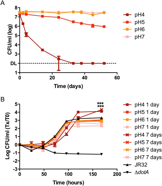
The survival of Lp in Fraquil at three different pH levels (4, 5 & 6) was tested in addition to the standard Fraquil whose pH hovers around 7.3. The pH experiment was conducted at 25°C since it is a permissive temperature for the survival of Lp in water (Fig 1A). Changing the pH had a much more drastic effect on the survivability of Lp compared to temperature variations. When the water medium was adjusted to a pH value of 4, CFU counts steadily declined to the detection limit within 33 days (Fig 3A). At a pH value of 5, the CFU counts started to visibly decrease after 24 days of incubation in the defined water medium. The higher pH values of 6 and 7.3 of normal Fraquil resulted in no loss of culturability over the course of the experiment (52 days).
Fig 3. Impact of pH on the short-term survival of Lp in Fraquil and intracellular multiplication (ICM) after exposure to different pH.
A) The JR32 strain of Lp was exposed to Fraquil at four different pH. Weekly CFU counts were performed to track survival. DL, detection limit. At pH 4 and pH 5, CFU counts from 2 days are statistically different (P≤0.05) than CFU counts at pH 7. At pH 6, CFU counts from 38 days are statistically different than CFU counts at pH 7. B) A. castellanii was infected with JR32 that had been exposed to the respective pH tested for either 1 day or 7 days at an MOI of 0.1. Daily CFU counts monitored the ICM inside amoeba and are presented as the ratio over CFU counts at day 0. JR32 from BCYE was used as the positive control and dotA - was used as the negative control. *** P≤ 0.005 versus control.
The virulence potential of Lp incubated in acidified Fraquil was tested by following the intracellular multiplication (ICM) in the same manner used for the temperature and trace metal variation experiments in A. castellanii. Since the CFU counts decreased dramatically in the first week of exposure to pH 4, we used Lp that had been exposed to water for 24 hours and for 7 days to test the ICM in amoeba. Interestingly, compared to samples at pH 7 and the positive control, incubation in Fraquil at pH 4 at both exposure times resulted in a statistically significant, 1 log increase of CFU counts at the end of a 168 hour infection cycle (Fig 3B). The ICM of Lp exposed to pH 5, pH 6 and standard Fraquil produced a similar increase in bacterial load upon infection of A. castellanii compared to Lp originating from rich media.
Long-term survival of Lp in water and subsequent effect on ICM
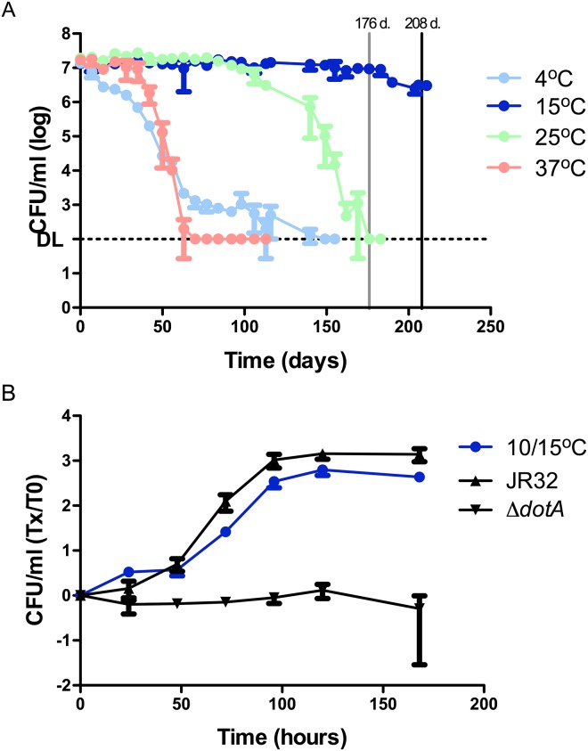
To test whether Lp was able to survive in Fraquil over a longer period of time, inoculums were tested at four temperatures (4°C, 15°C, 25°C and 37°C). The lower end of the temperature spectrum was used, since higher temperatures were found to be detrimental to Lp in the short-term (Fig 1A). We used 37°C as a short-term survival temperature control. CFU counts were tracked over a period of 211 days in total (Fig 4A). As was expected (Fig 1A), the fastest decline in the bacterial population was observed at 37°C, where no CFUs could be detected on agar plates after 70 days. At 25°C, inoculums were stable for 98 days after which the CFU counts started to decrease, reaching the detection limit by 176 days. At 4°C, the CFU counts reached the detection limit by 149 days. We tested long-term survival at 15°C instead of 10°C. Lp was initially exposed to 15°C for 176 days, but the flasks were incubated at 10°C for the following 32 days prior to testing ICM. On the long term, Lp survived at 15°C even better than at 25°C showing no significant decrease in CFU counts until 183 days.
Fig 4. Long-term survival of Lp in Fraquil and intracellular multiplication (ICM) after long-term exposure to a moderate temperature.
A) The JR32 strain was exposed to Fraquil and CFU counts were used to track survival over a 211-day time period. Weekly CFU counts were performed to track survival. DL, detection limit. The vertical lines indicate the time points at which the temperature of incubator was reduced from 15°C to 10°C (grey– 176 days) and when samples were harvested to infect amoeba (black– 208 days). At 4°C, CFU counts from 14 days are statistically different (P≤0.05) than CFU counts at 15°C. At 25°C, CFU counts from 113 days are statistically different than CFU counts at 15°C. At 37°C, CFU counts from 42 days are statistically different than CFU counts at 15°C. B) A. castellanii was infected with JR32 that had been exposed to 15°C for 176 days and 10°C for an additional 32 days at an MOI of 0.1. Daily CFU counts monitored the ICM inside amoeba and are presented as the ratio over CFU counts at day 0. JR32 from BCYE was used as the positive control and dotA - was used as the negative control.
To test whether the ICM capacity of Lp had been affected negatively or positively after a long incubation period in Fraquil, A. castellanii was infected with bacteria that had been incubated in water for a total of 208 days, 176 days at 15°C and another 32 days at 10°C. While the increase in bacterial counts during the infection is statistically lower than that of Lp originating from rich media, Lp seems to retain most of its capacity for ICM even after an extended time in water and in the absence of any additional nutrients (Fig 4B).
Discussion
A variety of water distribution systems can harbour Lp including shower heads, hot water tubs and hot water tanks [22, 42, 43]. In fact, the design of some systems can further favour Legionella contamination and persistence in water, either by cooler bodies of water within hot water systems or dead legs creating areas of stagnant water [25, 44]. For example, domestic water heating units in Quebec City, Canada, that are powered by electricity were shown to contain an area of water that was at a significantly lower temperature than the rest of the tank, thus, allowing the survival and growth of Legionella species in contaminated units [22, 25].
In this study, we have used Fraquil, to explore the effect of temperature, pH and trace metal concentration on the survival of Lp and on its subsequent capacity to grown inside amoeba. It is noteworthy that the JR32 laboratory strain was used for this study and that environmental isolates of Lp may behave differently in Fraquil. We are currently screening multiple clinical and environmental isolates.
Several studies report finding the bacterium at temperatures exceeding 50°C in the environment while others have shown that Lp is metabolically active above 45°C [26, 27, 40, 45–47]. In rich media, Lp grows optimally under laboratory conditions between 25°C and 37°C. Early studies also showed that Lp is able to multiply in unsterilized tap water containing amoeba between 25°C and 42°C over a period of 21 days, but that it could not replicate in the absence of amoeba at temperatures above 37°C [29, 31, 32]. Wadowsky et al. [29] demonstrated that an environmental isolate survived for 28 days in distilled water at 35°C. An early study by Dennis et al. [48] showed that Lp is more temperature tolerant on the short term than Pseudomonas and Micrococcus species, both found with Lp in water distribution systems. This characteristic has been taken advantage of when isolating Legionella spp. from environmental sources, using a mild heat treatment to increase isolation [49, 50]. Therefore, we expected to find that Lp would be relatively resistant to heat in Fraquil, the artificial freshwater medium used in this study. To our surprise, we found that Lp survive for only 6 and 3 weeks at 37°C and 42°C, respectively. Our study suggests that the reported persistence of Lp in water systems at high temperatures is positively affected by variables other than temperature; these variables may include protection inside thermophilic amoebal hosts, amoeba cysts or inside biofilm [31, 51, 52].
The steady decrease in CFUs observed at 4°C (Fig 4A) is consistent with recorded loss of culturability at 4°C, but the survival or culturablility periods vary according to different groups [28]. For example, Wadowsky et al. [29] observed that storing environmental samples at 5°C resulted in a decrease of CFUs. In addition, the earliest reports of viable-but-nonculturable (VBNC) Lp cells elude to a 4°C incubation temperature inducing this state [53]. We, therefore, suspect that Lp enters a VBNC state in water at 4°C; however, this will require further experimentation.
A moderate water temperature of 25°C allowed Lp to survive approximately three months in Fraquil (Fig 4A). At 15°C, Lp survive for at least 208 days. Similarly, Paszko-Kolva et al. [30] show that in drinking water samples, a clinical Lp strain has no significant difference in the CFU counts after incubation at 15°C for approximately 200 days, but that CFU counts decreased more significantly when using creek or estuarian waters. This ability to survive over a period of several months would allow Lp to persist in a water system and replicate in amoebal hosts when the latter arrive into the system. Most community outbreaks of Legionnaires’ disease occur in the late summer or early fall seasons [54, 55]. A gradual increase of Lp in a water system over this time frame will eventually allow the bacterium to attain a concentration sufficient for disease transmission in humans during the spring and summer, and survive over the winter at lower temperature.
We demonstrate that Lp incubated at a temperature ranging from 4°C to 42°C were able to replicate in A. castellanii and in cultured human macrophages after exposure to water for at least one week, while Lp at lower temperatures maintained intracellular multiplication (ICM) capacity for at least 28 days (Fig 1B and 1C). Temperature is known to regulate the expression of virulence factors in other pathogenic bacteria like Shigella, Streptococcus and P. aeruginosa [56–58]; however, the interplay between Lp virulence and temperature is yet to be clearly defined. A growth temperature of 37°C was shown to increase virulence of Lp while growth at 24°C results in an avirulent strain in a guinea pig model establishing a first link between temperature and virulence of Lp [23]. More importantly, the avirulence observed at 24°C was corrected when the temperature was shifted to 37°C [23]. In contrast, another study showed that Lp grown at 25°C is more lethal to guinea pig macrophages in vitro [24]. While contradictory, both studies show a definite effect of temperature on the virulence of Lp. In addition, pili and flagella are expressed in Lp in a temperature-dependent manner, and the expression of both structures have been linked to the bacterium’s virulence in host cells [59–63]. The type II secretion system is involved in the production of the type IV pili in Lp [59]. Soderberg et al. [64] reported that the type II secretion system of Lp allowed survival in water at 4°C and 17°C and also played a role in intracellular multiplication (ICM) in amoeba at 22°C-25°C. Moreover, pili were shown to play a role in adherence to both amoeba and human cells [61]. Flagella expression has also been demonstrated to be temperature regulated and is implicated in virulence [60, 62].
Moreover, we demonstrate that Lp incubated at 15°C were able to infect and kill amoeba after resting in Fraquil for approximately six months, albeit at a slightly lower rate than the positive control (Fig 4B). Our results suggest that, even in the absence of its natural hosts and lack of sufficient nutrients for growth (i.e. without the contamination of water systems by organic material), Lp may still pose a significant threat to public safety for a long period of time, as it remains virulent and competent for ICM. Therefore, Lp is able to easily linger in a clean water system for many months before coming into contact with its amoeba prey.
The presence of some metals has been linked to a decrease of Legionella in water systems while other metals are deemed as contributors to its survival [65]. Copper and silver ions are used to disinfect water distribution systems and have been studied for their negative effects on Legionella species, and they are known to decrease contamination levels in conjunction with other treatments [66, 67]. Indeed, a negative correlation is observed between the incidence of Lp and elevated trace levels of copper ions [13, 14]. In addition, as observed in the cases of many other bacteria, Lp requires iron for optimal growth on artificial media and in host cells as evidenced by its multiple iron acquisition systems [68–73]. In accordance, Lp contamination is positively correlated with higher concentrations of iron in water systems [12–14]. Furthermore, higher concentrations of manganese, zinc and cobalt have also been shown to correlate positively with Lp contamination of water systems [13, 36].
No survival defect (Fig 2A) was observed in any of the trace metal variations that were tested. While a positive relationship has been shown between the concentrations of manganese, iron and zinc, and the presence of Lp in hot water systems in some studies [13], no such correlation was found in others [12, 14]. There was no difference in ICM after 30 days of exposure to the respective trace metal concentrations (Fig 2B). The infection itself was performed in Ac buffer, which contains 5nM of iron. Therefore, any negative effects on ICM caused by the lack of iron in Fraquil without trace metals may have been rescued upon exposure of Lp to Ac buffer. The Ac buffer does not contain the other trace metals found in Fraquil; therefore, it is possible to conclude that the trace metals in Fraquil other than iron do not affect the ICM of Lp in the concentrations tested. Our results suggest that the impact of the concentration of metals on the survival and growth of Lp in water systems is linked to other conditions, potentially the presence of susceptible amoeba hosts.
Once inside a host cell, Lp grows within the L egionella containing vacuole (LCV) which evades the normal endocytic pathway by hijacking the host cell machinery [74]. While virulent Lp disturbs the acidification of the LCV, the recruitment of V-ATPases that carry out the acidification is not fully blocked in a small number of cases [75, 76]. Therefore, there is a biological need for Lp to have and to use mechanisms to survive pH stress. This may explain the relative hardiness of Lp toward varying pH that has been observed in nature and in man-made water systems. In fact, environmental sampling shows that Lp can be recovered at a wide range of pH (5.5 to 8.1) [26]. Wadowsky et al. [29] also found that an environmental isolate of Lp was able to replicate in filter-sterilized tap water from pH 5.5–9.2. Furthermore, isolating environmental strains of Legionella is known to be greatly enhanced by an acid treatment, suggesting that it is relatively more tolerant to acid than other bacteria found in water [77, 78]. Lp has been shown to tolerate a pH 2 treatment for at least 30 minutes [79]. More recently, the Lp genome was shown to encode carbonic anhydrases [80]. These enzymes are involved in pH regulation and may have a role in the bacterium’s survival inside the LCV, further supporting a relatively high level of tolerance to pH in Legionella species [80].
A low pH of 4 significantly affected the survival of Lp in the water environment, resulting in no CFU counts at the end of one month. It is conceivable that Lp, already under the stress of adapting to the nutritionally poor water environment, is unable to cope with the additional stress of an acidic pH. Variations in pH are known to cause the precipitation of some trace metals and have been used to evaluate trace metal contents in environmental samples [81, 82]. A more acute deprivation of metal cofactors caused by precipitation reactions may be responsible for the sensitivity of Lp at low pH, but this hypothesis is negated by Fig 3A showing no survival defect when no trace metals are added to Fraquil. Moreover, exposure to pH 4 for 24 hours or seven days resulted in higher ICM (Fig 3B). Another study demonstrated that a pH 6.5 acid treatment over a 24 hour time period provided increased resistance to a second pH stress, to oxidative stress, and induced virulence in previously non-virulent strains [83]. It is possible that prior exposure to pH 4 primes Lp for the intracellular environment that is reported to attain a pH value of 5.6 [75, 84]. This possibility will require further study.
Conclusions
We have successfully used Fraquil to investigate the survival of Lp in water. Temperature and pH were found to have a determinant effect on the survival, but the trace metal concentration does not impact survival of Lp. The ICM of Lp seems to increase in response to low pH, but the concentration of trace metals and temperature seem to have little effect on its ICM capacity. Importantly, our results show that Lp retains its ability to infect host cells after long-term survival in Fraquil. Our results support the use of Fraquil as a defined freshwater medium to study the biology of Lp in water systems, such as the transcriptomic response of Lp to Fraquil [85], and to facilitate the interpretation of datasets originating from different research groups.
Acknowledgments
This study was funded by Discovery Grant 418289–2012 from the National Sciences and Engineering Research Council of Canada (NSERC) and a John R. Evans Leaders Fund—Funding for research infrastructure from the Canadian Foundation for Innovation to SPF. NM is the recipient of a PhD scholarship from Fond de Recherche du Québec—Nature et Technologie. Peter McBride was the recipient of an Undergraduate Student Research Award from NSERC.
Data Availability
All relevant data are within the paper.
Funding Statement
This work was supported by the Natural Sciences and Engineering Research Council (NSERC, nserc-crsng.gc.ca) of Canada Discovery grant 418289-2012 and the John R. Evans Leaders Fund - Funding for research infrastructure from the Canadian Foundation for Innovation (CFI, innovation.ca/) to SPF. NM is supported by a PhD scholarship from Fonds de Recherche du Québec – Nature et Technologies (FRQNT, frqnt.gouv.qc.ca/accueil). PM was supported by a NSERC Undergraduate Student Research Award (nserc-crsng.gc.ca).
References
- 1. Fields BS, Benson RF, Besser RE. Legionella and Legionnaires' Disease: 25 Years of Investigation. Clin Microbiol Rev. 2002;15(3):506–26. [DOI] [PMC free article] [PubMed] [Google Scholar]
- 2. Diederen BMW. Legionella spp. and Legionnaires' disease. J Infect. 2008;56(1):1–12. 10.1016/j.jinf.2007.09.010. [DOI] [PubMed] [Google Scholar]
- 3. Glick TH, Gregg MB, Berman B, Mallison G, Rhodes WW, Kassanoff I. Pontiac Fever. An epidemic of unknown etiology in a health department: I. Clinical and epidemiological aspects. Am J Epidemiol. 1978;107(2):149–60. [DOI] [PubMed] [Google Scholar]
- 4. Tachibana M, Nakamoto M, Kimura Y, Shimizu T, Watarai M. Characterization of Legionella pneumophila Isolated from Environmental Water and Ashiyu Foot Spa. BioMed Res Int. 2013;2013(Article ID 514395). 10.1155/2013/514395 [DOI] [PMC free article] [PubMed] [Google Scholar]
- 5. Kuroki T, Ishihara T, Ito K, Kura F. Bathwater-Associated Cases of Legionellosis in Japan, with a Special Focus on Legionella Concentrations in Water. Jpn J Infect Dis. 2009;62:201–5. [PubMed] [Google Scholar]
- 6. Walczak M, Krawiec A, Lalke-Porczyk. Legionella pneumophila bacteria in a thermal saline bath. Ann Agric Environ Med. 2013;20(4):649–52. [PubMed] [Google Scholar]
- 7. Walczak M, Kletkiewicz H, Burkowska A. Occurrence of Legionella pneumophila in lakes serving as a cooling system of a power plant. Environ Sci Process Impacts. 2013;15(12):2273–8. 10.1039/c3em00452j [DOI] [PubMed] [Google Scholar]
- 8. Whiley H, Bentham R. Legionella longbeachae and legionellosis. Emerg Infect Dis 2011;17(4):579–83. 10.3201/eid1704.100446 [DOI] [PMC free article] [PubMed] [Google Scholar]
- 9. Yu VL, Plouffe JF, Pastoris MC, Stout JE, Schousboe M, Widmer A, et al. Distribution of Legionella Species and Serogroups Isolated by Culture in Patients with Sporadic Community-Acquired Legionellosis: An International Collaborative Survey. J Infect Dis. 2002;186(1):127–8. 10.1086/341087 [DOI] [PubMed] [Google Scholar]
- 10. Bartram J, Chartier Y, Lee JV, Pond K, Surman-Lee S. Legionella and the Prevention of Legionellosis. In: Organization WH, editor. Geneva: World Health Organization; 2007. [Google Scholar]
- 11. Lau HY, Ashbolt NJ. The role of biofilms and protozoa in Legionella pathogenesis: implications for drinking water. J Appl Microbiol. 2009;107(2):368–78. 10.1111/j.1365-2672.2009.04208.x [DOI] [PubMed] [Google Scholar]
- 12. Edagawa A, Kimura A, Tanaka H, Tomioka K, Sakabe K, Nakajima C, et al. Detection of culturable and nonculturable Legionella species from hot water systems of public buildings in Japan. J Appl Microbiol. 2008;105(6):2104–14. 10.1111/j.1365-2672.2008.03932.x [DOI] [PubMed] [Google Scholar]
- 13. Bargellini A, Marchesi I, Righi E, Ferrari A, Cencetti S, Borella P, et al. Parameters predictive of Legionella contamination in hot water systems: Association with trace elements and heterotrophic plate counts. Water Res. 2011;45(6):2315–21. 10.1016/j.watres.2011.01.009. 10.1016/j.watres.2011.01.009 [DOI] [PubMed] [Google Scholar]
- 14. Serrano-Suárez A, Dellundé J, Salvadó H, Cervero-Aragó S, Méndez J, Canals O, et al. Microbial and physicochemical parameters associated with Legionella contamination in hot water recirculation systems. Environ Sci Pollut Res Int. 2013;20(8):5534–44. 10.1007/s11356-013-1557-5 [DOI] [PubMed] [Google Scholar]
- 15. Mouchtouri VA, Goutziana G, Kremastinou J, Hadjichristodoulou C. Legionella species colonization in cooling towers: Risk factors and assessment of control measures. Am J Infect Control. 2010;38(1):50–5. 10.1016/j.ajic.2009.04.285. 10.1016/j.ajic.2009.04.285 [DOI] [PubMed] [Google Scholar]
- 16. Leoni E, De Luca G, Legnani PP, Sacchetti R, Stampi S, Zanetti F. Legionella waterline colonization: detection of Legionella species in domestic, hotel and hospital hot water systems. J Appl Microbiol. 2005;98(2):373–9. 10.1111/j.1365-2672.2004.02458.x [DOI] [PubMed] [Google Scholar]
- 17. Stewart CR, Muthye V, Cianciotto NP. Legionella pneumophila Persists within Biofilms Formed by Klebsiella pneumoniae, Flavobacterium sp., and Pseudomonas fluorescens under Dynamic Flow Conditions. PLoS ONE. 2012;7(11):e50560 10.1371/journal.pone.0050560 [DOI] [PMC free article] [PubMed] [Google Scholar]
- 18. Murga R, Forster TS, Brown E, Pruckler JM, Fields BS, Donlan RM. Role of biofilms in the survival of Legionella pneumophila in a model potable-water system. Microbiology. 2001;147(11):3121–6. [DOI] [PubMed] [Google Scholar]
- 19. Taylor M, Ross K, Bentham R. Legionella, Protozoa, and Biofilms: Interactions Within Complex Microbial Systems. Microb Ecol. 2009;58(3):538–47. 10.1007/s00248-009-9514-z [DOI] [PubMed] [Google Scholar]
- 20. Guerrieri E, Bondi M, Sabia C, de Niederhäusern S, Borella P, Messi P. Effect of Bacterial Interference on Biofilm Development by Legionella pneumophila . Curr Microbiol. 2008;57(6):532–6. 10.1007/s00284-008-9237-2 [DOI] [PubMed] [Google Scholar]
- 21. Messi P, Anacarso I, Bargellini A, Bondi M, Marchesi I, de Niederhäusern S, et al. Ecological behaviour of three serogroups of Legionella pneumophila within a model plumbing system. Biofouling. 2011;27(2):165–72. 10.1080/08927014.2010.551190 [DOI] [PubMed] [Google Scholar]
- 22. Dufresne SF, Locas MC, Duchesne A, Restieri C, Ismaïl J, Lefebvre B, et al. Sporadic Legionnaires' disease: the role of domestic electric hot-water tanks. Epidemiol Infect. 2012;140(01):172–81. 10.1017/S0950268811000355 [DOI] [PubMed] [Google Scholar]
- 23. Mauchline WS, James BW, Fitzgeorge RB, Dennis PJ, Keevil CW. Growth temperature reversibly modulates the virulence of Legionella pneumophila . Infect Immun. 1994;62(7):2995–7. [DOI] [PMC free article] [PubMed] [Google Scholar]
- 24. Edelstein P, Beer K, DeBoynton E. Influence of growth temperature on virulence of Legionella pneumophila . Infect Immun. 1987;55(11):2701–5. [DOI] [PMC free article] [PubMed] [Google Scholar]
- 25. Joly J. Legionella and domestic water heaters in the Quebec City area. CMAJ. 1985;132(2):160. [PMC free article] [PubMed] [Google Scholar]
- 26. Fliermans C, Cherry W, Orrison L, Smith S, Tison D, Pope D. Ecological distribution of Legionella pneumophila . Appl Environ Microbiol. 1981;41(1):9–16. [DOI] [PMC free article] [PubMed] [Google Scholar]
- 27. Moritz MM, Flemming H-C, Wingender J. Integration of Pseudomonas aeruginosa and Legionella pneumophila in drinking water biofilms grown on domestic plumbing materials. International Journal of Hygiene and Environmental Health. 2010;213(3):190–7. 10.1016/j.ijheh.2010.05.003. [DOI] [PubMed] [Google Scholar]
- 28. Borella P, Guerrieri E, Marchesi I, Bondi M, Messi P. Water ecology of Legionella and protozoan: environmental and public health perspectives In: El-Gewely MR, editor. Biotechnology Annual Review. Volume 11: Elsevier; 2005. p. 355–80. [DOI] [PubMed] [Google Scholar]
- 29. Wadowsky RM, Wolford R, McNamara AM, Yee RB. Effect of temperature, pH, and oxygen level on the multiplication of naturally occurring Legionella pneumophila in potable water. Appl Environ Microbiol. 1985;49(5):1197–205. [DOI] [PMC free article] [PubMed] [Google Scholar]
- 30. Paszko-Kolva C, Shahamat M, Colwell RR. Long-term survival of Legionella pneumophila serogroup 1 under low-nutrient conditions and associated morphological changes. FEMS Microbiol Lett. 1992;102(1):45–55. [Google Scholar]
- 31. Wadowsky R, Butler L, Cook M, Verma S, Paul M, Fields B, et al. Growth-supporting activity for Legionella pneumophila in tap water cultures and implication of hartmannellid amoebae as growth factors. Appl Environ Microbiol. 1988;54(11):2677–82. [DOI] [PMC free article] [PubMed] [Google Scholar]
- 32. Yee RB, Wadowsky RM. Multiplication of Legionella pneumophila in unsterilized tap water. Appl Environ Microbiol. 1982;43(6):1330–4. [DOI] [PMC free article] [PubMed] [Google Scholar]
- 33. Borella P, Montagna MT, Romano-Spica V, Stampi S, Stancanelli G, Triassi M, et al. Legionella infection risk from domestic hot water. Emerg Infect Dis. 2004;10(3):457 [DOI] [PMC free article] [PubMed] [Google Scholar]
- 34. Loret J, Robert S, Thomas V, Levi Y, Cooper A, McCoy W. Comparison of disinfectants for biofilm, protozoa and Legionella control. J Water Health. 2005;3:423–33. [DOI] [PubMed] [Google Scholar]
- 35. Trigui H, Dudyk P, Oh J, Hong J-I, Faucher SP. A regulatory feedback loop between RpoS and SpoT supports the survival of Legionella pneumophila in water. Appl Environ Microbiol. 2014:In press. Epub 21/11/2014. [DOI] [PMC free article] [PubMed] [Google Scholar]
- 36. Reeves M, Pine L, Hutner S, George J, Harrell W. Metal requirements of Legionella pneumophila . J Clin Microbiol. 1981;13(4):688–95. [DOI] [PMC free article] [PubMed] [Google Scholar]
- 37. Morel FMM, Westall JC, Ruerer JG, Chaplick JP. Descriptions of algal growth media AQUIL and FRAQUIL Technical Note, R M Parsons Laboratory for Water Resources and Hydrodynamics Massachusetts Institute of Technology, Cambridge, MA: 1975. [Google Scholar]
- 38. Sadosky AB, Wiater LA, Shuman HA. Identification of Legionella pneumophila genes required for growth within and killing of human macrophages. Infect Immun. 1993;61(12):5361–73. [DOI] [PMC free article] [PubMed] [Google Scholar]
- 39. Moffat JF, Tompkins LS. A quantitative model of intracellular growth of Legionella pneumophila in Acanthamoeba castellanii . Infect Immun. 1992;60(1):296–301. PMC257535. [DOI] [PMC free article] [PubMed] [Google Scholar]
- 40. Kusnetsov J, Ottoila E, Martikainen P. Growth, respiration and survival of Legionella pneumophila at high temperatures. J Appl Bacteriol. 1996;81(4):341–7. [DOI] [PubMed] [Google Scholar]
- 41. Rowbotham TJ. Preliminary report on the pathogenicity of Legionella pneumophila for freshwater and soil amoebae. J Clin Pathol. 1980;33(12):1179–83. 10.1136/jcp.33.12.1179 [DOI] [PMC free article] [PubMed] [Google Scholar]
- 42. Azuma K, Uchiyama I, Okumura J. Assessing the risk of Legionnaires’ disease: The inhalation exposure model and the estimated risk in residential bathrooms. Regul Toxicol Pharmacol. 2013;65(1):1–6. 10.1016/j.yrtph.2012.11.003. 10.1016/j.yrtph.2012.11.003 [DOI] [PubMed] [Google Scholar]
- 43. Roig J, Sabria M, Pedro-Botet ML. Legionella spp.: community acquired and nosocomial infections. Curr Opin Infect Dis. 2003;16(2):145–51. [DOI] [PubMed] [Google Scholar]
- 44. Thomas V, Bouchez T, Nicolas V, Robert S, Loret JF, Lévi Y. Amoebae in domestic water systems: resistance to disinfection treatments and implication in Legionella persistence. J Appl Microbiol. 2004;97(5):950–63. 10.1111/j.1365-2672.2004.02391.x [DOI] [PubMed] [Google Scholar]
- 45. Qin T, Yan G, Ren H, Zhou H, Wang H, Xu Y, et al. High Prevalence, Genetic Diversity and Intracellular Growth Ability of Legionella in Hot Spring Environments. PLoS ONE. 2013;8(3):e59018 10.1371/journal.pone.0059018 [DOI] [PMC free article] [PubMed] [Google Scholar]
- 46. Tison D, Pope D, Cherry W, Fliermans C. Growth of Legionella pneumophila in association with blue-green algae (Cyanobacteria). Appl Environ Microbiol. 1980;39(2):456–9. [DOI] [PMC free article] [PubMed] [Google Scholar]
- 47. Wadowsky RM, Yee RB, Mezmar L, Wing EJ, Dowling JN. Hot Water Systems as Sources of Legionella pneumophila in Hospital and Nonhospital Plumbing Fixtures. Appl Environ Microbiol. 1982;43(5):1104–10. [DOI] [PMC free article] [PubMed] [Google Scholar]
- 48. Dennis PJ, Green D, Jones BPC. A note on the temperature tolerance of Legionella . J Appl Bacteriol. 1984;56(2):349–50. 10.1111/j.1365-2672.1984.tb01359.x [DOI] [PubMed] [Google Scholar]
- 49. Guillemet TA, Lévesque B, Gauvin D, Brousseau N, Giroux J-P, Cantin P. Assessment of real-time PCR for quantification of Legionella spp. in spa water. Lett Appl Microbiol. 2010;51:639–44. 10.1111/j.1472-765X.2010.02947.x [DOI] [PubMed] [Google Scholar]
- 50.AFNOR. NF T90-471 Qualité de l’eau—Détection et quantification des Legionella et/ou Legionella pneumophila par concentration et amplification génique par réaction de polymérisation en chaîne en temps réel (RT-PCR): Association Française de Normalisation; 2010. Available: http://www.boutique.afnor.org. Accessed 2015 May 8.
- 51. Pryor M, Springthorpe S, Riffard S, Brooks T, Huo Y, Davis G, et al. Investigation of opportunistic pathogens in municipal drinking water under different supply and treatment regimes. Water Sci Technol. 2004;50(1):83–90. [PubMed] [Google Scholar]
- 52. Kilvington S, Price J. Survival of Legionella pneumophila within cysts of Acanthamoeba polyphaga following chlorine exposure. J Appl Bacteriol. 1990;68(5):519–25. 10.1111/j.1365-2672.1990.tb02904.x [DOI] [PubMed] [Google Scholar]
- 53. Hussong D, Colwell RR, O'Brien M, Weiss E, Pearson AD, Weiner RM, et al. Viable Legionella pneumophila Not Detectable by Culture on Agar Media. Nature Biotechnology. 1987;5(9):947–50. [Google Scholar]
- 54. Marston BJ, Lipman HB, Breiman RF. Surveillance for Legionnaires' Disease: Risk Factors for Morbidity and Mortality. Arch Intern Med. 1994;154(21):2417–22. [PubMed] [Google Scholar]
- 55. Carratala J, Garcia-Vidal C. An update on Legionella . Curr Opin Infect Dis. 2010;23(2):152–7. 10.1097/QCO.0b013e328336835b [DOI] [PubMed] [Google Scholar]
- 56. Grosso-Becerra MV, Croda-García G, Merino E, Servín-González L, Mojica-Espinosa R, Soberón-Chávez G. Regulation of Pseudomonas aeruginosa virulence factors by two novel RNA thermometers. Proc Natl Acad Sci U S A. 2014;111(43):15562–7. 10.1073/pnas.1402536111 [DOI] [PMC free article] [PubMed] [Google Scholar]
- 57. Smoot LM, Smoot JC, Graham MR, Somerville GA, Sturdevant DE, Migliaccio CAL, et al. Global differential gene expression in response to growth temperature alteration in group A Streptococcus . Proc Natl Acad Sci U S A. 2001;98(18):10416–21. 10.1073/pnas.191267598 [DOI] [PMC free article] [PubMed] [Google Scholar]
- 58. Maurelli A, Blackmon B, Curtiss R. Temperature-dependent expression of virulence genes in Shigella species. Infect Immun. 1984;43(1):195–201. [DOI] [PMC free article] [PubMed] [Google Scholar]
- 59. Liles MR, Viswanathan V, Cianciotto NP. Identification and temperature regulation of Legionella pneumophila genes involved in type IV pilus biogenesis and type II protein secretion. Infect Immun. 1998;66(4):1776–82. [DOI] [PMC free article] [PubMed] [Google Scholar]
- 60. Heuner K, Brand BC, Hacker J. The expression of the flagellum of Legionella pneumophila is modulated by different environmental factors. FEMS Microbiol Lett. 1999;175(1):69–77. 10.1111/j.1574-6968.1999.tb13603.x [DOI] [PubMed] [Google Scholar]
- 61. Stone BJ, Kwaik YA. Expression of multiple pili by Legionella pneumophila: identification and characterization of a type IV pilin gene and its role in adherence to mammalian and protozoan cells. Infect Immun. 1998;66(4):1768–75. [DOI] [PMC free article] [PubMed] [Google Scholar]
- 62. Heuner K, Steinert M. The flagellum of Legionella pneumophila and its link to the expression of the virulent phenotype. Int J Med Microbiol. 2003;293(2–3):133–43. 10.1078/1438-4221-00259. [DOI] [PubMed] [Google Scholar]
- 63. Molofsky AB, Shetron-Rama LM, Swanson MS. Components of the Legionella pneumophila Flagellar Regulon Contribute to Multiple Virulence Traits, Including Lysosome Avoidance and Macrophage Death. Infect Immun. 2005;73(9):5720–34. PMC1231111. [DOI] [PMC free article] [PubMed] [Google Scholar]
- 64. Söderberg MA, Dao J, Starkenburg SR, Cianciotto NP. Importance of type II secretion for survival of Legionella pneumophila in tap water and in amoebae at low temperatures. Appl Environ Microbiol. 2008;74(17):5583–8. 10.1128/AEM.00067-08 [DOI] [PMC free article] [PubMed] [Google Scholar]
- 65. Manske C, Hilbi H. Metabolism of the vacuolar pathogen Legionella and implications for virulence. Front Cell Infect Microbiol. 2014;4:125 10.3389/fcimb.2014.00125 [DOI] [PMC free article] [PubMed] [Google Scholar]
- 66. Landeen LK, Yahya MT, Gerba CP. Efficacy of copper and silver ions and reduced levels of free chlorine in inactivation of Legionella pneumophila . Appl Environ Microbiol. 1989;55(12):3045–50. [DOI] [PMC free article] [PubMed] [Google Scholar]
- 67. Cachafeiro SP, Naveira IM, García IG. Is copper—silver ionisation safe and effective in controlling legionella? J Hosp Infect. 2007;67(3):209–16. 10.1016/j.jhin.2007.07.017. [DOI] [PubMed] [Google Scholar]
- 68. Zheng H, Chatfield CH, Liles MR, Cianciotto NP. Secreted pyomelanin of Legionella pneumophila promotes bacterial iron uptake and growth under iron-limiting conditions. Infect Immun. 2013;81(11):4182–91. 10.1128/IAI.00858-13 [DOI] [PMC free article] [PubMed] [Google Scholar]
- 69. Cianciotto N. Iron Acquisition by Legionella pneumophila . Biometals. 2007;20(3–4):323–31. 10.1007/s10534-006-9057-4 [DOI] [PubMed] [Google Scholar]
- 70. Robey M, Cianciotto NP. Legionella pneumophila feoAB promotes ferrous iron uptake and intracellular infection. Infect Immun. 2002;70(10):5659–69. [DOI] [PMC free article] [PubMed] [Google Scholar]
- 71. Viswanathan V, Edelstein PH, Pope CD, Cianciotto NP. The Legionella pneumophila iraAB locus is required for iron assimilation, intracellular infection, and virulence. Infect Immun. 2000;68(3):1069–79. [DOI] [PMC free article] [PubMed] [Google Scholar]
- 72. Allard KA, Viswanathan V, Cianciotto NP. lbtA and lbtB are required for production of the Legionella pneumophila siderophore legiobactin. J Bacteriol. 2006;188(4):1351–63. [DOI] [PMC free article] [PubMed] [Google Scholar]
- 73. Ristroph JD, Hedlund KW, Gowda S. Chemically defined medium for Legionella pneumophila growth. J Clin Microbiol. 1981;13(1):115–9. [DOI] [PMC free article] [PubMed] [Google Scholar]
- 74. Horwitz MA. The Legionnaires' disease bacterium (Legionella pneumophila) inhibits phagosome-lysosome fusion in human monocytes. J Exp Med. 1983;158(6):2108–26. [DOI] [PMC free article] [PubMed] [Google Scholar]
- 75. Sturgill-Koszycki S, Swanson MS. Legionella pneumophila replication vacuoles mature into acidic, endocytic organelles. J Exp Med. 2000;192(9):1261–72. [DOI] [PMC free article] [PubMed] [Google Scholar]
- 76. Xu L, Shen X, Bryan A, Banga S, Swanson MS, Luo Z-Q. Inhibition of Host Vacuolar H+-ATPase Activity by a Legionella pneumophila Effector. PLoS Pathog. 2010;6(3):e1000822 10.1371/journal.ppat.1000822 [DOI] [PMC free article] [PubMed] [Google Scholar]
- 77. Buesching W, Brust R, Ayers L. Enhanced primary isolation of Legionella pneumophila from clinical specimens by low-pH treatment. J Clin Microbiol. 1983;17(6):1153–5. [DOI] [PMC free article] [PubMed] [Google Scholar]
- 78. Bopp C, Sumner J, Morris G, Wells J. Isolation of Legionella spp. from environmental water samples by low-pH treatment and use of a selective medium. J Clin Microbiol. 1981;13(4):714–9. [DOI] [PMC free article] [PubMed] [Google Scholar]
- 79. Wang WLL, Blaser MJ, Cravens J, Johnson MA. The Microorganism Growth, Survival, and Resistance of the Legionnaires' Disease Bacterium. Ann Intern Med. 1979;90(4):614–8. [DOI] [PubMed] [Google Scholar]
- 80. Nishimori I, Vullo D, Minakuchi T, Scozzafava A, Capasso C, Supuran CT. Sulfonamide inhibition studies of two β-carbonic anhydrases from the bacterial pathogen Legionella pneumophila . Bioorg Med Chem. 2014;22(11):2939–46. 10.1016/j.bmc.2014.04.006. 10.1016/j.bmc.2014.04.006 [DOI] [PubMed] [Google Scholar]
- 81. Lee G, Bigham JM, Faure G. Removal of trace metals by coprecipitation with Fe, Al and Mn from natural waters contaminated with acid mine drainage in the Ducktown Mining District, Tennessee. Appl Geochem. 2002;17(5):569–81. 10.1016/S0883-2927(01)00125-1 [DOI] [Google Scholar]
- 82. Tessier A, Campbell PGC, Bisson M. Sequential extraction procedure for the speciation of particulate trace metals. Anal Chem. 1979;51(7):844–51. 10.1021/ac50043a017 [DOI] [Google Scholar]
- 83. Bandyopadhyay P, Xiao H, Coleman HA, Price-Whelan A, Steinman HM. Icm/dot-independent entry of Legionella pneumophila into amoeba and macrophage hosts. Infect Immun. 2004;72(8):4541–51. [DOI] [PMC free article] [PubMed] [Google Scholar]
- 84. Allombert J, Lazzaroni J-C, Baïlo N, Gilbert C, Charpentier X, Doublet P, et al. Three Antagonistic Cyclic di-GMP-Catabolizing Enzymes Promote Differential Dot/Icm Effector Delivery and Intracellular Survival at the Early Steps of Legionella pneumophila Infection. Infect Immun. 2014;82(3):1222–33. 10.1128/IAI.01077-13 [DOI] [PMC free article] [PubMed] [Google Scholar]
- 85. Li L, Mendis N, Trigui H, Faucher SP. Transcriptomic changes of Legionella pneumophila in water. BMC Genomics. 2015;16:637 10.1186/s12864-015-1869-6 [DOI] [PMC free article] [PubMed] [Google Scholar]
Associated Data
This section collects any data citations, data availability statements, or supplementary materials included in this article.
Data Availability Statement
All relevant data are within the paper.


