Abstract
Botulinum neurotoxins (BoNTs) produced by Clostridium botulinum are the most poisonous substances known to mankind. It is essential to have a simple, quick and sensitive method for the detection and quantification of botulinum toxin in various media, including complex biological matrices. Our laboratory has developed a mass spectrometry-based Endopep-MS assay that is able to rapidly detect and differentiate all types of BoNTs by extracting the toxin with specific antibodies and detecting the unique cleavage products of peptide substrates. Botulinum neurotoxin type E (BoNT/E) is a member of a family of seven distinctive BoNT serotypes (A to G) and is the causative agent of botulism in both humans and animals. To improve the sensitivity of the Endopep-MS assay, we report here the development of novel peptide substrates for the detection of BoNT/E activity through systematic and comprehensive approaches. Our data demonstrate that several optimal peptides could accomplish 500-fold improvement in sensitivity compared to the current substrate for the detection of both not trypsin-activated and trypsin-activated BoNT/E toxin complexes. A limit of detection of 0.1 mouseLD50/mL was achieved using the novel peptide substrate in the assay to detect not trypsin-activated BoNT/E complex spiked in serum, stool and food samples.
Keywords: botulinum neurotoxin, botulism, mass spectrometry, peptide substrate
Introduction
The neurotoxins produced by Clostridium botulinum (Botulinum Neurotoxins, BoNT) are the most poisonous substances known to mankind. The life threatening diseases caused by these toxins include food-borne botulism, infant botulism, wound botulism, and adult intestinal colonization[1]. BoNTs also constitute a potential biological weapon as they are easy to produce[2]. On the other hand, botulinum toxins have been used for therapeutic or aesthetic applications[3]. For all these applications, it is essential to have a simple, quick and sensitive method for the detection and quantification of botulinum toxin in various media, including complex biological matrices.
The botulinum neurotoxins are synthesized as single chain polypeptides of 150 kDa which undergo proteolytic cleavage to generate active holotoxins constituted of two protein sub-units linked by a disulfide bond: a heavy chain (100 kDa) involved in target binding and a light chain (50 kDa) responsible for the toxicity through its peptidase activity[4]. In fact, the BoNTs belong to a family of zinc-dependent metallopeptidases. They cleave neuronal proteins involved in the exocytosis of neurotransmitters, such as SNAP-25, synaptobrevin and syntaxin, at the site specific to each toxin[5; 6]. This cleavage consequently blocks the release of neurotransmitter molecules at the neuromuscular junction ultimately leading to flaccid paralysis of muscle activity.
The neurotoxin type E (BoNT/E) forms part of a family of seven confirmed, related serotypes (botulinum toxins A to G) produced by different strains of Clostridium botulinum[7]. BoNT/E is a neurotoxin that causes botulism in both humans and animals. The most common intoxication by toxin type E is associated with eating contaminated fish [8; 9]. BoNT/E is unique because it is released from the bacterium as a single chain and cleaved into an active di-chain form by unidentified host cell proteases or other exogenous proteases such as trypsin [10; 11]. Activation of a single chain BoNT/E by trypsin leads to an approximately two orders of magnitude more potent neurotoxin than the single chain molecule [10; 12].
The mouse bioassay is the historic method for the detection of botulinum toxins[2]. It is very sensitive, detecting as little as approximately 10 picograms of active toxin which is defined as 1 mouse LD 50 (mLD50), but the assay can be slow in obtaining final results and requires the sacrifice of many animals. Therefore, much effort has been undertaken to develop alternative in vitro endopeptidase activity assays based on BoNT’s intrinsic enzymatic function. Several laboratories, including ours, have developed activity methods, by measuring the BoNTs’ cleavage products using synthetic peptide substrates with various detection platforms[13].
BoNT/E cleaves specifically one of the SNARE complex proteins, SNAP-25, at the Arg180-Ile181 bond [14]. Montecucco and coworkers revealed that the minimal length for proteolysis of SNAP-25 by BoNT/E includes a SNARE motif starting from Ala141 [15]. Binz and coworkers defined the minimal essential domain of SNAP-25 required for cleavage by BoNT/E as Ile156-Asp186 [16]. Through saturation mutagenesis and deletion mapping, Barbieri and Chen defined a short optimal cleavage domain of Met167-Asp186, where the subsite of Met167-Thr173 was considered as a binding domain contributing to substrate affinity [17; 18]. These findings led to the development of peptide substrates used in various in vitro activity assay platforms for the detection of the BoNT/E toxin. For instance, a fluorescence based assay uses a recombinant substrate consisting of the SNAP-25 sequence Ile134-Gly206 flanked by a green fluorescent protein (GFP) and a blue fluorescent protein (BFP)[19]. A 70-mer peptide of Val137-Gly206 as a substrate is included in an immuno-assay where the cleavage product was detected by a specific antibody [20]. The sequence of Ala141-Gly206 with a fluorescent tag on either terminus of the peptide formed a substrate included in the BoTestTM kit that uses Förster resonance energy transfer (FRET) technology to detect BoNT/E activity [21]. A 61-mer peptide consisting the sequence of Met146-Gly206 is reported in a capillary electrophoresis method [22]. The peptides of Ile156-Asp186 and Ile156-Thr190 are used in a mass spectrometry-based Endopep-MS assay developed in our laboratory[23; 24]. During the preparation of this manuscript, a new paper published claimed the peptide of Met167-Asp186 and its derivative with two Met replaced by Nle residues were effective substrates for the Endopep-MS platform[25]. This report described the development of a novel peptide substrate to improve the sensitivity of the Endopep-MS assay for the determination of BoNT/E catalytic activity. Through comprehensive optimization using approaches of truncation, deletion, single and multiple substitution and other modifications, we have developed several highly efficient peptides that showed more than a 500-fold improvement than the substrate currently used in the Endopep-MS assay.
Materials and method
All chemicals were obtained from Sigma–Aldrich (St. Louis, MO) except where indicated otherwise. Fmoc-amino acid derivatives and peptide synthesis reagents were purchased from EMD Chemicals, Inc. (Gibbstown, NJ) or Protein Technologies (Tucson, AZ). Isotopically labeled Fmoc-amino acid derivatives were purchased from Cambridge Isotope Laboratories (Tewksbury, MA). The complex forms of the botulinum neurotoxin without pre-activation and the trypsin activated BoNT/E toxin were obtained from Metabiologics (Madison, WI). Botulinum neurotoxin is highly toxic and appropriate safety measure is required. All BoNT neurotoxins were handled in a class 2 biosafety cabinet equipped with HEPA filters. Monoclonal antibodies were provided by Dr. James Marks at the University of California, San Francisco. Streptavidin coated Dynabeads were purchased from Invitrogen (Lake Success, NY). Serum and stool extracts were purchased from commercial source or collected from anonymous donors, and no demographic information was obtained (CDC IRB 4307).
Peptide synthesis
All peptides were prepared in house by a solid phase peptide synthesis method using Fmoc chemistry on a Liberty microwave peptide synthesizer (CEM, Matthews, NC, USA) or a Tribute peptide synthesizer (Protein Technologies, Tucson, AZ, USA). Peptides were cleaved and deblocked using a reagent mixture of 95% trifluroacetic acid :2% water: 2% anisole:1% ethanedithiol and purified by reversed-phase HPLC using a water:acetonitrile:0.1% TFA gradient (90-95% purity). Correct peptide structures were confirmed by MALDI mass spectrometry. All peptides were dissolved in deionized water as a 1 mM stock solution and were stored at −70°C until further use.
Endopep-MS assay
In-solution or on-bead Endopep-MS assays were carried out as previously described [26]. In brief, the reaction was conducted in a 20 μL reaction volume containing 0.1 mM peptide substrate, 10 μM ZnCl2, 1 mg/mL BSA, 10 mM dithiothreitol, and 200 mM HEPES buffer (pH 7.4) at 37°C for 1 or 4 hrs. For the in-solution assays without antibody-coated beads, various concentrations of BoNT/E, as indicated in the text, were directly added into the reaction mixture. For samples including complex matrices, the toxin spiked in matrix was first purified by antibodies immobilized on streptavidin beads followed by an activity assay as described [26].
After reaction, 2 μL of the supernatant was mixed with 20 μL of α-cyano-4-hydroxy cinnamic acid at 5 mg/mL in 50% acetonitrile/0.1% TFA/1 mM ammonium citrate; 2 μL of a 1 μM internal standard peptide (isotope labeled peptides resembling the sequence of either the C- or N-terminal cleavage product) was added to the solution. The formation of cleavage products was measured as the ratio of the isotope cluster areas of the cleavage product versus an internal standard.
MS detection
Each sample was spotted in triplicate on a MALDI plate and analyzed on a 5800 MALDI-TOF-MS instrument (Applied Biosystems, Framingham, MA). Mass spectra of each spot were obtained by scanning from 800 to 4000 m/z in MS-positive ion reflector mode. The instrument uses a Nd-YAG laser at 355 nm, and each spectrum is an average of 2400 laser shots.
Results and discussion
Optimal length of the peptide substrate of BoNT/E determined by truncation, deletion and mutation
Endopep-MS assay is a method using mass spectrometry, matrix assisted laser desorption ionization (MALDI) or electrospray ionization (ESI), to detect either one or two cleavage products hydrolyzed from a peptide substrate by an affinity enriched toxin. Therefore, assay sensitivity will not only depend on the hydrolysis efficiency (substrate binding and catalysis), but also rely on the ionization of cleaved peptide fragments, which is associated with their amino acid sequence. While different lengths of peptides including the essential elements for substrate binding and cleavage are applied in various in vitro BoNT activity assays as described above, a study on the optimal length of a peptide specifically suitable for the detection of BoNT/E by Endopep-MS was lacking. The peptide substrate (Pep-8 in Table 1) currently used in the Endopep-MS assay was derived from the partial sequence of SNAP-25, ranging from the amino acid residues Ile156 to Thr190 [27]. To examine whether further improvement can be achieved by optimizing peptide length for our Endopep-MS assay, we systematically prepared a series of peptides with extended or shortened sequences from either the N-terminal or C-terminal direction of the Pep-8, where the opposite end of the substrate remained the same (Table 1). In order to avoid bias caused by sequence-dependent ionization of cleavage fragments, the hydrolysis of the peptides with variable N-termini (Pep-1 to Pep-6) was compared by measuring the formation of C-terminal cleavage products by MALDI-TOF-MS, while that of the C-terminal varied peptides (Pep-7 to Pep-14) was compared with the production of N-terminal cleavage products. For those N-terminal truncated peptides, the longest peptide (Pep-1) with the SNAP-25 sequences of Ala141-Gly206 yielded the highest production of the cleavage product. When 10 or 15 N-terminal residues were removed (Pep-2 and Pep-3), the cleavage efficiency of these two substrates underwent a slight decrease. On the other hand, further deleting N-terminal resides (Pep-4 to Pep-6) led to a drastic reduction or non-detection of the cleavage products. For C-terminal truncated peptides, extending amino acid residues all the way to the last SNAP-25 residue at the position 206 (Pep-7) did not provide any benefit, compared to the activity of Pep-8 substrate which ends at position 190. Further C-terminal truncation of the peptides caused a steady decrease in their substrate hydrolysis (Pep-9 to Pep-14). In summary, Pep-1 performed the best among the tested BoNT/E substrate peptides of various lengths. This likely explained why the peptide itself, or with added fluoresence tags, was used as an efficient substrate in other studies and commercial kits (BoTest™ A/E Botulinum Neurotoxin Detection Kit, Biosentinel, Inc.). The large size of this peptide (7.5kDa), however, raised issues such as difficulty of peptide preparation and low solubility in assay buffers. In contrast, the molecular weight of Pep-8 (4.0kDa) is almost half of that of the Pep-1, but it has retained 90% of the substrate activity. For these reasons, we decided to use this peptide as a template for further optimization.
Table 1.
Hydrolysis of N- or C-terminal truncated peptide substrates by BoNT/E toxin.
| Peptide | SNAP-25 position |
Sequence | CT-prod (%)a |
NT- prod (%)b |
||
|---|---|---|---|---|---|---|
| Pep-1 | 141-206 | ARENEMDENLEQVSGIIGNLRHMALDMGNEIDTQNRQIDRIMEKADSNKTRIDEANQRATKMLGSGc | 100 | |||
| Pep-2 | 151-206 | EQVSGIIGNLRHMALDMGNEIDTQNRQIDRIMEKADSNKTRIDEANQRATKMLGSG | 75 | |||
| Pep-3 | 156-206 | IIGNLRHMALDMGNEIDTQNRQIDRIMEKADSNKTRIDEANQRATKMLGSG | 89 | |||
| Pep-4 | 161-206 | RHMALDMGNEIDTQNRQIDRIMEKADSNKTRIDEANQRATKMLGSG | 28 | |||
| Pep-5 | 166-206 | DMGNEIDTQNRQIDRIMEKADSNKTRIDEANQRATKMLGSG | 19 | |||
| Pep-6 | 171-206 | IDTQNRQIDRIMEKADSNKTRIDEANQRATKMLGSG | 0 | |||
| Pep-7 | 156-206 | IIGNLRHMALDMGNEIDTQNRQIDRIMEKADSNKTRIDEANQRATKMLGSG | 100 | |||
| Pep-8d | 156-190 | IIGNLRHMALDMGNEIDTQNRQIDRIMEKADSNKT | 100 | |||
| Pep-9 | 156-188 | IIGNLRHMALDMGNEIDTQNRQIDRIMEKADSN | 81 | |||
| Pep-10 | 156-186 | IIGNLRHMALDMGNEIDTQNRQIDRIMEKAD | 68 | |||
| Pep-11 | 156-185 | IIGNLRHMALDMGNEIDTQNRQIDRIMEKA | 77 | |||
| Pep-12 | 156-184 | IIGNLRHMALDMGNEIDTQNRQIDRIMEK | 38 | |||
| Pep-13 | 156-183 | IIGNLRHMALDMGNEIDTQNRQIDRIME | 15 | |||
| Pep-14 | 156-182 | IIGNLRHMALDMGNEIDTQNRQIDRIM | 10 | |||
Relative cleavage rate obtained from the analysis of C-terminal cleavage products (CT-prod). Condition: 37°C, 4 hour.
Relative cleavage rate obtained from the analysis of N-terminal cleavage products (CT-prod).
The cleavage site of BoNT/E substrate is depicted in bold.
Pep-8 is the BoNT/E substrate currently used in Endopep-MS assay.
The next modification to optimize the substrate focused on adding or deleting single amino acids to or from either end of the Pep-8 in order to examine whether smaller changes in peptide size impact the substrate activity. As shown in Table 2, removing one or two Ile residues from the N-terminus of the peptide resulted in a 30 to 40% decrease in cleavage efficiency of the newly formed peptides (Pep-15, Pep-16). In addition, extending the sequence (Pep-17) by adding a glycine (residue 155 of SNAP-25) to the N-terminus led to reduced production of enzyme cleavage products as well. The importance of two N-terminal hydrophobic residues led us to speculate that the side chains of these two Ile residues might have some direct contact with the enzyme through a hydrophobic-hydrophobic interaction. An increase in the cleavage product from Pep-18, after the third Ile residue was incorporated into the N-terminus of Pep-8, provided some supportive evidence for this hypothesis. More studies attempting to address this issue will be described below.
Table 2.
Relative production of the N-or C-terminal product cleaved from truncated or modified peptide substrates by BoNT/E toxin.
| Peptide | Sequence | CT-prod (%) |
NT- prod (%) |
||
|---|---|---|---|---|---|
| Pep-15 | GNLRHMALDMGNEIDTQNRQIDRIMEKADSNKT | 60 | |||
| Pep-16 | IGNLRHMALDMGNEIDTQNRQIRIIMEKADSNKT | 73 | |||
| Pep-8 | IIGNLRHMALDMGNEIDTQNRQIDRIMEKADSNKT | 100 | |||
| Pep-17 | GIIGNLRHMALDMGNEIDTQNRQIDRIMEKADSNKT | 81 | |||
| Pep-18 | IIIGNLRHMALDMGNEIDTQNRQIDRIMEKADSNKT | 123 | |||
| Pep-19 | IIIGNLRHMALDMGNEIDTQNRQIDRIMEKADSNKTRI | 2000 | 100 | ||
| Pep-20 | IIIGNLRHMALDMGNEIDTQNRQIDRIMEKADSNKTR | 2177 | 117 | ||
| Pep-18 | IIIGNLRHMALDMGNEIDTQNRQIDRIMEKADSNKT | 100 | 130 | ||
| Pep-21 | IIIGNLRHMALDMGNEIDTQNRQIDRIMEKADSN | 20 | 52 | ||
A remarkable improvement was obtained when one or two additional SNAP-25 residues were extended on the C-terminus of the Pep-18. Incorporation of Arg191 and Arg191-Ile192 into the Pep-19 and Pep-20, respectively, resulted in a 20-fold increase in the detection of the C-terminal cleavage products (CT-product) (Table 2). Since the N-terminal products (NT-product) cleaved from these peptides did not show significant changes, the elevated measurement of the CT-product should not come from altered cleavage efficiency of the new substrates, but must be due to the contribution of elevated ionization efficiency of the CT-products measured in positive ion mode by MALDI-TOF-MS, presumably directly associated with a positively charged Arg residue. In contrast, deletion of two C-terminal residues containing a positively charged Lysine from the Pep-18 led to a peptide (Pep-21) with reduced cleavage efficiency, indicated by a 52% production of the NT-product and decreased ionization of its CT-product (20%) as well.
In an attempt to shorten peptide size to improve synthesis yields and/or peptide solubility in the reaction buffer, internal deletions were applied on the N-terminal portion of the new template peptide, Pep-20. Table 3 shows that three peptides, Pep-22, Pep-23 and Pep-24, deleting 5 consecutive residues in different regions yielded different consequences. While less than 20% of the CT-product was detected from Pep-22 and Pep-23, removal of the area consisting of the sequence of RHMAL in Pep-24 retained 90% substrate activity, revealing that the chain of RHMAL did not play a critical role in peptide binding and/or substrate cleavage. Further deleting several residues sequentially, flanking either end of this 5-residue region, produced seven new peptides, Pep-25 through Pep-31. Among these peptides, Pep-29, with two more residues removed, maintained a relative production of the CT-product (88%) similar to that of the Pep-24. It was also interesting to see how a single residue difference significantly altered the cleavage efficiency of newly formed peptides by BoNT/E, for instance, Pep-28 (2%) versus Pep-29 (88%). In conclusion, this result demonstrated that seven internal residues (RHMALDM) within the BoNT/E peptide substrate seem to not participate in enzyme-substrate interaction and hence can be removed without significant negative impact on the substrate cleavage. To examine the viability of further shortening Pep-29, three new peptides (Pep-32, -33, and -34) were designed, where one to three C-terminal residues (T, KT and NKT) were removed but the terminal Arg residue was maintained. It was observed that the shortest Pep-34 turned out to be a poor BoNT/E substrate whereas the medium length substrate, Pep-33, retained the most substrate capability (Table 3). Pep-32, on the other hand, resulted in significant activity improvement compared to Pep-29. Pep-32 generated over 30% CT- and NT-products more than Pep-29, suggesting that Pep-32 possessed a higher BoNT/E cleavage efficiency, whereas the ionization efficiency of its CT-product remained unchanged. This peptide was then used as a new template for additional optimization discussed below.
Table 3.
Relative production of the N-or C-terminal product cleaved from internally deleted peptides by BoNT/E toxin.
| Peptide | Sequence | CT-prod (%) |
NT-prod (%) |
||
|---|---|---|---|---|---|
| Pep-20 | IIIGNLRHMALDMGNEIDTQNRQIDRIMEKADSNKTR | 100 | |||
| Pep-22 | IIIGNLRHMALDMGNE________RQIDRIMEKADSNKTR | 6 | |||
| Pep-23 | IIIGNLRHMAL________IDTQNRQIDRIMEKADSNKTR | 17 | |||
| Pep-24 | IIIGNL________DMGNEIDTQNRQIDRIMEKADSNKTR | 90 | |||
| Pep-25 | IIIGN__________DMGNEIDTQNRQIDRIMEKADSNKTR | 19 | |||
| Pep-26 | IIIG____________DMGNEIDTQNRQIDRIMEKADSNKTR | 43 | |||
| Pep-27 | III_____________DMGNEIDTQNRQIDRIMEKADSNKTR | 30 | |||
| Pep-28 | IIIGNL_____________NEIDTQNRQIDRIMEKADSNKTR | 2 | |||
| Pep-29 | IIIGNL____________GNEIDTQNRQIDRIMEKADSNKTR | 88 | |||
| Pep-30 | IIIGNL__________MGNEIDTQNRQIDRIMEKADSNKTR | 33 | |||
| Pep-31 | IIIGNL______LDMGNEIDTQNRQIDRIMEKADSNKTR | 53 | |||
| Pep-29 | IIIGNL____________GNEIDTQNRQIDRIMEKADSNKTR | 88 | 100 | ||
| Pep-32 | IIIGNL____________GNEIDTQNRQIDRIMEKADSNKR | 117 | 136 | ||
| Pep-33 | IIIGNL____________GNEIDTQNRQIDRIMEKADSNR | 91 | 84 | ||
| Pep-34 | IIIGNL____________GNEIDTQNRQIDRIMEKADSR | 18 | 36 | ||
Deleted amino acid residues from the Pep-20 are depicted in underscore.
Further improvement was accomplished by single or multiple substitutions
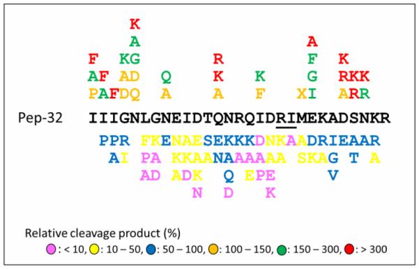
Based on the sequence of the best substrate, Pep-32, we carried out a thorough single mutation study where every single residue was substituted with selected amino acids and the peptide mutants were tested as BoNT/E substrates. While about two-third of the mutants tested produced less cleavage products than the wild-type did, another one-third of the single mutated peptides resulted in a higher substrate efficiency, some of those showed three-fold or higher improvement (Fig. 1), demonstrating the power of a mutation approach for substrate optimization.
Figure 1.

Effect of single amino acid mutations on the detection of cleavage product of mutated peptide-32 by BoNT/E. The residues at BoNT/E cleavage site are underlined. X represents norleucine.
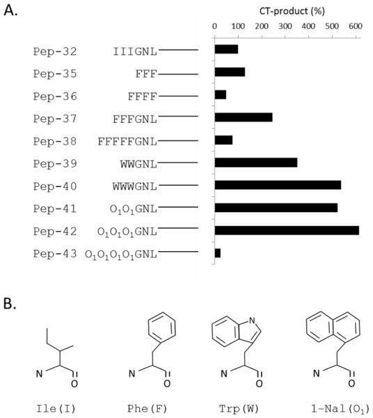
It was interesting to observe that a substantial improvement was accomplished when each of three N-terminal nonpolar Ile residues were replaced by Phe residues bearing a more hydrophobic side chain. This data emphasized our speculation described above that the N-terminal residues might be involved in direct contact with the catalytic domain of the toxin via hydrophobic-hydrophobic interactions. Substitution with even stronger hydrophobic residues probably enhanced such interaction and therefore increased substrate binding affinity. To explore whether those putative interactions can be further improved, a series of peptides with the modifications of hydrophobic residues in the N-terminal region of the Pep-32 were developed (Fig. 2A). A slight increase in detected CT-product was observed as the six N-terminal residues were replaced by three Phe residues in Pep-35. On the other hand, reduced detection of CT-product resulted when one more Phe was added in Pep-36. This suggested that three hydrophobic residues still retained the special enzyme-substrate interaction, even in a shorter peptide, but the contact might be impacted by a longer hydrophobic chain. When the three N-terminal Ile residues in Pep-32 were replaced by residues with a more hydrophobic structure, such as Phe in Pep-37, Trp in Pep-40, and 1-Nal in Pep-42, all new peptides acted as better substrates. In addition, the improvement degree, Pep-42 > Pep-40 > Pep-37, seems proportional to the size of the bulky side chain groups (1-Nal > Trp > Phe, Fig. 2B). This data provided additional supportive evidence for the suggestion of a hydrophobic interaction between the BoNT/E enzyme and the peptide substrates. Moreover, the hydrophobicity effect was also demonstrated by the fact that the double Trp substitution (Pep-39) displayed higher cleavage efficiency than the triple Phe substituted peptide (Pep-37) did, and the triple Trp replacement in Pep-40 resulted in better substrate efficiency than the double-Trp one in Pep-39, presumably due to the difference of their combined hydrophobicity. A similar effect was also observed by comparing the cleavage of the double 1-Nal peptide (Pep-41) with the triple Trp and triple 1-Nal ones (Pep-40 and Pep-42, respectively). Furthermore, significantly reduced substrate cleavage by BoNT/E on the peptides with a cluster of five Phe residues (Pep-38) or four 1-Nal residues (Pep-43) suggested that the size of the hydrophobic cluster on the N-terminus of a substrate was not unrestricted. In other words, the space of the putative hydrophobic pocket in the catalytic domain of the toxin was limited and it might not allow the placement of more than three very bulky side chain groups. More study is needed to further confirm or address this proposed hydrophobic interaction between BoNT/E protease and its peptide substrate.
Figure 2.
(A) Cleavage efficiency of the peptides modified with N-terminal hydrophobic residues. (B) the structure of some hydrophobic residues. Several N-terminal residues are represented by letters and other identical regions of the sequences are represented by lines. O1: 1-1-Naphthylalanine (Nal).
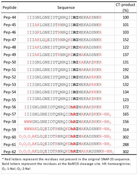
Additional effort on further substrate optimization was put on combining single mutations that showed enhanced detection of BoNT/E cleavage products. Since some single mutations may alter the conformation of mutated peptides or intra- and inter molecular interactions, and hence the property of their substrate binding and/or catalysis, it is not realistic to expect a best substrate can be obtained by simply placing together in a single sequence all good single substitutions derived from the studies described above. However, it is reasonable to believe that some combinations of the single mutations may achieve an augmented effect. For this purpose, new peptides were designed where different combinations of the amino acid substitutions, derived from the studies of single mutation and N-terminal modifications described above, were incorporated into their corresponding positions of the template peptide (Pep-32). In addition, some unnatural amino acids, such as homoarginine (hR), and terminal modifications, such as C-terminal amidation, were introduced in some peptides in order to increase peptide stability and reduce their susceptibility to non-specific cleavage by other proteases (e.g. Trypsin) present in biological samples. Table 4 lists some of such modified peptides (Pep-45 to Pep-62) that showed a comparable or better substrate performance than that of the peptide with a single internal substitution (Pep-44). While some multiple substituted peptides (e.g. Pep-45, Pep-46, Pep-48 or Pep-55) exhibited similar or slightly improved detection sensitivity, in terms of the detection of the C-terminal cleavage products, many of the novel peptides achieved a significant improvement with 50% to 300% increase in the CT-product detection, revealing that added benefit on assay sensitivity could be obtained by combining sound single mutations. Among four candidates displaying three-fold improvement over Pep-44, Pep-59 proved to be the best in solubility and the Pep-62 showed highest resistance toward undesired cleavage by nonspecific proteases present in clinical samples (Data not shown). The substitution of the arginine at the cleavage site with an unnatural homoarginine residue seems to contribute to improved resistance toward the cleavage by nonspecific proteases such as trypsin. Therefore, these two optimal peptides were selected to be used in further experiments and in routine analysis of biological samples for the BoNT/E detection by the Endopep-MS assay.
Table 4.
Effect of multiple mutations on the cleavage of modified peptides by BoNT/E toxin*.

|
Evaluation of optimized peptides as BoNT/E substrates in the Endopep-MS assay
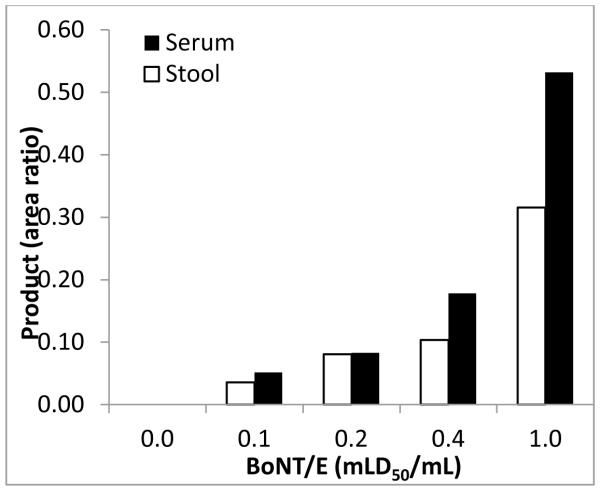
To evaluate the outcome of the optimization for the BoNT/E substrates, Pep-59, one of the four best optimized peptides, was compared to Pep-8, the substrate currently used in the Endopep-MS assay. The substrates were hydrolyzed by two forms of BoNT/E toxins: one is the single chain holotoxin without pre-activation as a complex with neurotoxin-associated proteins, and another is the BoNT/E di-chain complex that had been activated by exposing it to trypsin during the manufacturing process. As shown in Table 5, the optimal peptide was able to detect the cleavage products at a 500-fold lower level of BoNT/E compared to the old peptide substrate, for both not activated and trypsin-activated BoNT/E toxin complexes, under the same experimental conditions, demonstrating a dramatic improvement in the assay sensitivity using the new peptide substrate. When testing the sensitivity of the Endopep-MS assay using the newly developed optimal peptide, the limit of detection of 0.1 mLD50 (1pg/mL or 5.5 attomole/mL) was accomplished for the detection of not trypsin-activated BoNT/E toxin complex spiked in serum and stool extract, two common biological matrices used for botulism clinical samples, after a 4 hour cleavage reaction (Fig. 3, S/N > 3). This represent an assay sensitivity 10-fold lower than that measured by a traditional mouse bioassay. The specificity of the optimal peptides was examined by exposing them to other serotypes of botulinum neurotoxins including type A, B, and F, and no cleavage product was observed (data not shown). In addition to the specific BoNT/E subtype (E3) used in the experiments described above, the optimized peptides also proved to be effective substrates of other tested BoNT/E subtypes including E1, E2, E4, E7 and the most divergent E9 (Data not shown).
Table 5.
Comparison of the cleavage of currently used and newly developed peptide substrates by BoNT/E.
| Peptide | BoNT/E | Product (Area ratio) |
Relative product |
|
|---|---|---|---|---|
| Type | Activity (mLD50)* |
|||
| Pep-8 | not activated | 100 | 0.40 | 1 |
| Pep-59 | not activated | 1 | 2.35 | 581 |
| Pep-8 | activated | 0.16 | 1.07 | 1 |
| Pep-59 | activated | 0.0016 | 5.48 | 511 |
The specific activities of activated and not activated BoNT/E was provided by the manufacturer. Cleavage reactions were conducted at 37°C for 1 hour.
Figure 3.
Product response from the cleavage of Pep-59 by not trypsin-activated BoNT/E of various concentrations spiked in serum and stool matrices. Cleavage reaction condition: 37°C, 4 hours.
Conclusion
We developed novel peptide substrates for the mass spectrometry-based Endopep-MS assay for the detection of type E botulinum neurotoxin. The systematic and comprehensive optimization process included peptide terminal truncation, internal deletion, single and multiple substitution, terminal residue modification and incorporation of unnatural amino acid residues. Our data demonstrate that one of the four optimal peptides demonstrated a 500-fold improvement in assay sensitivity than the current substrate, used for the detection of both not activated and trypsin-activated BoNT/E toxin complexes. The limit of detection for the toxin complex without pre-activation in serum, stool and food samples using the new substrate is 0.1 mouseLD50/mL. In addition, the troublesome nonspecific cleavage in blank control samples was significantly improved by incorporating an unnatural homoarginine residue in the cleavage site of the optimal peptides. A patent application of these optimized peptides has been filed and the novel peptide substrates continue to be used in our laboratory for routine analysis of clinical samples.
Footnotes
Disclaimer: The findings and conclusions in this report are those of the authors and do not necessarily represent the official position of the Centers for Disease Control and Prevention.
References
- [1].Werner SB, Passaro D, McGee J, Schechter R, Vugia DJ. Wound botulism in California, 1951-1998: recent epidemic in heroin injectors. Clinical infectious diseases. 2000;31:1018–1024. doi: 10.1086/318134. [DOI] [PubMed] [Google Scholar]
- [2].Arnon SS, Schechter R, Inglesby TV, Henderson DA, Bartlett JG, Ascher MS, Eitzen E, Fine AD, Hauer J, Layton M, Lillibridge S, Osterholm MT, O'Toole T, Parker G, Perl TM, Russell PK, Swerdlow DL, Tonat K, Biodefense f.t.W.G.o.C. Botulinum Toxin as a Biological Weapon. JAMA: The Journal of the American Medical Association. 2001;285:1059–1070. doi: 10.1001/jama.285.8.1059. [DOI] [PubMed] [Google Scholar]
- [3].Ward AB, Barnes MP. Clinical Uses of Botulinum Toxins. Cambridge University Press; Cambridge, UK: 2007. [Google Scholar]
- [4].DasGupta BR, Dekleva ML. Botulinum neurotoxin type A: sequence of amino acids at the N-terminus and around the nicking site. Biochimie. 1990;72:661–664. doi: 10.1016/0300-9084(90)90048-l. [DOI] [PubMed] [Google Scholar]
- [5].Wictome M, Shone CC. Botulinum neurotoxins: mode of action and detection. Society for Applied Microbiology symposium series. 1998;27:87S–97S. doi: 10.1046/j.1365-2672.1998.0840s187s.x. [DOI] [PubMed] [Google Scholar]
- [6].Kalb S, Baudys J, Webb R, Wright P, Smith T, Smith L, Fernñ?ndez R, Raphael B, Maslanka S, Pirkle J, Barr J. Discovery of a novel enzymatic cleavage site for botulinum neurotoxin F5. FEBS letters. 2012;586:109–115. doi: 10.1016/j.febslet.2011.11.033. [DOI] [PMC free article] [PubMed] [Google Scholar]
- [7].Schiavo G, Matteoli M, Montecucco C. Neurotoxins Affecting Neuroexocytosis. Physiological Reviews. 2000;80:717–766. doi: 10.1152/physrev.2000.80.2.717. [DOI] [PubMed] [Google Scholar]
- [8].McLaughlin J. Botulism type E outbreak associated with eating a beached whale, Alaska. Emerging infectious diseases. 2004;10:1685–1687. doi: 10.3201/eid1009.040131. [DOI] [PMC free article] [PubMed] [Google Scholar]
- [9].Hannett G, Stone W, Davis S, Wroblewski D. Biodiversity of Clostridium botulinum type E associated with a large outbreak of botulism in wildlife from Lake Erie and Lake Ontario. Applied and environmental microbiology. 2011;77:1061–1068. doi: 10.1128/AEM.01578-10. [DOI] [PMC free article] [PubMed] [Google Scholar]
- [10].Simpson LL, Dasgupta BR. Botulinum neurotoxin type E: studies on mechanism of action and on structure-activity relationships. The journal of pharmacology and experimental therapeutics. 1983;224:135–140. [PubMed] [Google Scholar]
- [11].Sathyamoorthy V, DasGupta BR. Separation, purification, partial characterization and comparison of the heavy and light chains of botulinum neurotoxin types A, B, and E. Journal of biological chemistry. 1985;260:10461–10466. [PubMed] [Google Scholar]
- [12].Kukreja R, Sharma S, Singh B. Molecular basis of activation of endopeptidase activity of botulinum neurotoxin type E. Biochemistry. 2010;49:2510–2519. doi: 10.1021/bi902096r. [DOI] [PMC free article] [PubMed] [Google Scholar]
- [13].Singh A, Stanker L, Sharma S. Botulinum neurotoxin: where are we with detection technologies? Critical reviews in microbiology. 2013;39:43–56. doi: 10.3109/1040841X.2012.691457. [DOI] [PubMed] [Google Scholar]
- [14].Binz T, Blasi J, Yamasaki S, Baumeister A, Link E, Sñf¼dhof TC, Jahn R, Niemann H. Proteolysis of SNAP-25 by types E and A botulinal neurotoxins. Journal of biological chemistry. 1994;269:1617–1620. [PubMed] [Google Scholar]
- [15].Washbourne P, Pellizzari R, Baldini G, Wilson MC, Montecucco C. Botulinum neurotoxin types A and E require the SNARE motif in SNAP-25 for proteolysis. FEBS letters. 1997;418:1–5. doi: 10.1016/s0014-5793(97)01328-8. [DOI] [PubMed] [Google Scholar]
- [16].Vaidyanathan VV, Yoshino K, Jahnz M, Dñrries C, Bade S, Nauenburg S, Niemann H, Binz T. Proteolysis of SNAP-25 isoforms by botulinum neurotoxin types A, C, and E: domains and amino acid residues controlling the formation of enzyme-substrate complexes and cleavage. Journal of neurochemistry. 1999;72:327–337. doi: 10.1046/j.1471-4159.1999.0720327.x. [DOI] [PubMed] [Google Scholar]
- [17].Chen S, Barbieri J. Unique substrate recognition by botulinum neurotoxins serotypes A and E. Journal of biological chemistry. 2006;281:10906–10911. doi: 10.1074/jbc.M513032200. [DOI] [PubMed] [Google Scholar]
- [18].Chen S, Barbieri J. Multiple pocket recognition of SNAP25 by botulinum neurotoxin serotype E. Journal of biological chemistry. 2007;282:25540–25547. doi: 10.1074/jbc.M701922200. [DOI] [PubMed] [Google Scholar]
- [19].Gilmore M, Williams D, Okawa Y, Holguin B, James N, Ross J, Roger Aoki K, Jameson D, Steward L. Depolarization after resonance energy transfer (DARET): a sensitive fluorescence-based assay for botulinum neurotoxin protease activity. Analytical biochemistry. 2011;413:36–42. doi: 10.1016/j.ab.2011.01.043. [DOI] [PubMed] [Google Scholar]
- [20].Jones RGA, Ochiai M, Liu Y, Ekong T, Sesardic D. Development of improved SNAP25 endopeptidase immuno-assays for botulinum type A and E toxins. Journal of immunological methods. 2008;329:92–101. doi: 10.1016/j.jim.2007.09.014. [DOI] [PubMed] [Google Scholar]
- [21].Piazza T, Blehert D, Dunning FM, Berlowski Zier B, Zeytin F.s., Samuel M, Tucker W. In vitro detection and quantification of botulinum neurotoxin type e activity in avian blood. Applied and environmental microbiology. 2011;77:7815–7822. doi: 10.1128/AEM.06165-11. [DOI] [PMC free article] [PubMed] [Google Scholar]
- [22].Purcell A, Hoard Fruchey H. A capillary electrophoresis method to assay catalytic activity of botulinum neurotoxin serotypes: implications for substrate specificity. Analytical biochemistry. 2007;366:207–217. doi: 10.1016/j.ab.2007.04.048. [DOI] [PubMed] [Google Scholar]
- [23].Boyer A, Moura H, Woolfitt A, Kalb S, McWilliams L, Pavlopoulos A, Schmidt J, Ashley D, Barr J. From the mouse to the mass spectrometer: detection and differentiation of the endoproteinase activities of botulinum neurotoxins A-G by mass spectrometry. Analytical chemistry. 2005;77:3916–3924. doi: 10.1021/ac050485f. [DOI] [PubMed] [Google Scholar]
- [24].Kalb S, Garcia Rodriguez C, Lou J, Baudys J, Smith T, Marks J, Smith L, Pirkle J, Barr J. Extraction of BoNT/A, /B, /E, and /F with a single, high affinity monoclonal antibody for detection of botulinum neurotoxin by Endopep-MS. PLoS ONE. 2010;5:e12237–e12237. doi: 10.1371/journal.pone.0012237. [DOI] [PMC free article] [PubMed] [Google Scholar]
- [25].Rosen O, Feldberg L, Gura S, Zichel R. Improved detection of botulinum type-E by rational design of a new peptide-substrate for Endopep-MS assay. Analytical biochemistry. 2014 doi: 10.1016/j.ab.2014.03.024. [DOI] [PubMed] [Google Scholar]
- [26].Wang D, Baudys J, Kalb S, Barr J. Improved detection of botulinum neurotoxin type A in stool by mass spectrometry. Analytical biochemistry. 2011;412:67–73. doi: 10.1016/j.ab.2011.01.025. [DOI] [PubMed] [Google Scholar]
- [27].Gaunt P, Kalb S, Barr J. Detection of botulinum type E toxin in channel catfish with visceral toxicosis syndrome using catfish bioassay and endopep mass spectrometry. Journal of veterinary diagnostic investigation. 2007;19:349–354. doi: 10.1177/104063870701900402. [DOI] [PubMed] [Google Scholar]


