Abstract
MiR-184 was an important suppressor to tumor cells proliferation and invasion and some studies show that it was down-regulated in aggressive human tumor cells and a potential tumor therapy target through expression of miR-184 results in reduced tumor cell aggressiveness. In this study, miR-184 showed an inhibitive activity of glioma U87MG cell line and breast cancer MCF-7 cell line in proliferation and invasion by MTS and transwell assay. We found that the miR-184 also could arrest cell cycle and adhesion by up-regulating the expression of p53 and p21 and activity of caspase-3/8, suppressing the expression of SND1, MMP-2/9, CD44 and activity of AKT/NF-κB pathway. The results showed that miR-184 could be a potential target for glioma and breast cancer treatment.
Keywords: miR-184, proliferation, invasion
Introduction
All of gliomas and breast cancer were malignant tumor to threat to the people health [1,2]. However, malignant proliferation and invasion continues to be a major clinical obstacle to the successful treatment for cancer. Tumor proliferation and invasion was the complex process, involving in the interaction with oncogene and anti-oncogene.
MicroRNAs, a group of short non-coding RNAs, plays important roles in a variety of biologic processes and is shown to have both diagnostic and prognostic significance and to constitute a novel target for cancer treatment [3,4]. miR-184 (microRNA-184) was acted as a tumor suppressor gene in several tumor development process [5]. However, the role of miR-184 in gliomas and breast cancer was not full cleared.
Material and methods
Cell culture
Human glioma U87MG cell line and breast cancer MCF-7 cell line was cultured in DMEM complete medium (containing 10% FCS) under the conditions of 37°C, 5% CO2. Cells were cultured in 24-well plates for 24 h in DMEM with 10% FBS. miR-184 expression plasmid (GeneCopoeia) was transfected into 70-80% confluency cells for 12 h, then the medium was replaced by fresh complete medium and cells were plated in 6-well plates cultured for another 24 h for following experiments.
MTS assay
Cells (5 × 10³ cells/well) were cultured into a 96 well plate under the conditions of 37°C, 5% CO2 for 96 h. Then the medium was removed and MTS was added for another 4 h. Then the OD value was measured at 490 nm and growth inhibition rate was calculated. The tumor growth inhibition rate (%) = (1 - the OD value of the treatment group/the OD value of the negative control group) × 100%.
Cell invasion and adhesion assay
Cells (2 × 10³ cells/well) were cultured into a Matrigel coating transwell plate under the conditions of 37°C, 5% CO2 for 48 h. Then the membrane of lower surface were fixed with the methanol and stained with crystal violet, the cells were photographed by microscope to explore the effect of miR-184 on invasion in tumor cells.
Cells (1 × 10⁴ cells/well) were cultured into a Matrigel coating 96-well plate under the conditions of 37°C, 5% CO2 for 24 h. Then cells were washed by PBS and the absorbance was measured by MTS assay to explore the effect of miR-184 on adhesion in tumor cells.
Cell cycle assay
Cells (3 × 10⁵ cells/well) were cultured into 6-well plate under the conditions of 37°C, 5% CO2 for 48 h. Then cells were fixed with 70% ethanol at 4°C overnight and stained by PI for 30 min after PBS washing.
Real-time PCR assay
Total RNA was extracted from each group using Trizol method. Real-Time PCR Kit was used to carry out reverse transcription to obtain the cDNA, then SND1, MMP-2/9, CD44, p53 and p21 mRNA levels were detected; The PCR primers used were as follows: 5’-CCACATCGCTCAGACACCAT-3’ (sense) and 5’-ACCAGGCGCCCAATACG-3’ (antisense) for GAPDH; 5’-GCAGTGCAATACCTGAACACCTTC-3’ (sense) and 5’-CCATACTTCACACGGACCACTTG-3’ (antisense) for MMP-2; 5’-GACTCTACACCCGGGACGGCAATGCTG-3’ (sense) and 5’-CGTCCACCGGACTCAAAGGCACAGTAG-3’ (antisense) for MMP-9; 5’-TATCTAGAGCCGCCACCATGGACAAGTTTTGGTGG-3’ (sense) and 5’-TATCTAGAGCCATTCTGGAATTTGGGGTGT-3’ (antisense) for CD44; 5’-CGGCTCCTCCATGGCAGT-3’ (sense) and 5’-ACTGCCATGGAGGAGCCG-3’ (antisense) for p53; 5’-GGAGCAAAGTGTGCCGTTGTC-3’ (sense) and 5’-AGGAAGTACTGGGCCTCTTG-3’ (antisense) for p21. The real-time PCR reaction was conducted under the following conditions: 95°C for 30 s, 40 cycles of 95°C for 5 s and 60°C for 60 s.
Western blotting assay
Cells (3 × 10⁵ cells/well) were cultured into 6-well plate under the conditions of 37°C, 5% CO2 for 48 h. Then cells were digested and extracted the total protein. The protein was separated by 12% SDS polyacrylamide gel and transfer to PVDF membrane. The membrane was incubated with SND1 antibody (1:1500), MMP-2 antibody (1:1500), MMP-9 antibody (1:1500), CD44 antibody (1:1500), p53 antibody (1:1500), p21 antibody (1:1500), p-AKT antibody (1:800), and β-actin (1:5000) was added and incubated at 4°C overnight. Then IgG labeled with horseradish peroxidase (1:2000) was added and incubated at room temperature for 1 h. The protein was visualized with the HRP Western blot detection system.
ELIASA assay
Cells (3 × 10⁵ cells/well) were cultured into 6-well plate under the conditions of 37°C, 5% CO2 for 72 h and the activity of caspase-3/8 in cells was measured in accordance with operating manuals (Promega, USA).
Report gene assay
Cells (3 × 10⁵ cells/well) were cultured with NF-κB luciferase plasmid transfected into 6-well plate under the conditions of 37°C, 5% CO2 for 72 h. The luciferase fluorescence intensity was measured by ELIASA assay.
Statistical analysis
The data is analyzed by SPSS11.0. One-way ANOVA is adopted to compare the data, with considering P < 0.05 as the standard of statistically significant. Each experiment is repeated for three times.
Results
The effect of miR-184 on the proliferation, invasion, adhesion and cell cycle of human glioma and breast cancer cells
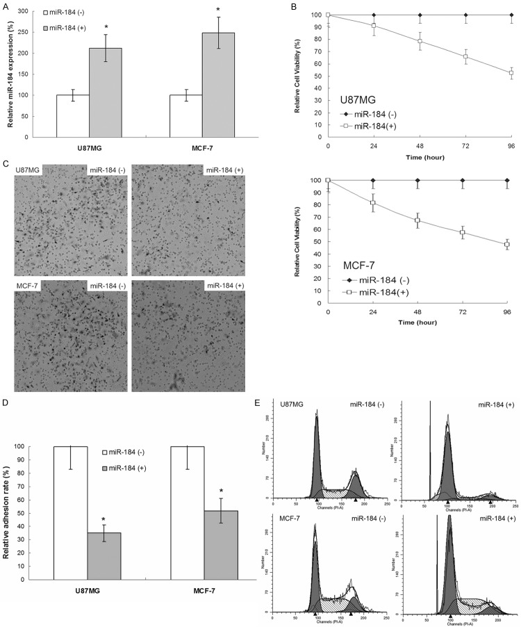
MTS assay was to explore the effect of miR-184 on the proliferation of human glioma U87MG and breast cancer MCF-7 cells in vitro. The experimental results showed that the potential of proliferation, invasion and adhesion was suppressed and the cell cycle was arrested in U87MG and MCF-7 cell line after miR-184 expression (Figure 1).
Figure 1.
The effect of miR-184 on the proliferation, invasion, adhesion and cell cycle of human glioma and breast cancer cells. A. Real-time PCR results showed that there was an increased expression of miR-184 in human glioma U87MG and breast cancer MCF-7 cell line after miR-184 expression plasmid transfected. B. The MTS results showed that there was an inhibitive effect on proliferation of human glioma U87MG and breast cancer MCF-7 cell line after miR-184 expression. Bars mean ± SD, n = 10. C. The transwell results showed that there was an inhibitive effect on invasion of human glioma U87MG and breast cancer MCF-7 cell line after -184 expression. D. The adhesion results showed that there was an inhibitive effect on adhesion of human glioma U87MG and breast cancer MCF-7 cell line after miR-184 expression. E. The flow cytometry results showed that there was an inhibitive effect on G0/G1 phase arrest of human glioma U87MG and breast cancer MCF-7 cell line after miR-184 expression.
The effect of miR-184 on the expression of tumor related gene and the activity of signal pathway of human glioma and breast cancer cells
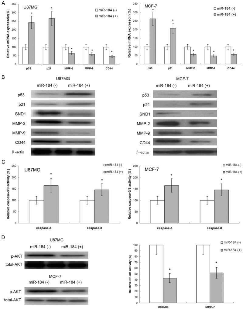
Western blotting and ELIASA assay showed that there was an increased expression of p53 and p21, and activity of caspase-3/8 in U87MG and MCF-7 cell line after miR-184 expression. Meanwhile, there was a decreased expression of SND1, MMP-2/9 and CD44. Further, western blotting assay showed that there was a decreased the phosphorylation of AKT and report gene assay showed that there was a decreased activity of NF-κB in U87MG and MCF-7 cell line after miR-184 expression (Figure 2).
Figure 2.

The effect of miR-184 on the expression of tumor related gene and the activity of signal pathway of human glioma and breast cancer cells. A. Real-time PCR assay results showed that there was an increased mRNA expression expression of p53 and p21 and there was a decreased mRNA expression of SND1, MMP-2/9 and CD44 in human glioma U87MG and breast cancer MCF-7 cell line after miR-184 expression. B. Western blotting assay results showed that there was an increased protein expression expression of p53 and p21 and there was a decreased protein expression of SND1, MMP-2/9 and CD44 in human glioma U87MG and breast cancer MCF-7 cell line after miR-184 expression. C. ELIASA assay results showed that there was an increased activity of caspase-3/8 in human glioma U87MG and breast cancer MCF-7 cell line after miR-184 expression. D. Western blotting assay showed that there was a decreased the phosphorylation of AKT and report gene assay showed that there was a decreased activity of NF-κB in human glioma U87MG and breast cancer MCF-7 cell line after miR-184 expression.
Discussion
In this study, we observed that miR-184 could suppress proliferation of U87MG and MCF-7 cell line compared with parent group. This is in agreement with miR-184 was required for tumor cell survival in tumor cells. At same time invasion and adhesion assay results showed that miR-184 also suppress the potential of invasion and adhesion of tumor cells. In order to explore the potential mechanism, we study the effect of miR-184 on cell cycle arrest in above mentioned cell lines and found that miR-184 could arrest the tumor cell cycle in G0/G1 phase.
Tumor process was involved multiplex gene, including oncogene and anti-oncogene. We also explored the effect of miR-184 on the expression of tumor related gene in U87MG and MCF-7 cell line. p53 and p21 were tumor suppressors which could suppress cell carcinomatous change, tumor proliferation, tumor invasion and so on [6-8]. p21 was considered an oncogene and low expressed in tumor. p21 could arrest the cell cycle in G0/G1 phase [9]. p53 could improve p21 expression, inducing to the cell apoptosis and proliferation inhibition [10]. Caspase-3/8 was a part of the cysteine-aspartic acid protease family. Sequential activation of caspase-3/8 could result to the execution-phase of cell apoptosis and cell death [11,12]. MMP-2/9 was belonging to matrix metalloproteinases family and there was a high risk for tumor invasion and heterogeneous adhesion in MMP-2/9 overexpressing tumor cells [13,14]. CD44 was an adhesion molecule which also could improve the invasion and heterogeneous adhesion [15].
AKT/NF-κB pathway attends to many process of organism and its overexpression could promote to cell carcinomatous change, tumor proliferation and invasion [16,17]. In this study, we found that miR-184 could inhibit to the activity of AKT/NF-κB pathway and improve the expression of p53/p21 and activity of caspase-3/8, suppress the expression of MMP-2/9 and CD44.
SND1 was original oncogene which overexpressed in many types tumor and regulated multiplex tumor biological process [18]. We found miR-184 could suppress the expression of miR-184, therefore, we considered that SND1 may be playing a key role in the anti-tumor activity of miR-184.
Disclosure of conflict of interest
None.
References
- 1.Rahman M, Reyner K, Deleyrolle L, Millette S, Azari H, Day BW, Stringer BW, Boyd AW, Johns TG, Blot V, Duggal R, Reynolds BA. Neurosphere and adherent culture conditions are equivalent for malignant glioma stem cell lines. Anat Cell Biol. 2015;48:25–35. doi: 10.5115/acb.2015.48.1.25. [DOI] [PMC free article] [PubMed] [Google Scholar]
- 2.Tabaczar S, Domeradzka K, Czepas J, Piasecka-Zelga J, Stetkiewicz J, Gwoździński K, Koceva-Chyła A. Anti-tumor potential of nitroxyl derivative Pirolin in the DMBA-induced rat mammary carcinoma model: A comparison with quercetin. Pharmacol Rep. 2015;67:527–34. doi: 10.1016/j.pharep.2014.12.010. [DOI] [PubMed] [Google Scholar]
- 3.Pang JC, Kwok WK, Chen Z, Ng HK. Oncogenic role of microRNAs in brain tumors. Acta Neuropathol. 2009;117:599–611. doi: 10.1007/s00401-009-0525-0. [DOI] [PubMed] [Google Scholar]
- 4.Hayes J, Thygesen H, Droop A, Hughes TA, Westhead D, Lawler SE, Wurdak H, Short SC. Prognostic microRNAs in high-grade glioma reveal a link to oligodendrocyte precursor differentiation. Oncoscience. 2014;2:252–62. doi: 10.18632/oncoscience.112. [DOI] [PMC free article] [PubMed] [Google Scholar]
- 5.Murad N, Kokkinaki M, Gunawardena N, Gunawan MS, Hathout Y, Janczura KJ, Theos AC, Golestaneh N. miR-184 regulates ezrin, LAMP-1 expression, affects phagocytosis in human retinal pigment epithelium and is downregulated in age-related macular degeneration. FEBS J. 2014;281:5251–64. doi: 10.1111/febs.13066. [DOI] [PubMed] [Google Scholar]
- 6.Kim T, Han W, Kim MK, Lee JW, Kim J, Ahn SK, Lee HB, Moon HG, Lee KH, Kim TY, Han SW, Im SA, Park IA, Kim JY, Noh DY. Predictive Significance of p53, Ki-67, and Bcl-2 Expression for Pathologic Complete Response after Neoadjuvant Chemotherapy for Triple-Negative Breast Cancer. J Breast Cancer. 2015;1:16–21. doi: 10.4048/jbc.2015.18.1.16. [DOI] [PMC free article] [PubMed] [Google Scholar]
- 7.Huang YD, Li P, Tong X, He Y, Zhuo Y, Xia SW, Luo XH. Effects of bleomycin A5 on caspase-3, P53, bcl-2 expression and telomerase activity in vascular endothelial cells. Indian J Pharmacol. 2015;47:55–8. doi: 10.4103/0253-7613.150337. [DOI] [PMC free article] [PubMed] [Google Scholar]
- 8.Kim HL, Ra H, Kim KR, Lee JM, Im H, Kim YH. Poly (ADP-ribosyl) ation of p53 Contributes to TPEN-Induced Neuronal Apoptosis. Mol Cells. 2015;38:312–7. doi: 10.14348/molcells.2015.2142. [DOI] [PMC free article] [PubMed] [Google Scholar]
- 9.Liu T, Qin W, Hou L, Huang Y. MicroRNA-17 promotes normal ovarian cancer cells to cancer stem cells development via suppression of the LKB1-p53-p21/WAF1 pathway. Tumour Biol. 2015;36:1881–93. doi: 10.1007/s13277-014-2790-3. [DOI] [PubMed] [Google Scholar]
- 10.Saf C, Gulcan EM, Ozkan F, Cobanoglu Saf SP, Vitrinel A. Assessment of p21, p53 expression, and Ki-67 proliferative activities in the gastric mucosa of children with Helicobacter pylori gastritis. Eur J Gastroenterol Hepatol. 2015;27:155–61. doi: 10.1097/MEG.0000000000000246. [DOI] [PubMed] [Google Scholar]
- 11.Qin FX, Shao HY, Chen XC, Tan S, Zhang HJ, Miao ZY, Wang L, Hui-Chen , Zhang L. Knockdown of NPM1 by RNA interference inhibits cells proliferation and induces apoptosis in leukemic cell line. Int J Med Sci. 2011;8:287–94. doi: 10.7150/ijms.8.287. [DOI] [PMC free article] [PubMed] [Google Scholar]
- 12.Yang D, Yaguchi T, Yamamoto H, Nishizaki T. Intracellularly transported adenosine induces apoptosis in HuH-7 human hepatoma cells by downregulating c-FLIP expression causing caspase-3/-8 activation. Biochem Pharmacol. 2007;73:1665–75. doi: 10.1016/j.bcp.2007.01.020. [DOI] [PubMed] [Google Scholar]
- 13.Xiao J, Liu L, Zhong Z, Xiao C, Zhang J. Mangiferin regulates proliferation and apoptosis in glioma cells by induction of microRNA-15b and inhibition of MMP-9 expression. Oncol Rep. 2015;33:2815–20. doi: 10.3892/or.2015.3919. [DOI] [PubMed] [Google Scholar]
- 14.Muluk NB, Arikan OK, Atasoy P, Kiliç R, Yalçinozan ET. The role of MMP-2, MMP-9, and TIMP-1 in the pathogenesis of nasal polyps: Immunohistochemical assessment at eight different levels in the epithelial, subepithelial, and deep layers of the mucosa. Ear Nose Throat J. 2015;94:E1–E13. [PubMed] [Google Scholar]
- 15.Zhang J, Dong J, Yang Z, Ma X, Zhang J, Li M, Chen Y, Ding Y, Li K, Zhang Z. Expression of ezrin, CD44, and VEGF in giant cell tumor of bone and its significance. World J Surg Oncol. 2015;13:168. doi: 10.1186/s12957-015-0579-5. [DOI] [PMC free article] [PubMed] [Google Scholar]
- 16.Lin CY, Chen JH, Fu RH, Tsai CW. Induction of Pi form of glutathione S-transferase by carnosic acid is mediated through PI3K/Akt/NF-κB pathway and protects against neurotoxicity. Chem Res Toxicol. 2014;27:1958–66. doi: 10.1021/tx5003063. [DOI] [PubMed] [Google Scholar]
- 17.Zhao M, Zhou A, Xu L, Zhang X. The role of TLR4-mediated PTEN/PI3K/AKT/NF-κB signaling pathway in neuroinflammation in hippocampal neurons. Neuroscience. 2014;269:93–101. doi: 10.1016/j.neuroscience.2014.03.039. [DOI] [PubMed] [Google Scholar]
- 18.Emdad L, Janjic A, Alzubi MA, Hu B, Santhekadur PK, Menezes ME, Shen XN, Das SK, Sarkar D, Fisher PB. Suppression of miR-184 in malignant gliomas upregulates SND1 and promotes tumor aggressiveness. Neuro Oncol. 2015;17:419–29. doi: 10.1093/neuonc/nou220. [DOI] [PMC free article] [PubMed] [Google Scholar]


