Abstract
Background:
Methicillin-resistant Staphylococcus aureus (MRSA) is known as a common pathogen in nosocomial and community-acquired infections. Sewage acts as an environmental reservoir and may have a significant role in development and dissemination of antibiotic resistance.
Objectives:
This study was undertaken to determine the epidemiological relatedness between the MRSA isolated from sewage and human infections.
Materials and Methods:
Samples were collected from a referral hospital and also a sewage treatment plant in Tehran, Iran, during 2010. All the MRSA isolates were identified at the species level and typed using Phene plate (PhP) system and SCCmec typing. Antibiotic susceptibility tests were also performed.
Results:
Of the 1142 isolates, 200 MRSA strains from the sewage (n = 100) and the clinic (n = 100) were isolated. Distinct PhP types, consisting of 16 common types and 13 single types, and also 3 different staphylococcal cassette chromosome mec (SCCmec) types (III, IVa and IVc) were found amongst the MRSA isolated from the two different sources. The results of antibiotic susceptibility testing showed an increased resistance to penicillin, ciprofloxacin, erythromycin, clindamycin and tetracycline. In addition, none of the isolates showed resistance to vancomycin, quinupristin -dalfopristin and linezolid.
Conclusions:
The presence of common PhP types and also SCCmec type III, as an indicator for hospital strains, among the isolates, may indicate an epidemiological link between clinical and sewage MRSA isolates in Tehran.
Keywords: Methicillin- Resistant Staphylococcus aureus, Sewage, Hospital, Staphylococcal Cassette Chromosome mec, Typing
1. Background
Staphylococcus aureus is a common cause of nosocomial and community-acquired infections, ranging from skin infections to necrotizing pneumonia (1, 2). Methicillin-resistant S. aureus (MRSA) isolates with high-level resistance to beta-lactam antibiotics have been reported to be spread and are epidemic in different hospitals in the world (3). The resistance to methicillin is mediated with mecA and novel mecC genes (3-5). Staphylococcal cassette chromosome mec (SCCmec) is the only vector for the mecA gene and its transfer occurs frequently (3, 4). Although hospital-acquired MRSA (HA-MRSA) is one of the most frequent causes of hospital-associated infections, community-acquired MRSA (CA-MRSA) has recently emerged as an additional threat, causing infections in healthy individuals with no healthcare-associated risk factors (3, 6).
As a large amount of antibiotics prescribed are introduced in the environment in an active form, sewage may act as an environmental reservoir and may have a significant role in the development and dissemination of antibiotic resistance, e.g. beta-lactam antibiotics which are detected in sewage systems and surface waters (3, 7). The role of horizontal gene transfer in the spread of antibiotic resistance in the environment has been documented (8). Ohlsen and colleagues (9) described the transfer of genes encoding antibiotic resistance by S. aureus in wastewater in vitro. Detection of the mecA gene has also been reported in hospitals and sewage (10-12). Using cultivation and molecular techniques, the presence of MRSA in municipal wastewater has been reported (10, 11). Therefore, sewage treatment plants (STPs) could be a potential source for dissemination and development of MRSA strains.
2. Objectives
This study was undertaken to determine the epidemiological relatedness between the MRSA strains isolated from sewage and human infections using a combination of different typing methods.
3. Materials and Methods
3.1. Sewage Sampling
In 2010, an urban STP located in the west of Tehran was selected for collecting the sewage samples. Three samples were collected in sterile bottles (250 mL) and transferred to the laboratory in a cold chain. After a five-fold diluting with phosphate buffered saline, the samples were filtered using a 0.45 µm membrane (Millipore Corporation, Bedford, MA) and were cultured in Baird Parker agar (Merck KGaA, Darmstadt, Germany) plates by the same procedure described in literature for enterococci (13-15).
3.2. Clinical Samples
Specimens (n = 489) were collected from patients in a referral hospital in Tehran during 2010. Using standard microbiological techniques such as catalase test, oxidase reaction, growth in 10 - 15% sodium chloride, mannitol fermentation, DNase and coagulase tests, the isolates were confirmed as S. aureus and were used for isolation of MRSA strains, as described previously (16-18).
3.3. Antibiotic Susceptibility Tests
According to the guidelines of Clinical and Laboratory Standards Institute (CLSI) (19), all the S. aureus isolates were examined for susceptibility to oxacillin (1 µg), using disc diffusion method. E-test (AB, Biomérieux, Marcy l'Etoile, France) was used to determine the minimum inhibitory concentrations (MICs) for oxacillin and vancomycin of all the collected MRSA isolates, according to the manufacturer's instructions. Sixteen common antibiotics were employed to determine the susceptibility of the MRSA isolates by disc diffusion method, as described by CLSI (19). These included amikacin (30 μg), chloramphenicol (30 μg), ciprofloxacin (5 μg), clindamycin (2 μg), erythromycin (15 μg), gentamicin (10 μg), kanamycin (30 μg), linezolid (30 μg), minocycline (30 μg), nitrofurantoin (300 μg), penicillin (5 μg), quinupristin-dalfopristin (15 μg), rifampicin (5 μg), sulfamethoxazole-trimethoprim (1.25 - 23.75 μg), tetracycline (30 μg), and tobramycin (10 μg) (Mast Diagnostics, Merseyside, UK).
3.4. DNA Extraction and Polymerase Chain Reaction
High Pure PCR Template Preparation Kit (Roche, Mannheim, Germany) was used to extract the DNA according to the manufacturer's guidelines with some modifications. PCR primers for nucA and mecA genes were prepared according to Du et al. (20). PCR conditions were as described previously (21).
3.5. Staphylococcal Cassette Chromosome mec and ccr Typing
A multiplex PCR typing assay was used for typing the SCCmec gene which contained eight pairs of primers including the unique and specific primers for SCCmec types and subtypes I, II, III, IVa, IVb, IVc, IVd, and V (22). Another multiplex PCR was used for characterization of ccr gene complexes which employed four sets of primers specific for each of the ccr genes, i.e. ccrAB-β2, ccrAB-α2, ccrAB-α3, and ccrAB-α4 (22). The multiplex PCR mixture and the PCR cycles were as previously described by Zhang et al. (22).
3.6. Detection of pvl Gene
For detection of pvl gene encoding PVL toxin among MRSA isolates, specific primers were used, as previously described by McClure and colleagues (23). The PCR cycles and mixture were according to McClure et al. (23).
3.7. Biochemical Fingerprinting
PhP-RF plates (PhPlate AB, Stockholm, Sweden) were used to type the 200 MRSA isolates. To measure the kinetics of bacterial metabolism of 23 substrates and a control, four sets of dehydrated reagents were used to differentiate the S. aureus strains (24). The absorbance values (A620) of each microplate wells that were incubated at 37°C, were measured at 16-, 40-, and 64-hour intervals (15). The diversity of the bacterial populations was calculated as Simpson’s index of diversity (Di) (15, 25). PhPWin software (PhPlate Microplates Techniques AB, Sweden) was used for optical readings, calculation of correlation coefficients, diversity indexes, and clustering (15).
4. Results
4.1. Prevalence of Methicillin-Resistant Staphylococcus aureus Isolates
Totally, 653 S. aureus strains were isolated from the sewage samples. One hundred (15.3%) MRSA strains were isolated from this STP. Of the total 489 clinical isolates, 100 (20.4%) strains were determined as MRSA. The frequencies of positive cases in clinical samples were as follows: 207 (42%) from wound, 96 (20%) urine, 69 (14%) sputum, 39 (8%) blood, 27 (6%) CSF, 26 (5%) nose, and 25 (5%) abscess.
4.2. Antibiotic Susceptibility Testing
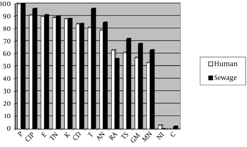
The percentages of antimicrobial resistance of the MRSA strains isolated from different sources are shown in Figure 1. High resistance to ciprofloxacin (91%), erythromycin (90%), tobramycin (89%), kanamycin (88%), clindamycin (84%) and tetracycline (81%) was observed in clinical cases. In contrast, low resistance to nitrofurantoin (3%) was detected. Furthermore, no resistance to vancomycin, synercid and linezolid was observed in clinical isolates. Concomitant resistance to Cip/E/Tn/K/CD/T was found in about 80% of the human MRSA isolates. This rate was somehow more for sewage isolates (Figure 1).
Figure 1. The Resistance Rate of Methicillin-Resistant Staphylococcus aureus Strains Isolated From Clinical and Sewage Samples Against the 14 Antibiotics Tested.
Abbreviations: AN, amikacin; C, chloramphenicol; CD, clindamycin; CIP, ciprofloxacin; E, erythromycin; GM, gentamicin; K, kanamycin; MN, minocycline; NI, nitrofurantoin; P, penicillin; RA, rifampicin; T, tetracycline; TN, tobramycin; TS, cotrimoxazole.
Susceptibility to all the antibiotics tested except for penicillin was observed in 7% and 6% of clinical and sewage isolates, respectively. Moreover, 18% and 13% of clinical and sewage isolates, respectively, showed resistance to all the different antibiotics except for vancomycin, synercid, nitrofurantoin, linezolid and chloramphenicol. The oxacillin MIC for all the MRSA isolated from humans and sewage showed that all the isolates were resistant to oxacillin (MIC ≥ 4 µg/mL). Moreover, 49% of clinical isolates and 58% of sewage isolates showed high-level resistance (MIC ≥ 256 µg/mL) to oxacillin. In addition, 7% of clinical isolates and 6% of sewage isolates showed low resistance to oxacillin (MIC = 4 µg/mL).
4.3. Biochemical Fingerprinting
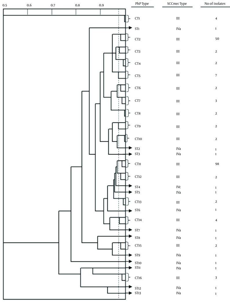
Diverse (diversity index, Di = 0.975) PhP types were detected among the MRSA strains isolated from clinical and sewage sources. The 200 isolates were discriminated into 29 types with 13 single types (6.5%) and 16 common types (CTs) (93.5%) (Figure 2). Each (C-BPT) (common biochemical phenotypes) included 2-98 strains. The highest number of MRSA strains was categorized in CT11 (98 isolates, 49%) and was considered as the dominant common type. In general, isolates in the same PhP type showed different antibiotic resistance patterns, indicating no relatedness between their clonal dissemination and also their antibiotic resistance patterns (i.e. ST2, ST3).
Figure 2. An Unweighted Pair Group With Arithmetic Averages Dendrogram, Showing the Methicillin-Resistant Staphylococcus aureus Strains Isolated From Different Sewage Treatment Plants and Clinical Samples in Tehran During 2010.
In this dendrogram, only two isolates of each CT have been included.
4.4. Staphylococcal Cassette Chromosome mec and ccr Typing
One hundred and eighty seven MRSA isolates were shown to carry SCCmec type III and were PCR-positive with the ccrAB-α4 specific primers indicating the presence of type 3 ccr. Moreover, 13 isolates that showed low resistance to oxacillin (MIC = 4 µg/mL) carried SCCmec type IV and also showed type 2 ccr. Moreover, all the seven clinical isolates shared SCCmec type IVa. However, one sewage MRSA isolate (6.7%) shared SCCmec type IVc.
4.5. Detection of pvl Gene
The results of the pvl gene detection among the MRSA isolates showed that only 13 (6.5%) MRSA isolates were PCR-positive with the pvl gene. These MRSA isolates showed susceptibility to all the antibiotics tested except for penicillin, as expected for MRSA isolates. The presence of pvl gene among the MRSA isolates was limited to isolates that shared type 2 ccr and also showed a low level of oxacillin resistance (MIC = 4 µg/mL).
5. Discussion
The frequency of the MRSA strains isolated from a referral hospital in this study was 100 out of 489 (20.4%), which was less than other reports from Iran (26-29). The differences observed could be due to the methods and techniques used, geographical locations, population, antibiotics prescription, and hygiene measures in different hospitals. Our study showed the presence of a high number and diverse PhP types of MRSA in sewage for the first time in Iran (Figure 2). This revealed that different lineages survive in this environment. These findings were not in agreement with other reports indicating the absence or the low prevalence or no survival of S. aureus in sewage (3, 12, 30, 31). The modified protocol for isolation of enterococci from sewage, used for isolation of S. aureus in this study, could be a possible explanation (13-15). Due to the high clinical prevalence of MRSA in Iran (1, 17, 18, 21, 26-29), its high prevalence in sewage could be expected. Therefore, this may indicate the role of sewage as a potential reservoir for MRSA.
Considering the high isolation of the PhP types from clinical sources, the origin of MRSA in sewage could be the resident population in the area studied (Figure 2). The presence of SCCmec type III, type 3 ccr and also resistance to different classes of antibiotics other than beta-lactam antibiotics indicated a genetic diversity among the isolated MRSA, which was somehow similar to the clinical isolates. This may indicate their hospital origin. On the other hand, the better adaptation of some PhP types (CT2 and CT11) to the sewage environment may indicate that some strains are resident in STP. While in the USA and Europe most of the MRSA strains have only been isolated from patients, there are also some reports of MRSA isolated from sewage in Sweden, Australia and the USA (3, 11, 32, 33). Our results were similar to the reports from Europe, Australia and the USA.
Extensive diversity was detected among the isolates by PhP analysis. Compared to clinical isolates, more homogeneity was observed among the sewage isolates. Due to high outbreaks of MRSA in Tehran and dissemination of dominant bacterial clones, a consistency was observed among the MRSA PhP types. The acquisition of oxacillin resistance genes by the majority of isolates from different sources may indicate the possibility of horizontal gene transfer. The recovery of PhP types 2 and 11 in different sources and also in different samplings supports the spread of these clonal types in Tehran and also indicates the genetic stability of PhP types in sewage and the clinic. The isolates in the same CT (ie, CT2 and CT11) had the same SCCmec type, which was similar to other report from Iran (29).
Similar to other studies in Iran (26, 27, 29), our findings showed that SCCmec type III was the dominant type in clinical and sewage MRSA isolates, which was followed by SCCmec type IV. All the MRSA isolates (clinical and sewage) that shared SCCmec type IV (a or c), showed susceptibility to all the classes of antibiotics tested except for penicillin. It was contrary to other reports suggesting that strains harboring SCCmec type IV can acquire resistance to other classes of antibiotics to survive in the hospital environment. This might in part be due to their new distribution from the community to hospital. The high prevalence of SCCmec type III and also type 3 ccr as indicators of HA-MRSA in sewage strains suggests the clinical origin of these isolates. The frequency of CA-MRSA in this study was higher than the report by Fatholahzadeh et al. (26), but is in agreement with our previous report (29). These findings revealed that frequency of CA-MRSA is increasing in Tehran. PVL is known as the indicator of CA-MRSA isolates, which was detected in all the MRSA strains that shared SCCmec type IV. These findings were similar to another report from Iran (29). The presence of PVL virulence factor, which is related to severe necrotizing pneumonia and necrotic skin infections, highlighted the important role of CA-MRSA isolates as serious health-threatening agents.
In conclusion, for the first time, we illustrated the presence and persistence of highly-resistant clonal groups of MRSA in sewage and in a clinic in Tehran, Iran, indicating the epidemiological link between the isolates from sewage and human infections. The spread of MRSA isolates via sewage to surface water could be a serious warning for public health, which emphasizes the importance of sewage treatment process.
Acknowledgments
Authors would like to thank Dr. Ali Jaralahi for providing the clinical strains.
Footnotes
Authors’ Contributions:Majid Bouzari supervised, developed the study concept, and design and critical revision of the manuscript; Fateh Rahimi researched and contributed to the development of drafting and critical revision of the manuscript.
Funding/Support:This research was funded by an operating grant from the Dean of Research and Graduate Studies at University of Isfahan.
References
- 1.Javidnia S, Talebi M, Saifi M, Katouli M, Rastegar Lari A, Pourshafie MR. Clonal dissemination of methicillin-resistant Staphylococcus aureus in patients and the hospital environment. Int J Infect Dis. 2013;17(9):e691–5. doi: 10.1016/j.ijid.2013.01.032. [DOI] [PubMed] [Google Scholar]
- 2.Lindsay JA, Holden MT. Staphylococcus aureus: superbug, super genome? Trends Microbiol. 2004;12(8):378–85. doi: 10.1016/j.tim.2004.06.004. [DOI] [PubMed] [Google Scholar]
- 3.Borjesson S, Matussek A, Melin S, Lofgren S, Lindgren PE. Methicillin-resistant Staphylococcus aureus (MRSA) in municipal wastewater: an uncharted threat? J Appl Microbiol. 2010;108(4):1244–51. doi: 10.1111/j.1365-2672.2009.04515.x. [DOI] [PubMed] [Google Scholar]
- 4.Hanssen AM, Ericson Sollid JU. SCCmec in staphylococci: genes on the move. FEMS Immunol Med Microbiol. 2006;46(1):8–20. doi: 10.1111/j.1574-695X.2005.00009.x. [DOI] [PubMed] [Google Scholar]
- 5.Monecke S, Gavier-Widen D, Mattsson R, Rangstrup-Christensen L, Lazaris A, Coleman DC, et al. Detection of mecC-positive Staphylococcus aureus (CC130-MRSA-XI) in diseased European hedgehogs (Erinaceus europaeus) in Sweden. PLoS One. 2013;8(6):e19760. doi: 10.1371/journal.pone.0066166. [DOI] [PMC free article] [PubMed] [Google Scholar]
- 6.Kluytmans-Vandenbergh MF, Kluytmans JA. Community-acquired methicillin-resistant Staphylococcus aureus: current perspectives. Clin Microbiol Infect. 2006;12 Suppl 1:9–15. doi: 10.1111/j.1469-0691.2006.01341.x. [DOI] [PubMed] [Google Scholar]
- 7.Watkinson AJ, Murby EJ, Kolpin DW, Costanzo SD. The occurrence of antibiotics in an urban watershed: from wastewater to drinking water. Sci Total Environ. 2009;407(8):2711–23. doi: 10.1016/j.scitotenv.2008.11.059. [DOI] [PubMed] [Google Scholar]
- 8.Allen HK, Donato J, Wang HH, Cloud-Hansen KA, Davies J, Handelsman J. Call of the wild: antibiotic resistance genes in natural environments. Nat Rev Microbiol. 2010;8(4):251–9. doi: 10.1038/nrmicro2312. [DOI] [PubMed] [Google Scholar]
- 9.Ohlsen K, Ternes T, Werner G, Wallner U, Loffler D, Ziebuhr W, et al. Impact of antibiotics on conjugational resistance gene transfer in Staphylococcus aureus in sewage. Environ Microbiol. 2003;5(8):711–6. doi: 10.1046/j.1462-2920.2003.00459.x. [DOI] [PubMed] [Google Scholar]
- 10.Borjesson S, Dienues O, Jarnheimer PA, Olsen B, Matussek A, Lindgren PE. Quantification of genes encoding resistance to aminoglycosides, beta-lactams and tetracyclines in wastewater environments by real-time PCR. Int J Environ Health Res. 2009;19(3):219–30. doi: 10.1080/09603120802449593. [DOI] [PubMed] [Google Scholar]
- 11.Borjesson S, Melin S, Matussek A, Lindgren PE. A seasonal study of the mecA gene and Staphylococcus aureus including methicillin-resistant S. aureus in a municipal wastewater treatment plant. Water Res. 2009;43(4):925–32. doi: 10.1016/j.watres.2008.11.036. [DOI] [PubMed] [Google Scholar]
- 12.Volkmann H, Schwartz T, Bischoff P, Kirchen S, Obst U. Detection of clinically relevant antibiotic-resistance genes in municipal wastewater using real-time PCR (TaqMan). J Microbiol Methods. 2004;56(2):277–86. doi: 10.1016/j.mimet.2003.10.014. [DOI] [PubMed] [Google Scholar]
- 13.Rahimi F, Talebi M, Saifi M, Pourshafie MR. Distribution of enterococcal species and detection of vancomycin resistance genes by multiplex PCR in Tehran sewage. Iran Biomed J. 2007;11(3):161–7. [PubMed] [Google Scholar]
- 14.Talebi M, Rahimi F, Katouli M, Kühn I, Möllby R, Eshraghi S, et al. Prevalence and Antimicrobial Resistance of Enterococcal Species in Sewage Treatment Plants in Iran. Water, Air, and Soil Pollution. 2007;185(1-4):111–9. doi: 10.1007/s11270-007-9435-8. [DOI] [Google Scholar]
- 15.Talebi M, Rahimi F, Katouli M, Mollby R, Pourshafie MR. Epidemiological link between wastewater and human vancomycin-resistant Enterococcus faecium isolates. Curr Microbiol. 2008;56(5):468–73. doi: 10.1007/s00284-008-9113-0. [DOI] [PubMed] [Google Scholar]
- 16.Kateete DP, Kimani CN, Katabazi FA, Okeng A, Okee MS, Nanteza A, et al. Identification of Staphylococcus aureus: DNase and Mannitol salt agar improve the efficiency of the tube coagulase test. Ann Clin Microbiol Antimicrob. 2010;9:23. doi: 10.1186/1476-0711-9-23. [DOI] [PMC free article] [PubMed] [Google Scholar]
- 17.Rahimi F, Bouzari M, Katouli M, Pourshafie M. Prophage Typing of Methicillin Resistant Staphylococcus aureus Isolated from a Tertiary Care Hospital in Tehran, Iran. Jundishapur J Microbiol. 2012;6(1):80–5. doi: 10.5812/jjm.4616. [DOI] [Google Scholar]
- 18.Rahimi F, Bouzari M, Katouli M, Pourshafie MR. Antibiotic Resistance Pattern of Methicillin Resistant and Methicillin Sensitive Staphylococcus aureus Isolates in Tehran, Iran. Jundishapur J Microbiol. 2013;6(2) doi: 10.5812/jjm.4896. [DOI] [Google Scholar]
- 19.Clinical and Laboratory Standard Institute. Performance standards for antimicrobial susceptibility testing, 16th informational supplement. Wayne: Clinical and Laboratory Standard Institute; 2006. [Google Scholar]
- 20.Du Z, Yang R, Guo Z, Song Y, Wang J. Identification of Staphylococcus aureus and determination of its methicillin resistance by matrix-assisted laser desorption/ionization time-of-flight mass spectrometry. Anal Chem. 2002;74(21):5487–91. doi: 10.1021/ac020109k. [DOI] [PubMed] [Google Scholar]
- 21.Rahimi F, Bouzari M, Katouli M, Pourshafie MR. Prophage and antibiotic resistance profiles of methicillin-resistant Staphylococcus aureus strains in Iran. Arch Virol. 2012;157(9):1807–11. doi: 10.1007/s00705-012-1361-4. [DOI] [PubMed] [Google Scholar]
- 22.Zhang K, McClure JA, Elsayed S, Louie T, Conly JM. Novel multiplex PCR assay for characterization and concomitant subtyping of staphylococcal cassette chromosome mec types I to V in methicillin-resistant Staphylococcus aureus. J Clin Microbiol. 2005;43(10):5026–33. doi: 10.1128/JCM.43.10.5026-5033.2005. [DOI] [PMC free article] [PubMed] [Google Scholar]
- 23.McClure JA, Conly JM, Lau V, Elsayed S, Louie T, Hutchins W, et al. Novel multiplex PCR assay for detection of the staphylococcal virulence marker Panton-Valentine leukocidin genes and simultaneous discrimination of methicillin-susceptible from -resistant staphylococci. J Clin Microbiol. 2006;44(3):1141–4. doi: 10.1128/JCM.44.3.1141-1144.2006. [DOI] [PMC free article] [PubMed] [Google Scholar]
- 24.Persson L, Strid H, Tidefelt U, Soderquist B. Phenotypic and genotypic characterization of coagulase-negative staphylococci isolated in blood cultures from patients with haematological malignancies. Eur J Clin Microbiol Infect Dis. 2006;25(5):299–309. doi: 10.1007/s10096-006-0129-8. [DOI] [PubMed] [Google Scholar]
- 25.Sneath P. H., Sokal RR. Numerical taxonomy. Taylor & Francis, Ltd; 1973. [Google Scholar]
- 26.Fatholahzadeh B, Emaneini M, Gilbert G, Udo E, Aligholi M, Modarressi MH, et al. Staphylococcal cassette chromosome mec (SCCmec) analysis and antimicrobial susceptibility patterns of methicillin-resistant Staphylococcus aureus (MRSA) isolates in Tehran, Iran. Microb Drug Resist. 2008;14(3):217–20. doi: 10.1089/mdr.2008.0822. [DOI] [PubMed] [Google Scholar]
- 27.Japoni A, Jamalidoust M, Farshad S, Ziyaeyan M, Alborzi A, Japoni S, et al. Characterization of SCCmec types and antibacterial susceptibility patterns of methicillin-resistant Staphylococcus aureus in Southern Iran. Jpn J Infect Dis. 2011;64(1):28–33. [PubMed] [Google Scholar]
- 28.Rahimi F, Bouzari M, Maleki Z, Rahimi F. Antibiotic susceptibility pattern among Staphylococcus spp. with emphasis on detection of mecA gene in methicillin resistant Staphylococcus aureus isolates. Clin Infect Dis. 2009;4(3):143–50. [Google Scholar]
- 29.Rahimi F, Katouli M, Pourshafie MR. Characteristics of hospital- and community-acquired meticillin-resistant Staphylococcus aureus in Tehran, Iran. J Med Microbiol. 2014;63(Pt 6):796–804. doi: 10.1099/jmm.0.070722-0. [DOI] [PubMed] [Google Scholar]
- 30.Savichtcheva O, Okayama N, Okabe S. Relationships between Bacteroides 16S rRNA genetic markers and presence of bacterial enteric pathogens and conventional fecal indicators. Water Res. 2007;41(16):3615–28. doi: 10.1016/j.watres.2007.03.028. [DOI] [PubMed] [Google Scholar]
- 31.Schwartz T, Kohnen W, Jansen B, Obst U. Detection of antibiotic-resistant bacteria and their resistance genes in wastewater, surface water, and drinking water biofilms. FEMS Microbiol Ecol. 2003;43(3):325–35. doi: 10.1111/j.1574-6941.2003.tb01073.x. [DOI] [PubMed] [Google Scholar]
- 32.Rosenberg Goldstein RE, Micallef SA, Gibbs SG, Davis JA, He X, George A, et al. Methicillin-resistant Staphylococcus aureus (MRSA) detected at four U.S. wastewater treatment plants. Environ Health Perspect. 2012;120(11):1551–8. doi: 10.1289/ehp.1205436. [DOI] [PMC free article] [PubMed] [Google Scholar]
- 33.Thompson JM, Gundogdu A, Stratton HM, Katouli M. Antibiotic resistant Staphylococcus aureus in hospital wastewaters and sewage treatment plants with special reference to methicillin-resistant Staphylococcus aureus (MRSA). J Appl Microbiol. 2013;114(1):44–54. doi: 10.1111/jam.12037. [DOI] [PubMed] [Google Scholar]


