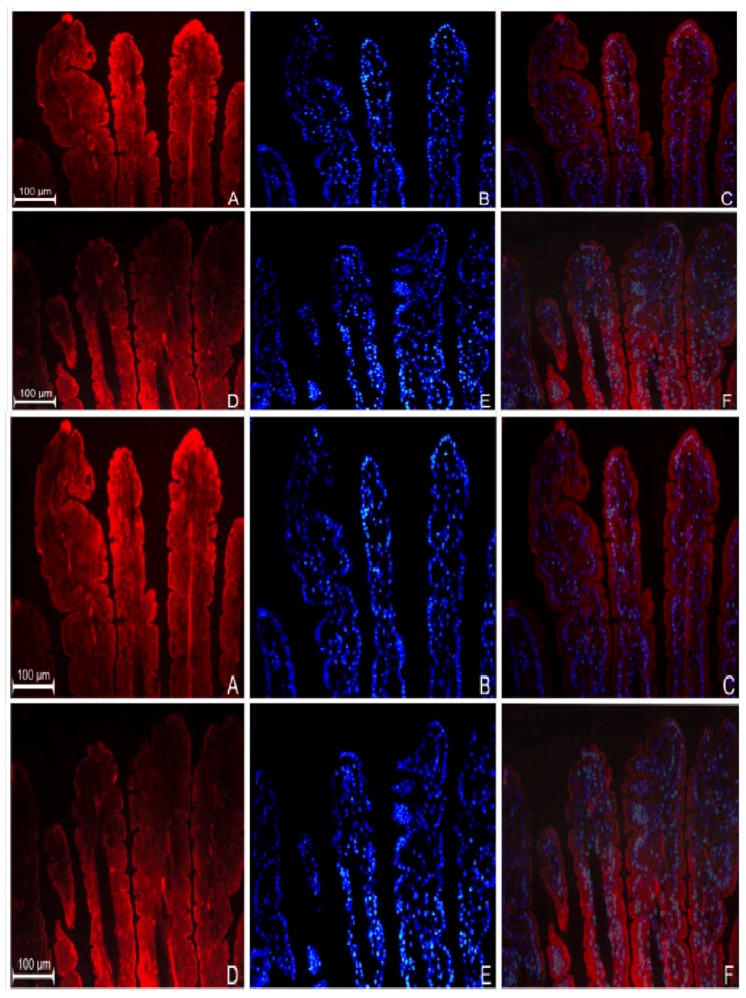
Figure 2.
Immunodetection of GPR109a in jejunum of 10-week-old m+/db mice (A) and 10-week-old db/db mice (D). (B,E) are the nuclear DAPI staining of (A,D) respectively; (C,F) are their corresponding merged images. Higher magnification of niacin receptor staining of m+/db (G) and db/db (H); (I) shows jejunal sections prepared in the absence of primary antibodies (negative control). Immunodetection of niacin receptor in Caco-2 cells grown in 5.6 mM (J) and 25.2 mM glucose (M); (K,N) show nuclear 4′,6-diamidino-2-phenylindole (DAPI) staining of (J) and (M) respectively; (L,O) are the corresponding merged images.

