Superconductivity in monolayer graphene
Monolayer graphene decorated with lithium atoms.
Graphene, a one-atom thick nanomaterial derived from graphite, exhibits a combination of remarkable mechanical, optical, and electronic properties. However, researchers have been thus far unable to induce superconductivity in graphene, despite well-documented examples of the property in certain graphite compounds. Bart Ludbrook et al. (pp. 11795–11799) demonstrate that attaching a layer of lithium (Li) atoms to monolayer graphene allows the material to achieve a stable, superconductive state at low temperatures. To apply Li through a process called decorating, the authors prepared the samples at 8 K in ultrahigh vacuum, guiding and verifying the process using high-resolution angle-resolved photoemission spectroscopy (ARPES). According to the authors, the Li atoms improve coupling between electrons and key vibrational states of the carbon atom lattice known as phonons, thus enhancing the quantum electron pairing that is ultimately responsible for superconductivity. Furthermore, the authors present ARPES measurements of a likely superconducting gap that suggests Li-decorated monolayer graphene shifts to a superconducting state at about 5.9 K. — T.J.
Human prion causes multiple system atrophy
Proteins that become self-propagating after adopting alternative shapes, prions are known to cause neurodegenerative diseases such as Creutzfeldt-Jakob disease. Stanley Prusiner et al. (pp. E5308–E5317) report the discovery of a human prion that causes multiple system atrophy (MSA), a progressive neurodegenerative disease. A distinguishing feature of MSA is the formation of cytoplasmic inclusions by the protein α-synuclein, and the newly discovered prion was found to be composed of this protein. The authors found that inoculating mice or cells genetically engineered to express human α-synuclein with brain extracts from 14 people who had died of MSA resulted in MSA transmission to both cells and mice. Nearly all the mice developed progressive neurological disease within about 120 days, accompanied by deposition of α-synuclein within neurons. In addition, brain extracts from the mice infected with MSA were also infectious to genetically engineered mice and cell cultures. In contrast, brain samples from six patients with Parkinson’s disease (PD) were unable to transmit prions to the mice or cells, suggesting that any putative α-synuclein prions that may be involved in PD are different from those causing MSA. The authors conclude that MSA is a transmissible neurodegenerative disease caused by α-synuclein prions, suggesting that α-synuclein is the first new prion to be discovered in the last 50 years. — S.R.
Ancient DNA links Basques to early farmers
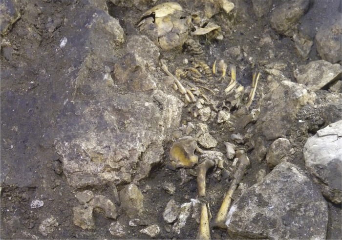
Burial of 6-year-old boy “Matojo” is rich with grave goods.
Compared with the richness of information regarding the Neolithic transition in other parts of Europe, little is known about this period of human history in Iberia. Torsten Günther et al. (pp. 11917–11922) examined ancient genomic DNA extracted from the remains of eight Chalcolithic humans in the El Portalón cave at the Sierra de Atapuerca, Spain, and found insight into the origin of modern Basques. The authors sequenced DNA extracted from bones and teeth that were determined through radiocarbon dating to be 3,500–5,500 years old, a period after the human transition to farming. The authors compared the genomes with those of ancient Europeans from other regions as well as with those of modern-day Europeans. The results suggest that the Iberian farmers, like other early European farmers, may have emerged from a common ancestral group, as migrating farming populations intermixed with local hunter–gatherer groups. Next, the authors used population genetics methods to infer the relationship between early Iberian farmers and modern-day Europeans. According to the authors, the results challenge the assumption that Basques are continuous descendants from a remnant Mesolithic population, and suggest instead that modern-day Basques are most closely related to early farmers on the Iberian Peninsula who remained relatively isolated through modern times. — T.H.D.
How ApoE4 heightens Alzheimer’s disease risk
ApoE4 and risk of AD. Image courtesy of iStockphoto/dra_schwartz.
Carriers of the Apolipoprotein (Apo) E4 allele, found in one-fifth of the general population, face an increased risk of developing sporadic Alzheimer’s disease (AD). However, the mechanism by which ApoE4 confers heightened risk of AD remains unclear. Li Zhu et al. (pp. 11965–11970) tested the hypothesis that ApoE4 is tied to a loss of brain phospholipid balance. Analysis of postmortem brain tissues from ApoE4 carriers revealed reduced levels of the brain metabolite phosphoinositol biphosphate (PIP2), reflecting progressively declining brain PIP2 levels with normal human aging, mild cognitive impairment, and AD. Compared with mice carrying the ApoE3 allele, mice engineered to express ApoE4 displayed reduced PIP2 levels in hippocampal neurons and astrocytes. Synaptojanin 1, a brain enzyme that breaks down PIP2, was correspondingly elevated in brain tissues from mouse and human ApoE4 carriers. When levels of the enzyme were genetically tamped down in ApoE4 mice, cognitive deficits previously observed in behavioral tests, including memory impairment, failure to recognize familiar objects, and abnormal fear conditioning, were reversed, suggesting that restoring normal PIP2 levels can ameliorate AD-associated cognitive dysfunction. Thus, the inability of ApoE4 carriers to maintain normal brain PIP2 levels might underlie their increased risk of sporadic AD. According to the authors, synaptojanin 1 might serve as a potential drug target for treating AD. — P.N.
Childhood malnutrition and gut viruses
Child treated for SAM.
Development of the human gut microbial community is typically completed within the first few years after birth. However, the assembly of the bacterial component of the gut microbiome in children with severe acute malnutrition (SAM) is perturbed, resulting in a seemingly immature gut bacterial community, compared with healthy children. Through DNA sequencing of fecal samples collected from 20 Malawian twin pairs during the first 3 years after birth, Alejandro Reyes et al. (pp. 11941–11946) explored the gut microbiome’s viral component, known as the virome. In eight of the twin pairs, both siblings were healthy, whereas one sibling was healthy and the other developed SAM in 12 so-called discordant pairs. The authors identified a number of known and unknown viruses that helped chart the normal course of gut virome maturation. Both twins in pairs discordant for SAM had less diverse and more immature viromes, compared with age-matched children in healthy pairs, raising the possibility of a familial risk of malnutrition. The presence of certain DNA viruses from the Anelloviridae and Circoviridae families as well as certain bacteriophages distinguished the two groups of twins. Giving children with SAM a peanut-based food supplement used to treat SAM failed to restore a normal gut virome. According to the authors, the findings may have diagnostic and therapeutic implications for malnutrition-related disorders. — P.N.
Visualizing spatial relationships between genomic loci
Visualizing the 3D locations of genomic loci in the nuclei of cells can help elucidate how the spatial organization of the genome affects gene expression. Current fluorescent DNA probes require harsh heat and chemical treatments to denature DNA, potentially affecting the genome’s natural 3D organization. Wulan Deng et al. (pp. 11870–11875) developed a specific and efficient technique to label DNA in fixed cells and tissues while preserving cell shape and DNA structure. The authors repurposed the bacterial clustered regularly interspaced short palindromic repeats (CRISPR)/CRISPR-associated caspase 9 (Cas9) system—commonly used for genome editing—to fluorescently label DNA sequences without denaturing DNA. The authors combined fluorescently labeled Cas9 protein with various RNA sequences as probes, and found that the resulting complex could specifically label target genomic DNA sequences. The authors used the technique to visualize both repetitive and nonrepetitive sequences in different genomic regions. The Cas9–RNA complex was stable and could be used for sequential or simultaneous staining of multiple genomic targets. This rapid technique, which took only 15 minutes under optimized conditions, could be used directly on primary tissues. The authors suggest that the assay could be used to rapidly visualize genomic elements while preserving their spatial relationship, potentially serving as a tool for rapid genetic diagnosis and detection of infectious agents. — S.R.


