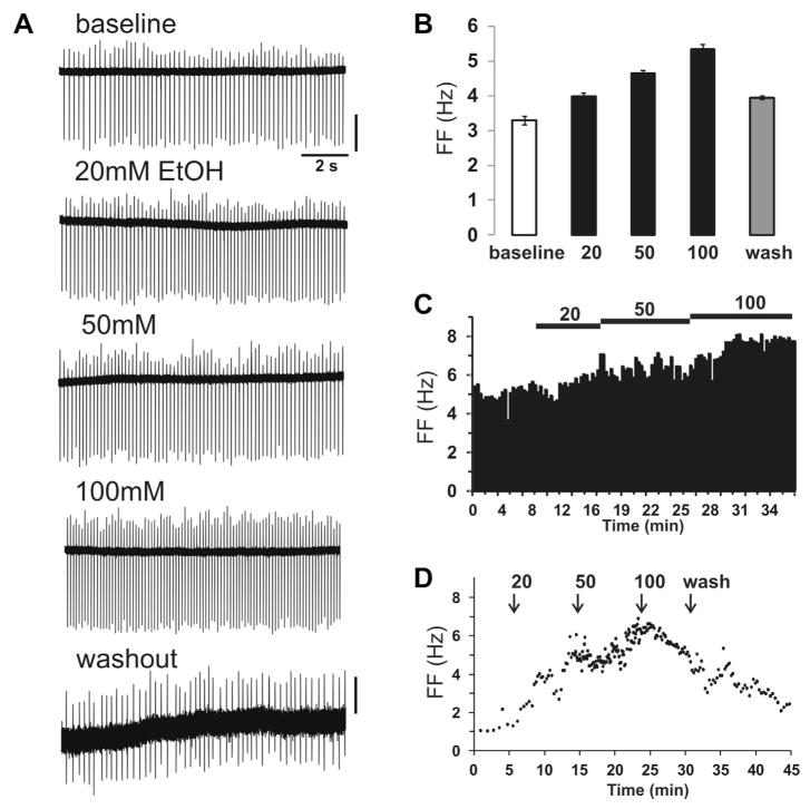
Fig. 4.
Acute EtOH responses in VTA DA cells. (A) Representative trace of extracellular recording from DA neuron in medial VTA (IF nucleus, quinpirole-sensitive cell) with a baseline FF 3.3 Hz. (Scale bars, top: 40 pA, bottom: 20 pA). (B) This cell had a robust concentration-dependent increase of FF (21%, 41%, and 62%) with increasing [EtOH] (20, 50, 100 mM), respectively. Washout completely reversed this effect. (C) Spike ratemeter showing entire recording from baseline to EtOH application. (D) Spontaneous firing of a medial VTA DA cell with baseline FF of 1.3 Hz. EtOH increased the FF to 3.9 Hz at 20 mM, 5.5 Hz at 50 mM, and 5.1 Hz at 100 mM. Washout restored FF to 2.3 Hz but was incomplete after 15 min.

