Abstract
An unusual rotavirus strain, SKT-27, with the G6P[14] genotypes (RVA/Human-wt/THA/SKT-27/2012/G6P[14]), was identified in a stool specimen from a hospitalized child aged eight months with severe diarrhea. In this study, we sequenced and characterized the complete genome of strain SKT-27. On whole genomic analysis, strain SKT-27 was found to have a unique genotype constellation: G6-P[14]-I2-R2-C2-M2-A3-N2-T6-E2-H3. The non-G/P genotype constellation of this strain (I2-R2-C2-M2-A3-N2-T6-E2-H3) is commonly shared with rotavirus strains from artiodactyls such as cattle. Phylogenetic analysis indicated that nine of the 11 genes of strain SKT-27 (VP7, VP4, VP6, VP2-3, NSP1, NSP3-5) appeared to be of artiodactyl (likely bovine) origin, while the remaining VP1 and NSP2 genes were assumed to be of human origin. Thus, strain SKT-27 was found to have a bovine rotavirus genetic backbone, and thus is likely to be of bovine origin. Furthermore, strain SKT-27 appeared to be derived through interspecies transmission and reassortment events involving bovine and human rotavirus strains. Of note is that the VP7 gene of strain SKT-27 was located in G6 lineage-5 together with those of bovine rotavirus strains, away from the clusters comprising other G6P[14] strains in G6 lineages-2/6, suggesting the occurrence of independent bovine-to-human interspecies transmission events. To our knowledge, this is the first report on full genome-based characterization of human G6P[14] strains that have emerged in Southeast Asia. Our observations will provide important insights into the origin of G6P[14] strains, and into dynamic interactions between human and bovine rotavirus strains.
Introduction
Group A rotavirus (RVA), a member of the Reoviridae family, is the most important etiological agent of severe gastroenteritis in the young of humans and many animal species worldwide. In humans, RVA infections are associated with high morbidity and mortality, being responsible for an estimated 453,000 deaths per year in children <5 years of age [1]. More than half of these deaths were estimated to occur in developing countries in Asia and Africa [1, 2].
The RVA virion is a triple-layered, non-enveloped icosahedron enclosing an 11-segment genome of double-stranded (ds)RNA [3]. RVA has two outer capsid proteins, VP7 and VP4, which are implicated independently in neutralization, and define the G and P genotypes, respectively. To date, RVA has been classified into at least 27 G and 37 P genotypes [4, 5]. Among them, some specific G and P genotypes are dominant in individual host species [6]. In human RVAs, 6 G genotypes (G1-4, G9, and G12) and 3 P genotypes (P[4], P[6], and P[8]) are commonly associated with human infections [7, 8]. In addition, several G genotypes (G5-6, G8, G10-11, and G20) and P genotypes (P[1]-[3], P[5], P[7], P[9]-[11], P[14], P[19], and P[25]) have been detected sporadically in humans [9–11]. Many of these unusual genotypes are believed to have originated from animal RVA strains that were introduced into the human population through interspecies transmission and/or reassortment events, one of the major ways in which the genetic diversity of RVAs is generated [6, 12, 13].
G6 strains, one of the most common G genotypes found in cattle [6, 13], have been identified in diarrheic children in combination with either a P[4], P[6], P[8], P[9], P[11], or P[14] genotype, most notably a P[14] genotype [14–17]. P[14] strains, the P genotype commonly found in rabbit and artiodactyls such as cattle, have also been found in children with diarrhea in combination with a G6 genotype [18]. The first G6P[14] strain, PA169, was identified in a child with acute gastroenteritis in Italy in 1988 [19], and subsequently in Australia [20–22], Belgium [18], Egypt [11], Hungary [23–25], India [26], Italy [15, 27], and Spain [28]. In host species other than humans, G6P[14] strains have been detected only in artiodactyls (antelope and goat) in South Africa [18]. In 2012, we detected the first Thai G6 strain from a diarrheic child in a total of 685 RVA-positive stool specimens by PCR-based genotyping (Tacharoenmuang et al., in preparation).
A whole genome-based genotyping system was recently proposed for RVAs based on assignment of all the 11 gene segments (i.e., G/P and non-G/P genotypes) [29]. In the new genotyping system, the acronym Gx-P[x]-Ix-Rx-Cx-Mx-Ax-Nx-Tx-Ex-Hx, where x is an integer, defines the genotype of the VP7-VP4-VP6-VP1-VP2-VP3-NSP1-NSP2-NSP3-NSP4-NSP5 genes, respectively, of a given RVA strain. Most human RVA strains have genes similar in sequence to those of prototype human strains Wa (RVA/Human-tc/USA/Wa/1974/G1P[8]), DS-1 (RVA/Human-tc/USA/DS-1/1976/G2P[4]), or AU-1 (RVA/Human-tc/JPN/AU-1/1982/G3P[9]) [30]. The Wa-like strains are characterized by non-G/P genotypes (I1-R1-C1-M1-A1-N1-T1-E1-H1), and tend to have G/P genotypes G1P[8], G3P[8], G4P[8], and G9P[8]. In contrast, the DS-1-like strains are characterized by non-G/P genotypes (I2-R2-C2-M2-A2-N2-T2-E2-H2), and tend to have G/P genotype G2P[4]. The third minor AU-1-like strains are characterized by non-G/P genotypes (I3-R3-C3-M3-A3-N3-T3-E3-H3), and tend to have G/P genotype G3P[9]. Whole genome-based analysis is a reliable method for obtaining conclusive data on the origin of an RVA strain, and for tracing its evolutionary pattern [29, 31]. To date, however, the whole genomes of some G6P[14] strains from Europe, Australia, and Africa have been analyzed, which provided evidence of their artiodactyl (likely bovine) origin [18, 25, 29]. G6P[14] strains are speculated to be the result of reassortment between strains from cattle and other member(s) of the Artiodactyla [18]. Partial-length genomic analyses of G6P[14] strains from Africa and Asia have also shown the presence of a unique non-G/P genotype constellation (I2-R2-C2-M2-(A3/A11)-N2-T6-E2-H3), commonly shared with RVA strains from artiodactyls including cattle [17, 26]. However, the overall genomic constellation and the exact evolutionary patterns of G6P[14] strains remain to be elucidated. Although only one Asian G6P[14] strain, Indian human N-1, has been studied, its genomic analysis was based on partial-length genome sequences. Thus, we analyzed the whole genome of Thai strain SKT-27 with the G6P[14] genotypes, to gain a more precise understanding of the evolutionary patterns of G6P[14] strains in Asia. In the present study, we analyzed the whole genome of the first Thai strain, SKT-27, with the G6P[14] genotypes isolated from a diarrheic child. Moreover, in this study, deep sequencing with the next generation sequencing (NGS) Illumina MiSeq platform was carried out to obtain the complete nucleotide sequence of the whole genome of strain SKT-27.
Materials and Methods
Ethics statement
The study was approved by the Ethical Review Committee for Research in Human Subjects of the Ministry of Public Health, Thailand (Ref. no. 10/2555). In this study, written informed consent for the testing of stool samples for RVAs and characterization of identified RVA strains was obtained from the children’s parents/guardians.
Virus strains
The full-genomic sequence was determined for strain SKT-27, which was identified as the sole pathogen causing diarrhea in stool specimens from a hospitalized child aged eight months with severe diarrhea in Sukhothai Province in November 2012 during the RVA strain surveillance program in Phetchaboon and Sukhothai Provinces, Thailand in 2012–2014, which involved a total of 685 RVA-positive fecal samples (Tacharoenmuang et al., in preparation). Stool specimens were collected from hospital-based surveillance activities under the RVA vaccine effectiveness evaluation study in Thailand. Stool samples containing strain SKT-27 were kept at −30°C until use.
Viral dsRNA extraction
The viral dsRNAs were extracted from stool suspensions using a QIAamp Viral RNA Mini Kit (Qiagen). The extracted dsRNAs were used for (i) polyacrylamide gel electrophoresis (PAGE) analysis, and (ii) whole genomic analysis. For PAGE analysis, the dsRNAs were electrophoresed in a 10% polyacrylamide gel for 16 h at 20 mA at room temperature, followed by silver staining [32] to determine the genomic dsRNA profile. For whole genomic analysis, viral dsRNAs were subjected to Illumina MiSeq sequencing as described below.
cDNA library building and Illumina MiSeq sequencing
Preparation of a cDNA library and Illumina MiSeq sequencing were carried out as described previously [30, 33]. Briefly, a 200 bp fragment library ligated with bar-coded adapters was constructed for strain SKT-27 using an NEBNext Ultra RNA Library Prep Kit for Illumina v1.2 (New England Biolabs) according to the manufacturer’s instructions. Library purification was performed using Agencourt AMPure XP magnetic beads (Beckman Coulter). The quality of the purified cDNA library was assessed on an Agilent 2100 Bioanalyzer (Agilent Technologies). Nucleotide sequencing was performed on an Illumina MiSeq sequencer (Illumina) using a MiSeq Reagent Kit v2 (Illumina) to generate 151 paired-end reads. Data analysis was carried out using CLC Genomics Workbench v8.0.1 (CLC Bio). Contigs were assembled from the obtained sequence reads by de novo assembly. Using the assembled contigs as query sequences, the Basic Local Alignment Search Tool (BLAST) non-redundant nucleotide database was searched to obtain the full-length nucleotide sequence of each gene segment of strain SKT-27. The nucleotide sequences were translated into amino acid sequences using GENETYX v11 (GENETYX).
Determination of RVA genotypes
The genotype of each of the 11 gene segments of strain SKT-27 was determined using the RotaC v2.0 automated genotyping tool (http://rotac.regatools.be/) [34] according to the guidelines proposed by the Rotavirus Classification Working Group (RCWG).
Phylogenetic analyses
Sequence comparisons were performed as described previously [33, 35]. Briefly, multiple alignment of each viral gene was performed using CLUSTAL W [36]. Phylogenetic trees were constructed using the maximum likelihood method and the Tamura-Nei substitution model using MEGA6.06 [37]. The reliability of the branching order was estimated from 1000 bootstrap replicates [38]. The results of phylogenetic analyses were validated using several other genetic distance models, such as the Jukes-Cantor, Kimura 2-parameter, Tamura 3-parameter, and Hasegawa-Kishino-Yano ones (data not shown).
Nucleotide sequence accession numbers
The nucleotide sequence data presented in this manuscript have been deposited in the DDBJ and EMBL/GenBank data libraries. The accession numbers for the nucleotide sequences of the VP1-4, VP6-7, and NSP1-5/6 genes of strain SKT-27 are LC055547-LC055557, respectively.
Results and Discussion
Extraction of genomic dsRNAs of strain SKT-27
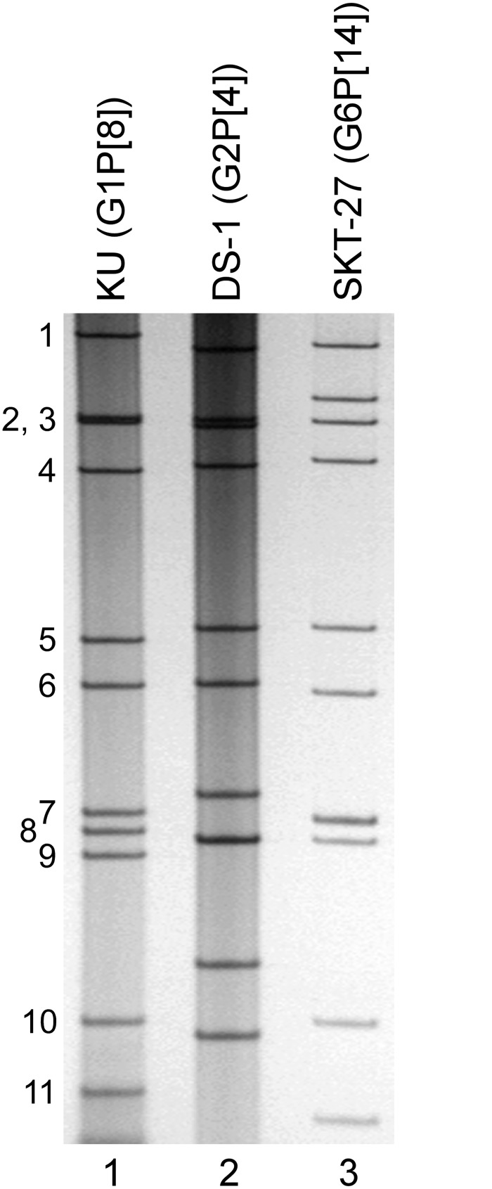
Virion dsRNAs of strain SKT-27 were extracted from stool specimens and then analyzed by PAGE. Fig 1 shows the profiles of viral dsRNAs from human strains KU (RVA/Human-tc/JPN/KU/1974/G1P[8]) [39] (lane 1) and DS-1 (G2P[4]) [40] (lane 2), as references, extracted from the cell cultures, and strain SKT-27 (lane 3) from a stool sample. Strain SKT-27 showed a long electropherotype.
Fig 1. Genomic dsRNA profile of strain SKT-27.
Lanes 1–2, dsRNAs of strains KU (G1P[8]) (lane 1) and DS-1 (G2P[4]) (lane 2), respectively, extracted from the cell cultures; lane 3, dsRNAs of strain SKT-27 extracted from a stool sample. The numbers on the left indicate the order of the genomic dsRNA segments of strain KU.
Nucleotide sequencing and whole-genome-based genotyping of strain SKT-27
PCR-based genotyping [41] showed that strain SKT-27 has the G6 genotype (Tacharoenmuang et al., in preparation). Because G6 strains have been detected almost exclusively in artiodactyls such as cattle [6, 18], the detection of a G6 strain in the human population was suggestive of zoonotic transmission. The whole genome of strain SKT-27 was amplified using a sequence-independent primer set and then sequenced successfully. Illumina MiSeq sequencing yielded 6.8 x 10⁵ reads (~145 bp average length) for strain SKT-27. Complete or nearly complete nucleotide sequences of all the 11 gene segments of strain SKT-27 could be obtained.
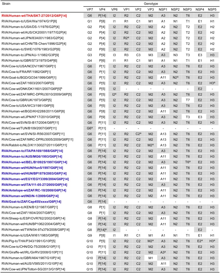
The 11 genes of strain SKT-27 were assigned as G6-P[14]-I2-R2-C2-M2-A3-N2-T6-E2-H3 (Fig 2). Strain SKT-27 was confirmed to have the G6 genotype as determined by PCR-based genotyping. Furthermore, the P genotype of strain SKT-27 was successfully determined to be P[14] using the Illumina MiSeq sequencing approach, although the VP4 gene of this strain could not be genotyped by PCR-based genotyping (Tacharoenmuang et al., in preparation). Thus, strain SKT-27 was named RVA/Human-wt/THA/SKT-27/2012/G6P[14] according to the guidelines for the uniformity of RVAs proposed by the RCWG. Comparison of the complete genotype constellation of strain SKT-27 with those of other G6 and non-G6 strains is shown in Fig 2. Strain SKT-27 exhibited a unique non-G/P genotype constellation (I2-R2-C2-M2-A3-N2-T6-E2-H3), which is commonly found in RVA strains from artiodactyls such as cattle [17, 18, 42]. Strain SKT-27 was found to share the same non-G/P genotype constellation with bovine strains (NCDV (RVA/Cow-tc/USA/NCDV/1967/G6P[1]), RF (RVA/Cow-tc/FRA/RF/1982/G6P[1]), 1603 (G6P[5]), MRC-DPRU3010 (G6P[5]), WC3 (RVA/Cow-tc/USA/WC3/1981/G6P[5]), SI-B17 (G6P[11]), 1604 (G8P[1]), DQ-75 (G10P[11]), and Tottori-SG (G15P[14])), bovine-like human strains (B12 (G8P[1]), PA169 (G6P[14]), B10925 (G6P[14]), and 111-05-27 (G6P[14])), bovine-like porcine strain P343 (G10P[5]), and bovine-like giraffe strain GirRV (G10P[11]), although some bovine and bovine-like strains have been found to have other NSP1 (A11 or A13 instead of A3) and NSP3 (T3 or T7 instead of T6) genotypes. Human strains (B12, PA169, B10925, and 111-05-27), porcine strain P343, and giraffe strain GirRV have been shown to have bovine backbones, and to be likely of bovine origin through their full genomic analyses [12, 29, 43, 44]. Thus, the genotype constellation of strain SKT-27 is mostly identical to those of bovine and bovine-like strains.
Fig 2. Genotype natures of the 11 gene segments of strain SKT-27 compared with those of selected human and animal strains.
Strain SKT-27 is shown in red, while other G6P[14] strains are shown in blue. Gray shading indicates the gene segments with a genotype identical to that of strain SKT-27. aThe gene segments that are most similar to those of strain SKT-27. “−” indicates that no sequence data were available in the DDBJ and EMBL/GenBank data libraries. bGenotype assignment of strain DK11601 based on a report by Midgley et al. (2012). To our knowledge, to date, nucleotide sequence accession numbers for the VP7 and VP4 genes of strain DK11601 are not available in the DDBJ and EMBL/GenBank data libraries.
Phylogenetic analyses
We next constructed phylogenetic trees using the full-genome sequence for each of the 11 gene segments because phylogenetic analysis of RVA nucleotide sequences provides direct evidence of their relatedness to those of other strains, even within the same genotype [29].
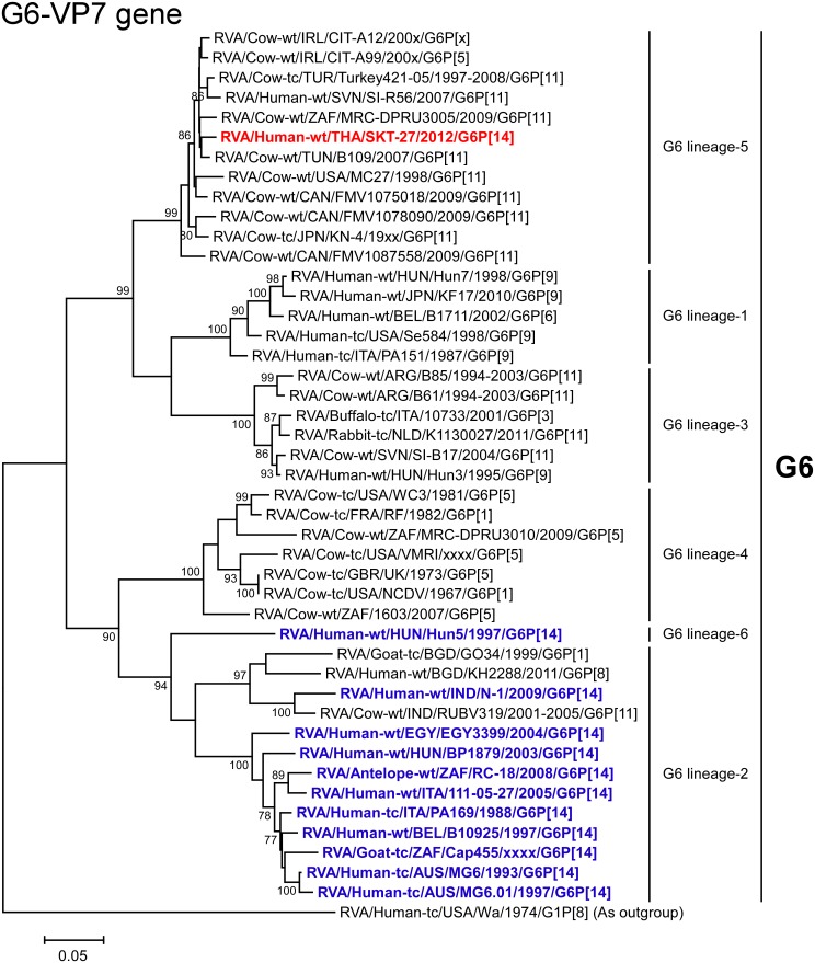
The VP7 gene of strain SKT-27 exhibited the maximum nucleotide sequence identity (98.4%) with that of Tunisian bovine strain B109 (G6P[11]) [45] (Fig 2), and comparable identities (98.1–98.2%) with Irish bovine strains CIT-A12 (G6P[x]) and CIT-A99 (G6P[5]) [46]. On phylogenetic analysis, strain SKT-27 was found to form a cluster with the above-mentioned bovine strains, and several bovine and bovine-like strains with G6P[11] genotypes in G6 lineage-5, away from the clusters comprising other G6P[14] strains in G6 lineages-2/6 (Fig 3).
Fig 3. Phylogenetic tree constructed from the nucleotide sequences of the VP7 genes of strain SKT-27 and representative RVA strains.
In the tree, the position of strain SKT-27 is shown in red, while other G6P[14] strains are shown in blue. Bootstrap values of <75% are not shown. Scale bars: 0.05 substitutions per nucleotide.
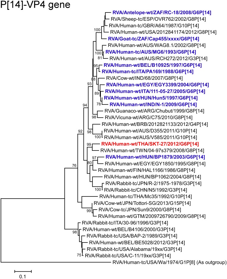
The VP4 gene of strain SKT-27 showed the highest nucleotide sequence identity (92.4%) with the cognate gene of Taiwanese bovine-like human strain 04-97s379 (G8P[14]) [47] (Fig 2), and somewhat lower identity (90.0%) with Finnish bovine-like human strain HAL1166 (G8P[14]) [48–50]. On phylogenetic analysis, strain SKT-27 was shown to be closely related with strain 04-97s379 in a common branch with strain HAL1166 (Fig 4).
Fig 4. Phylogenetic tree constructed from the nucleotide sequences of the VP4 genes of strain SKT-27 and representative RVA strains.
See legend of Fig 3. Scale bars: 0.1 substitutions per nucleotide.
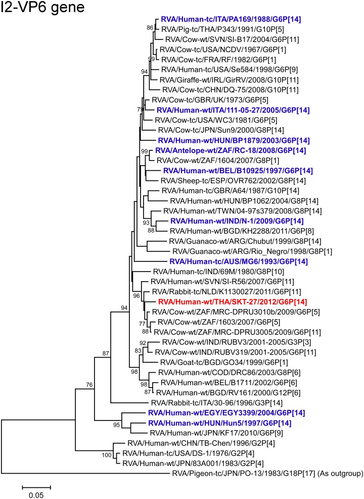
The VP6 gene of strain SKT-27 exhibited the highest nucleotide sequence identity (97.8%) with the VP6 gene of South African bovine strain MRC-DPRU3010b (G6P[5]) [51] (Fig 2), and comparable identities (97.5–97.7%) with South African bovine strains 1603 (G6P[5]) [52] and MRC-DPRU3005 (G6P[11]). Phylogenetically, strain SKT-27 was closely related with these South African bovine strains in a common branch with Dutch bovine-like lapine strain K1130027 (G6P[11]) [53] (Fig 5).
Fig 5. Phylogenetic tree constructed from the nucleotide sequences of the VP6 genes of strain SKT-27 and representative RVA strains.
See legend of Fig 3. Scale bars: 0.05 substitutions per nucleotide.
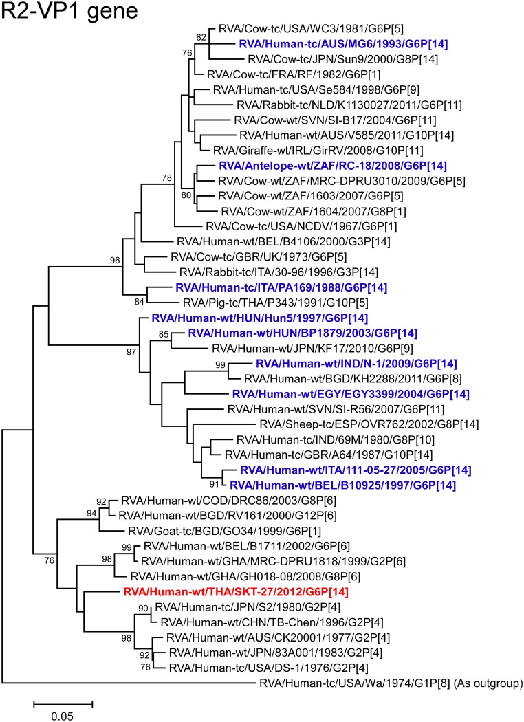
The VP1 gene of strain SKT-27 showed the maximum nucleotide sequence identity (92.7%) with the cognate gene of Japanese human strain 83A001 (G2P[4]) [54] (Fig 2), and comparable identities (92.3–92.6%) with reference human strains DS-1 (G2P[4]), S2 (RVA/Human-tc/JPN/S2/1980/G2P[4]), and TB-Chen (RVA/Human-wt/CHN/TB-Chen/1996/G2P[4]) [40, 55, 56]. On phylogenetic analysis, strain SKT-27 was shown to cluster near the prototype human G2P[4] strains DS-1 and S2 (Fig 6).
Fig 6. Phylogenetic tree constructed from the nucleotide sequences of the VP1 genes of strain SKT-27 and representative RVA strains.
See legend of Fig 3. Scale bars: 0.05 substitutions per nucleotide.
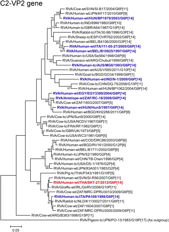
The VP2 gene of strain SKT-27 showed the highest nucleotide sequence similarity (96.7%) with the VP2 gene of Slovene bovine-like human strain SI-R56 (G6P[11]) [57] (Fig 2), and comparable identity (96.5%) with Irish bovine-like giraffe strain GirRV (G10P[11]) [44]. Phylogenetically, strain SKT-27 was shown to be closely related with strain SI-R56 in a common branch with strain GirRV (Fig 7).
Fig 7. Phylogenetic tree constructed from the nucleotide sequences of the VP2 genes of strain SKT-27 and representative RVA strains.
See legend of Fig 3. Scale bars: 0.05 substitutions per nucleotide.
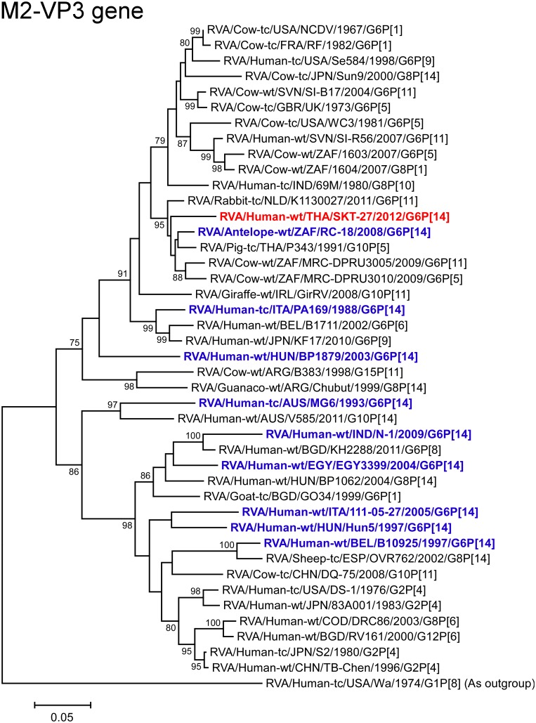
The VP3 gene of strain SKT-27 showed the maximum nucleotide sequence identity (95.4%) with those of Thai bovine-like porcine strain P343 (G10P[5]) [43] and Dutch bovine-like lapine strain K1130027 (G6P[11]) (Fig 2), and comparable similarity (95.3%) with South African bovine-like antelope strain RC-18 (G6P[14]) [18]. On phylogenetic analysis, strain SKT-27 was closely related with these bovine-like strains, and South African bovine strains MRC-DPRU3010 (G6P[5]) and MRC-DPRU3005 (G6P[11]) (Fig 8).
Fig 8. Phylogenetic tree constructed from the nucleotide sequences of the VP3 genes of strain SKT-27 and representative RVA strains.
See legend of Fig 3. Scale bars: 0.05 substitutions per nucleotide.
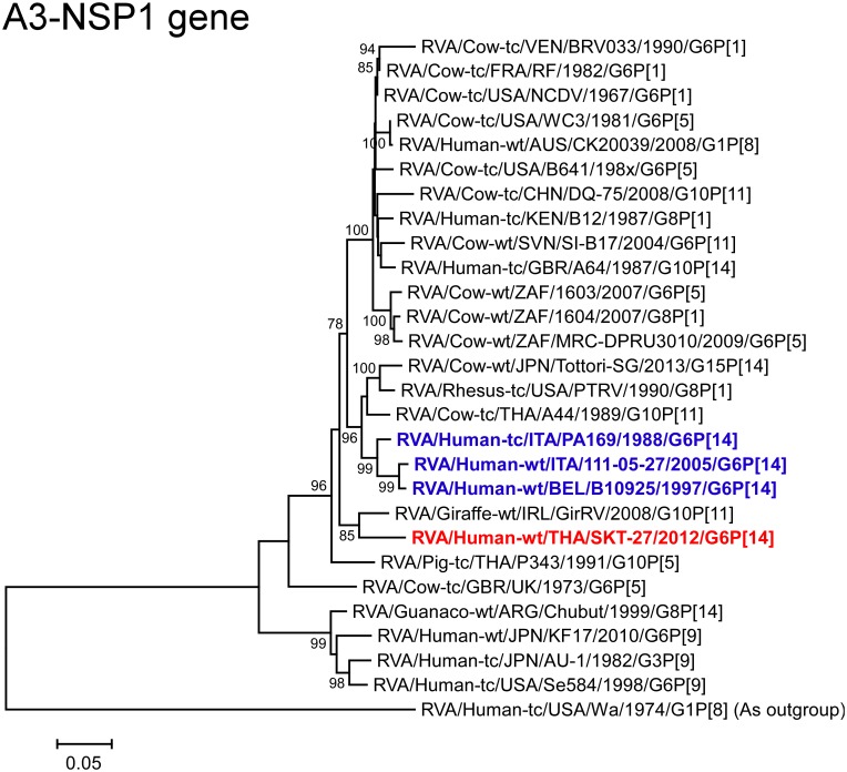
The NSP1 gene of strain SKT-27 exhibited the highest nucleotide sequence identity (93.7%) with the cognate gene of Irish bovine-like giraffe strain GirRV (G10P[11]) (Fig 2). On phylogenetic analysis, strain SKT-27 was shown to be closely related with strain GirRV near the clusters formed by several bovine and bovine-like strains (Fig 9).
Fig 9. Phylogenetic tree constructed from the nucleotide sequences of the NSP1 genes of strain SKT-27 and representative RVA strains.
See legend of Fig 3. Scale bars: 0.05 substitutions per nucleotide.
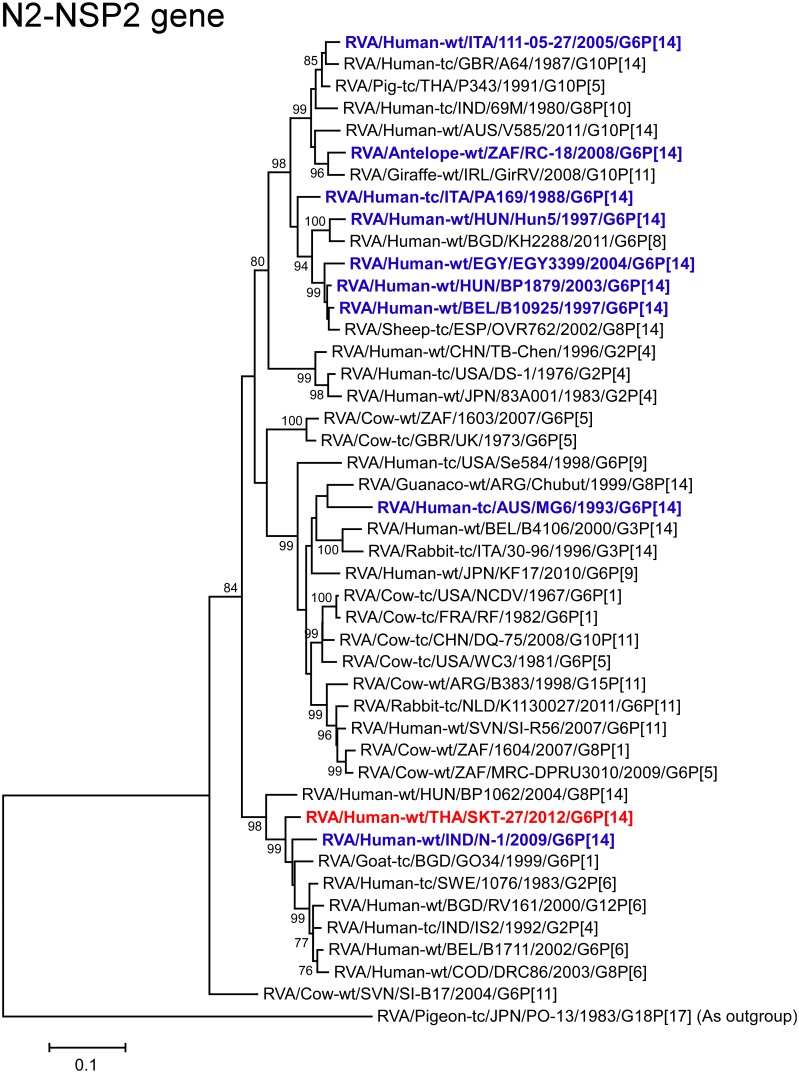
The NSP2 gene of strain SKT-27 exhibited the maximum nucleotide sequence identity (95.4%) with that of caprine strain GO34 (RVA/Goat-tc/BGD/GO34/1999/G6P[1]) from Bangladesh [58] (Fig 2), and comparable identity (95.3%) with Swedish human strain 1076 (G2P[6]) [59]. Phylogenetically, strain SKT-27 was found to cluster near these, and several human G2, G6, G8, and G12 strains from different parts of the world (Fig 10).
Fig 10. Phylogenetic tree constructed from the nucleotide sequences of the NSP2 genes of strain SKT-27 and representative RVA strains.
See legend of Fig 3. Scale bars: 0.1 substitutions per nucleotide.
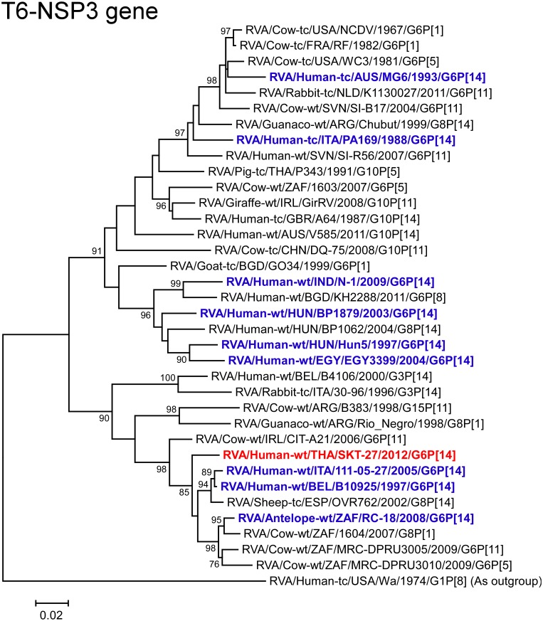
The NSP3 gene of strain SKT-27 showed the highest nucleotide sequence identity (97.2%) with the NSP3 gene of Belgian bovine-like strain human B10925 (G6P[14]) [18] (Fig 2), and somewhat lower identities (96.6–96.8%) with Italian bovine-like human strain 111-05-27 (G6P[14]) [18] and Spanish bovine-like ovine strain OVR762 (G8P[14]) [18]. On phylogenetic analysis, strain SKT-27 was shown to cluster near these bovine-like P[14] strains (Fig 11).
Fig 11. Phylogenetic tree constructed from the nucleotide sequences of the NSP3 genes of strain SKT-27 and representative RVA strains.
See legend of Fig 3. Scale bars: 0.02 substitutions per nucleotide.
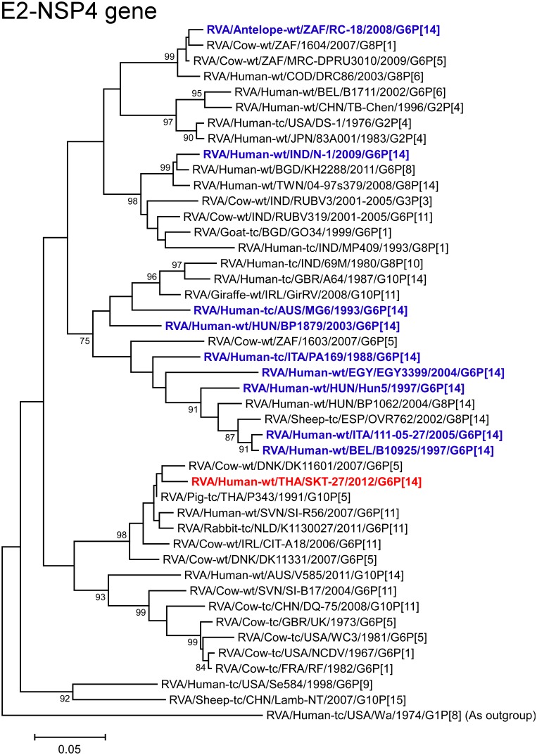
The NSP4 gene of strain SKT-27 showed the maximum nucleotide sequence similarity (98.5%) with the cognate gene of Thai bovine-like porcine strain P343 (G10P[5]) (Fig 2), and somewhat lower identity (97.4%) with Danish bovine strain DK11601 (G6P[5]) [60]. On phylogenetic analysis, strain SKT-27 was found to be closely related with strain DK11601 in a common branch with strain P343, Slovene bovine-like human strain SI-R56 (G6P[11]), and Dutch bovine-like lapine strain K1130027 (G6P[11]) (Fig 12).
Fig 12. Phylogenetic tree constructed from the nucleotide sequences of the NSP4 genes of strain SKT-27 and representative RVA strains.
See legend of Fig 3. Scale bars: 0.05 substitutions per nucleotide.
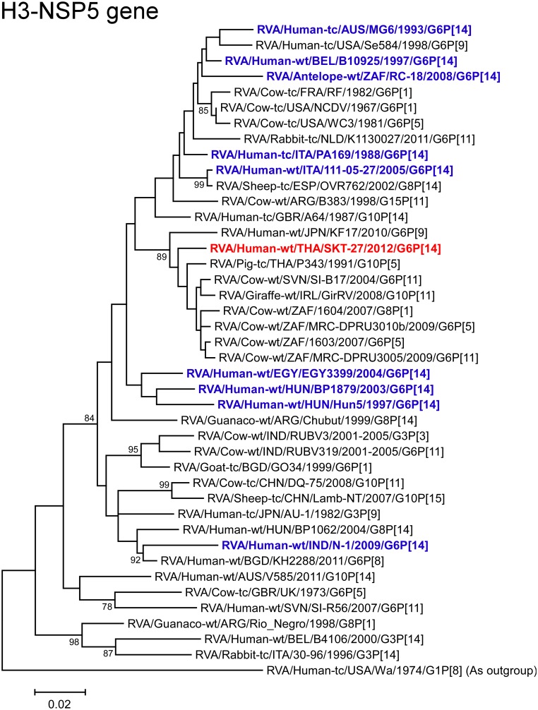
The NSP5 gene of strain SKT-27 exhibited the maximum nucleotide sequence identity (98.1%) with those of South African bovine strain 1603 (G6P[5]) and Thai bovine-like porcine strain P343 (G10P[5]) (Fig 2), and comparable identities (97.8–97.9%) with South African bovine strains (MRC-DPRU3010b (G6P[5]) and MRC-DPRU3005 (G6P[11])) and Slovene bovine strain SI-B17 (G6P[11]) [57]. On phylogenetic analysis, strain SKT-27 was shown to be clustered near these, and several bovine and bovine-like strains (Fig 13).
Fig 13. Phylogenetic tree constructed from the nucleotide sequences of the NSP5 genes of strain SKT-27 and representative RVA strains.
See legend of Fig 3. Scale bars: 0.02 substitutions per nucleotide.
In the present study, we analyzed the whole genome of a Thai strain, SKT-27, with the G6P[14] genotypes (RVA/Human-wt/THA/SKT-27/2012/G6P[14]) from a child with acute gastroenteritis. Strain SKT-27 showed a unique genotype constellation: G6-P[14]-I2-R2-C2-M2-A3-N2-T6-E2-H3. The non-G/P genotype constellation of this strain (I2-R2-C2-M2-A3-N2-T6-E2-H3) is commonly shared with RVA strains from artiodactyls such as cattle. Phylogenetic analysis revealed that nine of the 11 genes of strain SKT-27 (VP7, VP4, VP6, VP2-3, NSP1, and NSP3-5) appeared to be of artiodactyl (likely bovine) origin. On the other hand, the remaining VP1 and NSP2 genes of strain SKT-27 were assumed to be of human origin. However, the exact origins of the VP1 and NSP2 genes of strain SKT-27 could not be ascertained due to a lack of a sufficient number of representative strains as references. Therefore, we could not determine if the suspected ancestral G6P[14] strain of strain SKT-27 had human-like VP1 and NSP2 genes in its genome before it was transmitted from cattle to humans. In any case, strain SKT-27 was assumed to be derived through interspecies transmission and/or reassortment events involving bovine and human RVA strains.
Notably, the VP7 gene of strain SKT-27 was located in G6 lineage-5, away from the clusters comprising other G6P[14] strains in G6 lineages-2/6. This could support the hypothesis that distinct human G6P[14] strains are more likely the result of individual interspecies transmissions from artiodactyls rather than the result of human-to-human transmission after interspecies transmission [18, 20]. To our knowledge, this is the first description of full genome-based characterization of human G6P[14] strains that have emerged in Southeast Asia.
Our findings add to the increasing evidence supporting animal-to-human interspecies transmission and reassortment events. The bovine origin of strain SKT-27 also suggests interspecies transmission due to the close proximity of humans to livestock, especially in developing countries in Asia where there is intimate contact between humans and livestock such as cattle. Because G6P[14] strains have been able to cause constant human infections worldwide, continuing RVA surveillance of this G-P combination in the human population may be required. Simultaneous monitoring of RVA strains in humans and animals is also essential for a better understanding of RVA ecology. Furthermore, whole genome-based analyses are essential to understand the evolutionary dynamics of novel RVA strains such as G6P[14] strains.
Acknowledgments
We wish to thank staffs of the original rotavirus vaccine effectiveness study program for their great support.
Data Availability
The nucleotide sequence data presented in this manuscript have been deposited in the DDBJ and EMBL/GenBank data libraries. The accession numbers for the nucleotide sequences of the VP1-4, VP6-7, and NSP1-5 genes of strain SKT-27 are LC055547-LC055557, respectively.
Funding Statement
This study was supported in part by the MEXT-Supported Program for the Strategic Research Foundation at Private Universities, 2010-2014 (KT), Grants-in-Aid for Research on Emerging and Re-emerging Infectious Diseases from the Ministry of Health, Labor, and Welfare of Japan (KT), and the Original Rotavirus Vaccine Effectiveness Study Program from the Ministry of Public Health of Thailand (SS). The funders had no role in study design, data collection and analysis, decision to publish, or preparation of the manuscript.
References
- 1. Tate JE, Burton AH, Boschi-Pinto C, Steele AD, Dugue J, Parashar UD, et al. 2008 estimate of worldwide rotavirus-associated mortality in children younger than 5 years before the introduction of universal rotavirus vaccination programmes: a systemic review and meta-analysis. Lancet Infect Dis. 2012;12: 136–141. 10.1016/S1473-3099(11)70253-5 [DOI] [PubMed] [Google Scholar]
- 2. Kahn G, Fitzwater S, Tate J, Kang G, Ganguly N, Nair G, et al. Epidemiology and prospects for prevention of rotavirus disease in India. Indian Pediatr. 2012;49: 467–474. [DOI] [PubMed] [Google Scholar]
- 3. Estes MK, Greenberg HB. Rotaviruses In: Knipe DM, Howley PM, editors. Fields Virology. 6th ed Philadelphia: Lippincott Williams & Wilkins; 2013. pp. 1347–1401. [Google Scholar]
- 4. Matthijnssens J, Ciarlet M, McDonald SM, Attoui H, Bányai K, Brister JR, et al. Uniformity of rotavirus strain nomenclature proposed by the Rotavirus Classification Working Group (RCWG). Arch Virol. 2011;156: 1397–1413. 10.1007/s00705-011-1006-z [DOI] [PMC free article] [PubMed] [Google Scholar]
- 5. Trojnar E, Sachsenröder J, Twardziok S, Reetz J, Otto PH, Johne R. Identification of an avian group A rotavirus containing a novel VP4 gene with a close relationship to those of mammalian rotaviruses. J Gen Virol. 2013;94: 136–142. 10.1099/vir.0.047381-0 [DOI] [PubMed] [Google Scholar]
- 6. Martella V, Bányai K, Matthijnssens J, Buonavoglia C, Ciarlet M. Zoonotic aspects of rotaviruses. Vet Microbiol. 2010;140: 246–255. 10.1016/j.vetmic.2009.08.028 [DOI] [PubMed] [Google Scholar]
- 7. Santos N, Hoshino Y. Global distribution of rotavirus serotypes/genotypes and its implication for the development and implementation of an effective rotavirus vaccine. Rev Med Virol. 2005;15: 29–56. [DOI] [PubMed] [Google Scholar]
- 8. Matthijnssens J, Heylen E, Zeller M, Rahman M, Lemey P, Van Ranst M. Phylodynamic analyses of rotavirus genotypes G9 and G12 underscore their potential for swift global spread. Mol Biol Evol. 2010;27: 2431–2436. 10.1093/molbev/msq137 [DOI] [PubMed] [Google Scholar]
- 9. Gentsch JR, Laird AR, Bielfelt B, Griffin DD, Banyai K, Ramachandran M, et al. Serotype diversity and reassortment between human and animal rotavirus strains: implications for rotavirus vaccine programs. J Infect Dis. 2005;192: S146–S159. [DOI] [PubMed] [Google Scholar]
- 10. Solberg OD, Hasing ME, Trueba G, Eisenberg JN. Characterization of novel VP7, VP4, and VP6 genotypes of a previously untypeable group A rotavirus. Virology. 2009;385: 58–67. 10.1016/j.virol.2008.11.026 [DOI] [PMC free article] [PubMed] [Google Scholar]
- 11. E1 Sherif M, Esona MD, Wang Y, Gentsch JR, Jiang B, Glass RI, et al. Detection of the first G6P[14] human rotavirus strain from a child with diarrhea in Egypt. Infect Genet Evol. 2011;11: 1436–1442. 10.1016/j.meegid.2011.05.012 [DOI] [PubMed] [Google Scholar]
- 12. Ghosh S, Kobayashi N. Whole-genomic analysis of rotavirus strains: current status and future prospects. Future Microbiol. 2011;6: 1049–1065. 10.2217/fmb.11.90 [DOI] [PubMed] [Google Scholar]
- 13. Papp H, László B, Jakab F, Ganesh B, De Grazia S, Matthijnssens J, et al. Review of group A rotavirus strains reported in swine and cattle. Vet Microbiol. 2013;165: 190–199. 10.1016/j.vetmic.2013.03.020 [DOI] [PMC free article] [PubMed] [Google Scholar]
- 14. Rahman M, De Leener K, Goegebuer T, Wollants E, Van der Donck I, Van Hoovels L, et al. Genetic characterization of a novel, naturally occurring recombinant human G6P[6] rotavirus. J Clin Microbiol. 2003;41: 2088–2095. [DOI] [PMC free article] [PubMed] [Google Scholar]
- 15. De Grazia S, Martella V, Rotolo V, Bonura F, Matthijnssens J, Bányai K, et al. Molecular characterization of genotype G6 human rotavirus strains detected in Italy from 1986 to 2009. Infect Genet Evol. 2011;11: 1449–1455. 10.1016/j.meegid.2011.05.015 [DOI] [PubMed] [Google Scholar]
- 16. Iturriza-Gómara M, Dallman T, Bányai K, Böttiger B, Buesa J, Diedrich S, et al. Rotavirus genotypes co-circulating in Europe between 2006 and 2009 as determined by EuroRotaNet, a pan-European collaborative strain surveillance network. Epidemiol Infect. 2011;139: 895–909. 10.1017/S0950268810001810 [DOI] [PubMed] [Google Scholar]
- 17. Afrad MH, Matthijnssens J, Moni S, Kabir F, Ashrafi A, Rahman MZ, et al. Genetic characterization of a rare bovine-like human VP4 mono-reassortant G6P[8] rotavirus strain detected from an infant in Bangladesh. Infect Genet Evol. 2013;19: 120–126. 10.1016/j.meegid.2013.06.030 [DOI] [PubMed] [Google Scholar]
- 18. Matthijnssens J, Potgieter CA, Ciarlet M, Parreño V, Martella V, Bányai K, et al. Are human P[14] rotavirus strains the result of interspecies transmissions from sheep or other ungulates that belong to the mammalian order Artiodactyla? J Virol. 2009;83: 2917–2929. 10.1128/JVI.02246-08 [DOI] [PMC free article] [PubMed] [Google Scholar]
- 19. Gerna G, Sarasini A, Parea M, Arista S, Mirandra P, Brussow H, et al. Isolation and characterization of two distinct human rotavirus strains with G6 specificity. J Clin Microbiol. 1992;30: 9–16. [DOI] [PMC free article] [PubMed] [Google Scholar]
- 20. Palombo EA, Bishop RF. Genetic and antigenic characterization of a serotype G6 human rotavirus isolated in Melbourne, Australia. J Med Virol. 1995;47: 348–354. [DOI] [PubMed] [Google Scholar]
- 21. Cooney MA, Gorrell RJ, Palombo EA. Characterisation and phylogenetic analysis of the VP7 proteins of serotype G6 and G8 human rotaviruses. J Med Microbiol. 2001;50: 462–467. [DOI] [PubMed] [Google Scholar]
- 22. Diwakarla S, Clark R, Palombo EA. Expanding distribution of human serotype G6 rotaviruses in Australia. Microbiol Immunol. 2002;46: 499–502. [DOI] [PubMed] [Google Scholar]
- 23. Bányai K, Gentsch JR, Glass RI, Szucs G. Detection of human rotavirus serotype G6 in Hungary. Epidemiol Infect. 2003;130: 107–112. [DOI] [PMC free article] [PubMed] [Google Scholar]
- 24. Bányai K, Gentsch JR, Glass RI, Uj M, Mihály I, Szücs G. Eight-year survey of human rotavirus strains demonstrates circulation of unusual G and P types in Hungary. J Clin Microbiol. 2004;42: 393–397. [DOI] [PMC free article] [PubMed] [Google Scholar]
- 25. Bányai K, Bogdán A, Domonkos G, Kisfali P, Molnár P, Tóth A, et al. Genetic diversity and zoonotic potential of human rotavirus strains, 2003–3006, Hungary. J Med Virol. 2009;81: 362–370. 10.1002/jmv.21375 [DOI] [PubMed] [Google Scholar]
- 26. Mullick S, Mukherjee A, Ghosh S, Pazhani GP, Sur D, Manna B, et al. Genomic analysis of human rotavirus strains G6P[14] and G11P[25] isolated from Kolkata in 2009 reveals interspecies transmission and complex reassortment events. Infect Genet Evol. 2013;14: 15–21. 10.1016/j.meegid.2012.11.010 [DOI] [PubMed] [Google Scholar]
- 27. Arista S, Vizzi E, Alaimo C, Palermo D, Cascio A. Identification of human rotavirus strains with the P[14] genotype by PCR. J Clin Microbiol. 1999;37: 2706–2708. [DOI] [PMC free article] [PubMed] [Google Scholar]
- 28. Cilla G, Montes M, Gomariz M, Piñeiro L, Pérez-Trallero E. Rotavirus genotypes in children in the Basque Country (northern Spain) over a 13-year period (July 1996-June 2009). Eur J Clin Microbiol Infect Dis. 2010;29: 955–960. 10.1007/s10096-010-0951-x [DOI] [PubMed] [Google Scholar]
- 29. Matthijnssens J, Ciarlet M, Heiman E, Arijs I, Delbeke T, McDonald SM, et al. Full genome-based classification of rotaviruses reveals a common origin between human Wa-like and porcine rotavirus strains and human DS-1-like and bovine rotavirus strains. J Virol. 2008;82: 3204–3219. 10.1128/JVI.02257-07 [DOI] [PMC free article] [PubMed] [Google Scholar]
- 30. Dennis FE, Fujii Y, Haga K, Damanka S, Lartey B, Agbemabiese CA, et al. Identification of novel Ghanaian G8P[6] human-bovine reassortant rotavirus strain by Next Generation Sequencing. PLoS One. 2014;9: e100699 10.1371/journal.pone.0100699 [DOI] [PMC free article] [PubMed] [Google Scholar]
- 31. Ghosh S, Gatheru Z, Nyangao J, Adachi N, Urushibara N, Kobayashi N. Full genomic analysis of a G8P[1] rotavirus strain isolated from an asymptomatic infant in Kenya provides evidence for an artiodactyl-to-human interspecies transmission event. J Med Virol. 2011;83: 367–376. 10.1002/jmv.21974 [DOI] [PubMed] [Google Scholar]
- 32. Komoto S, Sasaki J, Taniguchi K. Reverse genetics system for introduction of site-specific mutations into the double-stranded RNA genome of infectious rotavirus. Proc Natl Acad Sci USA. 2006;103: 4646–4651. [DOI] [PMC free article] [PubMed] [Google Scholar]
- 33. Ide T, Komoto S, Higo-Moriguchi K, Htun KW, Myint YY, Myat TW, et al. Whole genomic analysis of human G12P[6] and G12P[8] rotavirus strains that have emerged in Myanmar. PLoS One. 2015;10: e0124965 10.1371/journal.pone.0124965 [DOI] [PMC free article] [PubMed] [Google Scholar]
- 34. Maes P, Matthijnssens J, Rahman M, Van Ranst M. RotaC: a web-based tool for the complete genome classification of group A rotaviruses. BMC Microbiol. 2009;9: 238 10.1186/1471-2180-9-238 [DOI] [PMC free article] [PubMed] [Google Scholar]
- 35. Komoto S, Apondi EW, Shah M, Odoyo E, Nyangao J, Tomita M, et al. Whole genomic analysis of human G12P[6] and G12P[8] rotavirus strains that have emerged in Kenya: identification of porcine-like NSP4 genes. Infect Genet Evol. 2014;27: 277–293. 10.1016/j.meegid.2014.08.002 [DOI] [PubMed] [Google Scholar]
- 36. Thompson JD, Higgins DG, Gibson TJ. CLUSTAL W: improving the sensitivity of progressive multiple sequence alignment through sequence weighting, position-specific gap penalties and weight matrix choice. Nucleic Acids Res. 1994;22: 4673–4680. [DOI] [PMC free article] [PubMed] [Google Scholar]
- 37. Tamura K, Stecher G, Peterson D, Filipski A, Kumar S. MEGA6: molecular evolutionary genetics analysis version 6.0. Mol Biol Evol. 2013;30: 2725–2729. 10.1093/molbev/mst197 [DOI] [PMC free article] [PubMed] [Google Scholar]
- 38. Felsenstein J. Confidence limits on phylogenies: an approach using the bootstrap. Evolution. 1985;39: 783–791. [DOI] [PubMed] [Google Scholar]
- 39. Urasawa S, Urasawa T, Taniguchi K, Chiba S. Serotype determination of human rotavirus isolates and antibody prevalence in pediatric population in Hokkaido, Japan. Arch Virol. 1984;81: 1–12. [DOI] [PubMed] [Google Scholar]
- 40. Wyatt RG, James HD Jr, Pittman AL, Hoshino Y, Greenberg HB, Kalica AR, et al. Direct isolation in cell culture of human rotaviruses and their characterization into four serotypes. J Clin Microbiol. 1983;18: 310–317. [DOI] [PMC free article] [PubMed] [Google Scholar]
- 41. Komoto S, Maeno Y, Tomita M, Matsuoka T, Ohfu M, Yodoshi T, et al. Whole genomic analysis of a porcine-like human G5P[6] rotavirus strain isolated from a child with diarrhoea and encephalopathy in Japan. J Gen Virol. 2013;94: 1568–1575. 10.1099/vir.0.051011-0 [DOI] [PubMed] [Google Scholar]
- 42. Matthijnssens J, Van Ranst M. Genotype constellation and evolution of group A rotaviruses infecting humans. Curr Opin Virol. 2012;2: 426–433. 10.1016/j.coviro.2012.04.007 [DOI] [PubMed] [Google Scholar]
- 43. Komoto S, Pongsuwanna Y, Ide T, Wakuda M, Guntapong R, Dennis FE, et al. Whole genomic analysis of porcine G10P[5] rotavirus strain P343 provides evidence for bovine-to-porcine interspecies transmission. Vet Microbiol. 2014;174: 577–583. 10.1016/j.vetmic.2014.09.033 [DOI] [PubMed] [Google Scholar]
- 44. O’Shea H, Mulherin E, Matthijnssens J, McCusker MP, Collins PJ, Cashman O, et al. Complete genomic sequence analyses of the first group A giraffe rotavirus reveals close evolutionary relationship with rotaviruses infecting other members of the Artiodactyla. Vet Microbiol. 2014;170: 151–156. 10.1016/j.vetmic.2014.01.012 [DOI] [PubMed] [Google Scholar]
- 45. Hassine-Zaafrane M, Ben Salem I, Sdiri-Loulizi K, Kaplon J, Bouslama L, Aouni Z, et al. Distribution of G (VP7) and P (VP4) genotypes of group A bovine rotaviruses from Tunisian calves with diarrhoea. J Appl Microbiol. 2014;116: 1387–1395. 10.1111/jam.12469 [DOI] [PubMed] [Google Scholar]
- 46. Cashman O, Lennon G, Sleator RD, Power E, Fanning S, O’Shea H. Changing profile of the bovine rotavirus G6 population in the south of Ireland from 2002 to 2009. Vet Microbiol. 2010;146: 238–244. 10.1016/j.vetmic.2010.05.012 [DOI] [PubMed] [Google Scholar]
- 47. Wu FT, Bányai K, Wu HS, Yang DC, Lin JS, Hsiung CA, et al. Identification of a G8P[14] rotavirus isolate obtained from a Taiwanese child: evidence for a relationship with bovine rotaviruses. Jpn J Infect Dis. 2012;65: 455–457. [PMC free article] [PubMed] [Google Scholar]
- 48. Browning GF, Snodgrass DR, Nakagomi O, Kaga E, Sarasini A, Gerna G. Human and bovine serotype G8 rotaviruses may be derived by reassortment. Arch Virol. 1992;125: 121–128. [DOI] [PubMed] [Google Scholar]
- 49. Gerna G, Sears J, Hoshino Y, Steele AD, Nakagomi O, Sarasini A, et al. Identification of a new VP4 serotype of human rotaviruses. Virology. 1994;200: 66–71. [DOI] [PubMed] [Google Scholar]
- 50. Gerna G, Steele AD, Hoshino Y, Sereno M, Garcia D, Sarasini A, et al. A comparison of the VP7 gene sequences of human and bovine rotaviruses. J Gen Virol. 1994;75: 1781–1784. [DOI] [PubMed] [Google Scholar]
- 51. Nyaga MM, Jere KC, Esona MD, Seheri ML, Stucker KM, Halpin RA, et al. Whole genome detection of rotavirus mixed infections in human, porcine and bovine samples co-infected with various rotavirus strains collected from sub-Saharan Africa. Infect Genet Evol. 2015;31: 321–334. 10.1016/j.meegid.2015.02.011 [DOI] [PMC free article] [PubMed] [Google Scholar]
- 52. Jere KC, Mlera L, O’Neill HG, Peenze I, van Dijk AA. Whole genome sequence analyses of three African bovine rotaviruses reveal that they emerged through multiple reassortment events between rotaviruses from different mammalian species. Vet Microbiol. 2012;159: 245–250. 10.1016/j.vetmic.2012.03.040 [DOI] [PubMed] [Google Scholar]
- 53. Schoondermark-van de Ven E, Van Ranst M, de Bruin W, van den Hurk P, Zeller M, Matthijnssens J, et al. Rabbit colony infected with a bovine-like G6P[11] rotavirus strain. Vet Microbiol. 2013;166: 154–164. 10.1016/j.vetmic.2013.05.028 [DOI] [PubMed] [Google Scholar]
- 54. Doan YH, Nakagomi T, Nakagomi O. Repeated circulation over 6 years of intergenogroup mono-reassortant G2P[4] rotavirus strains with genotype N1 of the NSP2 gene. Infect Genet Evol. 2012;12: 1202–1212. 10.1016/j.meegid.2012.04.023 [DOI] [PubMed] [Google Scholar]
- 55. Urasawa S, Urasawa T, Taniguchi K. Three human rotavirus serotypes demonstrated by plaque neutralization of isolated strains. Infect Immun. 1982;38: 781–784. [DOI] [PMC free article] [PubMed] [Google Scholar]
- 56. Chen Y, Wen Y, Liu X, Xiong X, Cao Z, Zhao Q, et al. Full genomic analysis of human rotavirus strain TB-Chen isolated in China. Virology. 2008;375: 361–373. 10.1016/j.virol.2008.01.003 [DOI] [PubMed] [Google Scholar]
- 57. Steyer A, Sagadin M, Kolenc M, Poljšak-Prijatelj M. Whole genome sequence analysis of bovine G6P[11] rotavirus strain found in a child with gastroenteritis. Infect Genet Evol. 2013;13: 89–95. 10.1016/j.meegid.2012.09.004 [DOI] [PubMed] [Google Scholar]
- 58. Ghosh S, Alam MM, Ahmed MU, Talukdar RI, Paul SK, Kobayashi N. Complete genome constellation of a caprine group A rotavirus strain reveals common evolution with ruminant and human rotavirus strains. J Gen Virol. 2010;91: 2367–2373. 10.1099/vir.0.022244-0 [DOI] [PubMed] [Google Scholar]
- 59. Ghosh S, Urushibara N, Chawla-Sarkar M, Krishnan T, Kobayashi N. Whole genomic analyses of asymptomatic human G1P[6], G2P[6] and G3P[6] rotavirus strains reveal intergenogroup reassortment events and genome segments of artiodactyl origin. Infect Genet Evol. 2013;16: 165–173. 10.1016/j.meegid.2012.12.019 [DOI] [PubMed] [Google Scholar]
- 60. Midgley SE, Hjulsager CK, Larsen LE, Falkenhorst G, Böttiger B. Suspected zoonotic transmission of rotavirus group A in Danish adults. Epidemiol Infect. 2012;140: 1013–1017. 10.1017/S0950268811001981 [DOI] [PubMed] [Google Scholar]
Associated Data
This section collects any data citations, data availability statements, or supplementary materials included in this article.
Data Availability Statement
The nucleotide sequence data presented in this manuscript have been deposited in the DDBJ and EMBL/GenBank data libraries. The accession numbers for the nucleotide sequences of the VP1-4, VP6-7, and NSP1-5 genes of strain SKT-27 are LC055547-LC055557, respectively.


