Abstract
Though numerous polymorphisms have been associated with risk of developing lymphoma, how these variants function to promote tumorigenesis is poorly understood. Here, we report that lymphoma risk SNPs, especially in the non-Hodgkin’s lymphoma subtype chronic lymphocytic leukemia, are significantly enriched for co-localization with epigenetic marks of active gene regulation. These enrichments were seen in a lymphoid-specific manner for numerous ENCODE datasets, including DNase-hypersensitivity as well as multiple segmentation-defined enhancer regions. Furthermore, we identify putatively functional SNPs that are both in regulatory elements in lymphocytes and are associated with gene expression changes in blood. We developed an algorithm, UES, that uses a Monte Carlo simulation approach to calculate the enrichment of previously identified risk SNPs in various functional elements. This multiscale approach integrating multiple datasets helps disentangle the underlying biology of lymphoma, and more broadly, is generally applicable to GWAS results from other diseases as well.
Introduction
Lymphoma, including the non-Hodgkin’s lymphoma subtype chronic lymphocytic leukemia (CLL), was responsible for more than 130,000 new cases of cancer and 44,000 deaths in 2014 [1]. Both CLL and Hodkin’s lymphoma are of B-cell origin and have been shown to have a high heritable component [2, 3]. In an effort to understand this effect and the overall etiology of these diseases, numerous genome-wide association studies (GWAS) have been performed and have identified common genetic variants associated with the risk of developing lymphoma [4–16]. Collectively, these studies have identified loci across more than half of the human autosomes that contain low-risk variants that associate with these diseases. Some of the reported risk alleles are found at loci that begin to suggest how this risk is conferred, for example being located in a region containing genes involved in apoptosis [4], a member of the NFκB transcription factor family [9], and the multi-cancer associated locus 8q24 [7, 9]. However, most of these reported GWAS hits are non-coding and the direct mechanism by which these allele lead to increased in risk is yet unresolved.
We have previously observed that single nucleotide polymorphisms (SNPs) found in evolutionary conserved regions and in regions epigenetically marked for transcriptional regulation are more likely to be under negative selection in humans, suggesting biological function [17]. Others have shown that risk variants are enriched in particular epigenomic marks of transcriptional regulatory regions[18, 19] and that trait-associated SNPs, including GWAS-identified risk SNPs, are often found in expression quantitative trait loci (eQTL) that affect nearby gene expression[20]. Furthermore, recent studies have shown the etiologic nature of transcription factors themselves in some diseases[21]. Taken together, these data suggest the hypothesis that for many GWAS-identified risk lock, the functional variant may modulate disease risk through alteration of gene regulation rather than coding sequence.
To test the hypothesis that lymphoma risk SNPs, or their LD partners, tend to alter regulatory elements, we interrogated the functional genomics data from ENCODE (Encyclopedia of DNA Elements) [22, 23] and the Roadmap Epigenomics [24]consortia. The large amount of data available for the lymphoblastoid cell line GM12878 and other hematologically derived cells allows integrative analysis to give more accurate representation of the segments of the genome that are active regulatory elements[25, 26]. To test our hypothesis, we developed a computational pipeline, UES (Uncovering Enrichment through Simulation), that uses a Monte Carlo approach to test whether a set of SNPs is significantly enriched for a particular functional genomic annotation of the genome, taking linkage disequilibrium (LD) patterns into account. We demonstrate a significant enrichment of these lymphoma risk SNPs in regulatory marks specific for lymphoid tissue.
Results
Enrichment of CLL & Lymphoma Risk SNPs in GM12878 Regulatory Tracks
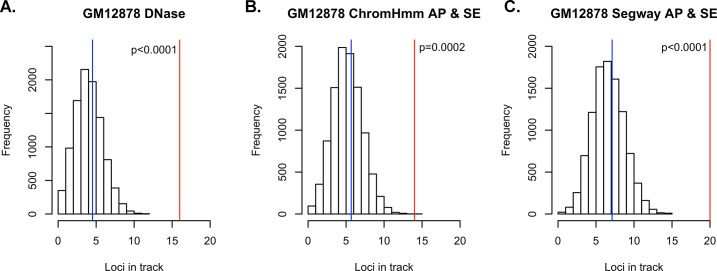
We first asked if lymphoma risk SNPs are enriched in regions annotated as putatively regulatory in GM12878 using our novel method, UES (Fig 1). Using the NHGRI GWAS catalog [27], we identified 56 risk SNPs for lymphoma, including both the Hodgkin’s lymphoma (HD) and non-Hodgkin’s lymphoma (NHL) types. Once the list was pruned to ensure the SNPs were independent and the HLA region was excluded, the resultant list contained 36 risk SNPs (S1 Table) [4–16]. We confirmed that the minor allele distribution of our random SNPs were similar to the original input (input SNPs MAF mean = 0.277; random SNP sets mean = 0.259, median = 0.259, min = 0.176, 1st qu = 0.244, 3rd qu = 0.274, max = 0.347). We first looked at the Deoxyribonuclease I (DNase I) hypersensitivity sites (DHSs) for GM12878, since genomic regions open to DNase digestion have been shown to be accurate markers of regulatory DNA[28]. We queried the ENCODE “unified DNase” track for GM12878, which identifies regions of open chromatin regardless of the particular factors that bind. The lymphoma risk-SNPs were significantly enriched in GM12878 DNase hypersensitivity sites (p < 0.0001), with 16 distinct regions containing risk SNPs potentially explainable by a variant in a DNase hypersensitive site. The 10,000 control sets of randomly selected SNPs with similar characteristics only showed an average of 4.5 regions potentially explainable by variants overlapping a DNase hypersensitive site (Fig 2A and S2 Table). The lymphoma risk-SNPs showed equal enrichment (p<0.0001) in the Roadmap Epigenomics DNase data for GM12878 (S3 Table).
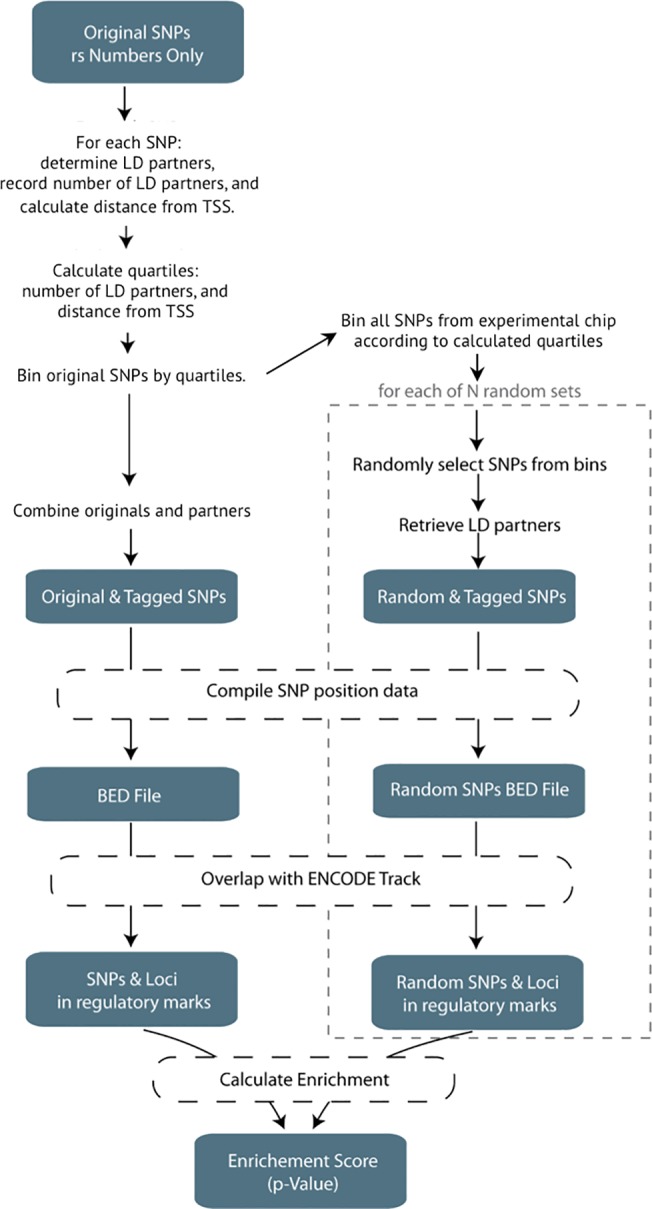
Fig 1. UES algorithm visualization.

This represents the generalized workflow to determine the SNP enrichment in an ENCODE track. A full description and details of the algorithm can be found in the Materials and Methods.
Fig 2. Overlap of lymphoma risk SNPs with regulatory regions in GM12878.
The histograms represent the distribution of how many random loci overlap a specific annotation. The blue represents the mean of the empirical null distribution while the red line represents the real number of loci from the lymphoma and CLL GWAS that overlap the specific regulatory annotation. A, Overlap of SNPs with DNase hypersensitivity regions in GM12878. B, Overlap of SNPs with active promoters and strong enhancers as annotated by ChromHMM in GM12878. C, Overlap of SNPs with active promoters and strong enhancers as annotated by Segway in GM12878.
As some physical regions of the genome harbor more than one independent risk SNP, we were concerned this could lead to oversampling of a given region and false positives. To test this, we location pruned our input SNPs, ensuring that none of the SNPs tested were within one megabase of one another, reducing the input set from 36 initial SNPs down to 30 SNPs. We observed nearly identical results between the location pruned dataset and the original dataset (S2 Table) and concluded that these 6 extra SNPs were not falsely inflating the observed statistical result.
We next asked if enrichment could be observed in regulatory elements predicted by genome segmentation of integrated functional genomics data. Rather than querying individual histone modifications, we chose to examine the results from two different segmentation algorithms, ChromHMM[25] and Segway[26], since both algorithms use a combination of multiple histone modification datasets to determine the segmentation calls. We asked if lymphoma risk SNPs are enriched in regions identified as active promoters or strong enhancers for GM12878 and observed a significant enrichment of the lymphoma SNPs in regulatory regions as defined by both ChromHMM and Segway with p = 0.0002 and p<0.0001, respectively (Fig 2B–2C). Upon looking deeper at the ChromHMM data for GM12878, we observed the risk SNPs were enriched in each of the four classes of enhancers (2 strong enhancer classes and 2 weak enhancer classes) with p< = 0.0002 when analyzed separately. When combined into a separate strong enhancer set and a weak enhancer set, we saw a significant enrichment (p<0.0001) when compared to random controls for both (S4 Table). Interestingly, the “Active Promoter” state, by itself showed no significant enrichment (p = 0.3845). We observed similar results when performing the enrichment analysis for the Segway segmentation track of GM12878 (S4 Table): strong enhancers were the most enriched (p < 0.0001); weak enhancers and active promoters did not achieve significance at the Bonferoni threshold (p = 0.0031 and p = 0.0419, respectively).
We originally perfomed the analysis with the LD threshold cutoff at r2 > 0.8 since this was the threshold during the design of the GWAS chips. In order to test whether our cutoff was appropriate, we performed the enrichment analysis using additional r2 thresholds of r2>0.2, r2>0.4, r2>0.6, and r2 = 1. The results showed nearly identical enrichment of the same cell lines and tissue specificity for both DNase hypersensitivity marks and segmentation data (S2 and S6 Tables) for every LD threshold, except for r2 = 1. Similar levels of enrichment within specific segmentation datatypes of GM12878 are also observed at the other LD threshold levels.
We hypothesized that functional SNPs may be those localized at transcription factor binding sites (TFBS). Using the same SNPs, we interrogated the set of ENCODE ChIP-Seq data for GM12878 (January 2011 data freeze). We created a master dataset consisting of the union of 75 GM12878 transcription factor ChIP-Seq data and observed a significant enrichment of the lymphoma SNPs in the peaks when compared to the random controls (p < 0.0001; S4 Table). We created additional sets containing the union of all of the transcription factor peaks with the Gm12878 DNase hypersensitivity and a union of all the LCL DNase hypersensitivity and observed similar enrichment in both (p < 0.0001, S4 Table). In order to identify specific transcription factors that colocalize with lymphoma risk SNPs or their LD partners, we ran the enrichment analysis pipeline for each factor in the ChIP-Seq dataset, 4 of which reached the significance threshold once corrected for multiple testing: NFIC, RUNX3, NFκB, and TBLR1 (p< 0.0001; p<0.0001; p = 0.0002; p = 0.0005; S5 Table).
To test the validity of our findings and verify our approach, we performed the same analysis with a set of breast cancer risk SNPs identified from the NHGRI GWAS catalogue as there were a similar number of SNPs in the database at the time (n = 31). Our hypothesis is that since the diseases do not share a tissue of origin, there should be no enrichment of the breast cancer SNPs in the GM12878 annotations. We did not observe statistical enrichment (random n = 10,000) of the breast cancer risk loci in DNase hypersensitivity, ChromHMM enhancer, Segway enhancer data, or TF union data for GM12878 (S6 Table). We expanded our scope to see whether or not these tissue-specific observations held true when we used a different disease, prostate cancer, with roughly double the number of input SNPs (n = 62). As seen with the breast cancer SNPs, there was no statistical enrichment of the prostate cancer SNPs in any of the datasets for GM12878 after correcting for multiple testing (S6 Table). These results suggest that the observed signal is specific to the lymphoma and are not generalizable to cancer risk as a whole.
Tissue Specificity of CLL & Lymphoma Risk SNPs
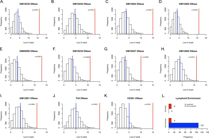
We next asked if the observed enrichments were specific to cells of the lymphoid lineage. First, we interrogated the other 124 unified DNase tracks from ENCODE with the same GWAS and random SNP sets used for the GM12878 analysis and observed enrichment of 8 additional cell lines that achieved Bonferoni-corrected significance with p<0.0004. Interestingly, all of the lines that showed enrichment at that level were of the lymphoid lineage (Fig 3A–3E, 3J and 3K and S2 Table). When we relax the stringency and expand the scope to any cell lines with p<0.01, we see that 15 out of the 18 cell lines which surpassed that threshold were of the lymphoid lineage. All of the LCLs in the ENCODE database were below this threshold and had a p< = 0.0065 (Fig 3F–3I). There was only one cell line, HRE (renal epithelial cells), which was not from the lymphoid lineage that almost met the statistical threshold once corrected for multiple testing (p = 0.0004). These data show that the previously reported lymphoma risk SNPs are enriched in DNase hypersensitivity regions in a tissue-specific manner (Fig 3l). Similar tissue-specific enrichment results were seen in the Roadmap Epigenomics data (S3 Table).
Fig 3. Enrichment of lymphoma and CLL risk SNPs in DNase-hypersensitive sites of lymphoblastoid cell lines.
(A-I) These histograms represent the distribution of how many random loci overlap a specific annotation. The blue represents the mean of the empirical null distribution while the red line represents the real number of loci from the lymphoma and CLL GWAS that overlap the DNase hypersensitive site in the specified cell line: (A) GM19238 (B) GM19240 (C) GM12864 (D) GM12865 (E) GM06990 (F) GM19239 (G) GM18507 (H) GM12892 (I) GM12891. (J) Th0 (K) CD20+ (L) Summary of distribution of tissue of origin for cell lines in which lymphoma and CLL risk SNPs are either enriched (p<0.0004) in DNase hypersensitive sites or not enriched.
We next performed a similar analysis on the chromatin segmentation data. We analyzed eight additional cell lines with ChromHMM segmentation data and five additional lines with Segway segmentation data for enhancer classes. For both the ChromHMM and Segway strong enhancer classifications, GM12878 was the only cell line that showed strong, significant enrichment with p = 0.0002 and p<0.0001 for each dataset, respectively (S7 Table). When looking at the weak enhancer classifications, again, GM12878 was the only cell type to demonstrate any significance with p<0.0001 and p = 0.0031 for the ChromHMM and Segway data, respectively (S7 Tables). Similarly, tissue-specific enhancer enrichment were also observed in the Roadmap Epigenomics data while the enrichment calculations for the transcription start site segmentation failed to reach significance (S8 and S9 Tables).
Estimating enrichment by local annotation shifts
It has been previously noted that, under some models in which the functional variants underlying GWAS are not regulatory, enrichment of GWAS-identified SNPs in regulatory regions could occur if proper controls are not used. While our method controls for the major factors that need to be controlled for (LD patterns and distance from transcription start), we nevertheless asked if a similar enrichment could be observed with an alternative approach, GoShifter, that shifts annotations at the associated loci to test the significance of enrichment. This approach identified 5 cell lines that showed enrichment for the risk SNPs. Notably, 4 of these 5 lines that showed enrichment at p < 0.05– GM19238, Th0, Cd20, and GM06990 –are from the lymphoid lineage (S10 Table).
Lymphoma & CLL SNPs as eQTLs
Another prediction of our hypothesis that lymphoma risk SNPs alter regulatory regions is that these SNPs will be associated with expression changes in nearby genes. To test this hypothesis, we asked how may of the published lymphoma risk SNPs are expression quantitative trait loci (eQTLs) using a recently published set of eQTLs in blood[29]. We observed that 21 of the original loci have at least 1 cis eQTL, (S11 Table). Furthermore, most of these eQTLs are tissue specific when queried against the GTEx eQTL database. One example of the power of this overall approach is evidenced by rs7097. This SNP was initially defined as a lymphoma risk SNP[10] but did not intersect with any LCL DNase hypersensitivity sites, nor with promoter or enhancer regions of chromatin segmentation data. However, one of the SNPs it tags, rs694609 (r2 = 0.978, D’ = 0.996), was found in a DNase hypersensitivity site of GM12878, which was categorized as falling in an “Active Promoter” by ChromHMM or a “Transcription Start Site” by Segway. Even as both the original SNP, rs7097, and the tag, rs694609, showed evidence of being cis-eQTLs for the same genes (POLR1D, LNX2, and GTF3A), our pipeline would suggest that the tagged SNP is the more likely candidate to be the functionally relevant SNP as it is found in open chromatin and in functional marks (S11 Table).
Discussion
We have shown significant enrichment of previously reported lymphoma and CLL risk SNPs in regulatory elements in lymphoblastoid cell lines, pointing to the tissue specific manner through which these genetic loci may confer increased risk for lymphomagenesis. Looking specifically at the analyses in GM12878, we saw this enrichment in DNase hypersensitivity loci as well as numerous enhancer sites; we did not observe enrichment when we looked at risk loci for other cancer types. We have also identified candidate functional SNPs that co-localize with these genomic marks and have also been shown to be eQTLs in published blood datasets.
We were able to perform these analyses because there are significant amounts of functional genomic data available for the cell line GM12878. Taking a deeper at those results, we note a similar number of loci for which a candidate functional SNP can be found in DNase regions, ChrommHMM-Strong Enhancers, and Segway-Strong Enhancers (n = 16, 12, and 17, respectively). While none of these 3 datasets were complete subsets of each other, there is significant overlap (S11 Table). However, as DNase hypersensitivity data can be obtained from a single assay as opposed to a combination of multiple assays for the segmentation data, in the case where data on relevant cell types do not yet exist, DNase data may be sufficient to identify putatively functional SNPs before investing the time and resources to generate all the assays needed for segmentation analysis.
We believe that these enrichment studies can provide valuable insight into the potential etiology of the disease of interest. For example, looking at the enrichment of lymphoma SNPs in ChIP-Seq data, we see an enrichment of risk SNPs in RUNX3 binding sites(p<0.0001). RUNX3 is a gene which is highly expressed in LCLs [30] and has been shown, paradoxically, to act both in promoting and suppressing tumor growth [31]. We also observed enrichment of risk SNPs in binding sites for NfκB and TNF (p<0.0001); variation within these two pathways have also been shown to associate with non-Hodgkin’s lymphoma risk [32]. Lymphoma risk SNPs are also enriched at binding sites of TBLR1 (p = 0.0005); disruptions at the TBLR1 locus in diffuse large B-cell lymphoma have been seen through a deletion of the locus [33] and the identification of a novel fusion between it and TP63, a paralogue of TP53 [34].
It should be no surprise that we observed an enrichment of lymphoma risk-SNPs in the DNase hypersensitivity sites for multiple B-cells lines, as a majority of the reported SNPs are from studies on CLL. However, we also see an enrichment in lymphoid but non-B cell types such as adult CD4+ Th0 cells (p < 0.0001). If we slightly relax the stringent definition of significance and look to those lines that just failed to meet statistical significance, such as p < 0.001, the vast majority of those cells are also T-cells. These data makes sense since the B-cell and T-cells lines share a common lineage. This hypothesis is further supported as a hematopoietic progenitor cell line, CD34+ Mobilized cells, was approaching significance in the DNase hypersensitivity enrichment analysis (p = 0.016). This type of analysis could also reveal novel insight into how different cell-types work in concert to lead towards the progression of a disease. For example, in our own results we saw an enrichment of lymphoma risk-SNPs in the DNase hypersensitivity track of human renal epithelial cells (DNase: p = 0.0004). A literature search revealed that there is a known, rare disease–primary renal lymphoma–with 31 reported cases through 1991[35]. Little is known beyond that is a non-Hodgkin’s lymphoma affecting large B-cells; our results suggest that this rare condition may share commonalities between itself and more familiar lymphomas.
We acknowledge that there are pipelines similar to our UES algorithm that are already deployed and perform in similar ways. However, the UES pipeline makes improvements on those methods through the controlling for the input SNPs based on the initial SNP chip the study was done on, the distance a SNP is from a TSS, and the number of LD partners to compute an empirical p-value. Though Maurano et al.[18] were able to show a distinct enrichment of GWAS SNPs in DNase hypersensitivity sites, by accounting for the particular genotyping platform used in a GWAS we reduce the risk of spurious enrichment signals due to a nonrandom distribution of SNPs from the GWAS platform with respect to genomic features. The strength of both RegulomeDB[36] and HaploReg[37] is in their ability to quickly and thoroughly annotate the input SNPs with genomic regulatory data. The UES method differs from them in its ability to perform an enrichment analysis for a specific genomic interval track. RegulomeDB does not provide a formal statistical test of enrichment. HaploReg does provide enrichment scores, however these results are limited to pre-calculated scores for specific enhancer tracks and cannot be calculated on the fly for a specific track of interest. FunciSNP [38] is an excellent resource to identify additional, putatively functional SNPs, though it must be run in the context of a cell line of interest. UES differs in that it would provide researchers with the relevant cell line and epigenomic marks in which to search for these SNPs. Trynka et al.[19] also used a permutation approach to test for significance of enrichment, but their approach focuses on the question of tissue specificity rather than a general test of enrichment. Our decisions to calculate the specific enrichment scores for each run leads to the biggest limitation of our pipeline: time. A single run can take a few hours to complete, though performing the enrichment over a distributed cluster can mitigate this effect. We believe that the sacrifice in speed of performance is acceptable in order to allow for better-controlled enrichment scores for specific tracks of interest.
More recently, Trynka et al. [39] discuss the pitfalls of a SNP-matching approach which lead to an over-inflation of the significance scores. However, their study also demonstrates that a large amount of the over-inflation of results can be corrected when taking into account the LD structure of the input SNPs, which UES does. Both approaches agree with each other and show significant enrichment of the lymphoma SNPs in the DNase hypersensitivity sites for numerous LCLs. While acknowledging the strength of GoShifter, UES provides an improvement over the other SNP-matching algorithms in the field today and can provide useful insights that were not captured by GoShifter.
Overall, the data presented support the hypothesis that regulatory variants that influence transcription in cells of the lymphoid lineage contribute to inherited risk of lymphoma and chronic lymphocytic leukemia. These results validate our computational approach that, moving forward, could provide novel insight into disease etiology when applied to other diseases.
Methods
Pipeline Construction and Workflow
We developed a computational pipeline entitled “Uncovering Enrichment through Simulation” (UES) to test if GWAS-identified SNPs are enriched in particular functional annotations through use of Monte Carlo simulations. The pipeline (Fig 1) is written predominantly in Perl and accepts 3 parameters: a text file containing the input set of SNPs, the genotyping platform from which to choose the random sets, and the number of random sets to be constructed. SNPs that had been identified at the HLA region–defined as chr6:29570005–33377658 (build 37)–were removed due to the high amount of variability and linkage disequilibrium at that region. LD was calculated using European populations of the 1000 Genomes database, phase 3. We chose the European population specifically since most of the GWAS that were included in our study were of individuals of European descent. The number of LD partners for each SNP was calculated and recorded for various r2 thresholds: r2>0.2, r2>0.4, r2>0.6, r2>0.8, and r2 = 1. Furthermore, LD was calculated between both SNPs and indels in the 1000genomes database. We excluded 79364 SNPs or indels that had duplicate start positions, and all the remaining variants were used in the simulations. Each of the initial SNPs is then categorized by its distance from the nearest transcription start site (TSS) and its number of LD partners. Quartiles for both the TSS distance and LD partner count are calculated separately, and the initial SNPs are binned accordingly. The number of each of the initial SNPs contained in each bin (characterized by distance from TSS and LD partner count) is recorded and used for subsequent random SNP set selection. Upon completion of this step, all of the SNPs from the appropriate genotyping platform are loaded (excluding the HLA region) and binned according to the initial SNP criteria. Since it has been shown that disease-associated SNPs have a higher MAF than expected by chance[40], we filter the platform SNPs and keep only those with a MAF > = 5% keeping in concert with the common filter steps when performing GWAS. Random SNP sets are chosen, matching the original bin frequencies, and LD partners are retrieved (r2>0.8). All the data have been pre-calculated and are retrieved using Tabix[41]. The script executes an instance of BedTool’s intersectBed[42] in order to determine which SNPs fall directly in a given track. Those resultant SNPs are then collapsed into loci that co-localize with marks based on LD structure. Finally, the empirical p-value for a specific track is calculated by the following formula:
where r loci = the number of instances when the frequency of co-localization of the random SNP sets with the feature > = the number of loci that co-localize with the feature for the initial input set of SNPs, and n = the number of random-SNP sets chosen. Using the Bonferoni method of multiple test correction, the p-value was considered significant if p < 0.05/(number of tracks tested for enrichment). The current pipeline and subsequent versions are available for download from the Klein lab’s website http://research.mssm.edu/kleinlab/ues/
CLL & Lymphoma Risk SNPs
We manually queried the NHGRI GWAS Catalog and selected a master list of CLL/lymphoma SNPs that had been reported as having a significant association. To ensure independence, for any SNPs that were correlated (r2 > 0.8), the SNP with the lower reported p-value was kept. Initially, 56 CLL & lymphoma SNPs were entered into the pipeline, and once the HLA region was excluded, there were 36 SNPs used for the remainder of the analysis (S1 Table). Next the LD partners were found, resulting in 591 SNPs used for analysis of the original lymphoma and CLL data. The enrichment pipeline produced 10,000 sets consisting of 36 matched random SNPs. Once LD partners were included, the sets used for analysis range in size from 331 to 4028 SNPs.
Location Pruning of CLL & Lymphoma Risk SNPs
In order to ensure that the observed signal was not due to oversampling of a region, we pruned SNPs from the input set so that SNPs were separated by at least a megabase. For those SNPs in close proximity, we retained the SNP that had the lowest reported p-value, resulting in a set of 30 input SNPs.
Pipeline Run Parameters
Since our analysis was run on a collection of SNPs from multiple studies, we provided the parameter that chose the matched-random SNPs from a union set of both Illumina and Affymetrix genotyping chips. The pipeline outputted 10,000 sets of feature-matched random SNPs.
Regulatory Track Data
The ENCODE datasets were obtained directly from the ENCODE Consortium’s website. The DNase hypersensitivity analysis was performed using the ENCODE Consortium’s “unified DNase hypersensitivity” tracks (http://hgdownload-test.cse.ucsc.edu/goldenPath/hg19/encodeDCC/wgEncodeAwgDnaseUniform/). The ChromHMM track was also downloaded from ENCODE (http://hgdownload-test.cse.ucsc.edu/goldenPath/hg19/encodeDCC/wgEncodeAwgSegmentation/), after which a Perl script was used to extract the active promoter, strong enhancer, and weak enhancer regions, or combine the active promoter and strong enhancer regions into a combination track. The Segway segmentation was downloaded directly from the Noble lab’s website and was modified in the same way as described for the ChromHMM data (http://noble.gs.washington.edu/proj/segway/).
The Roadmap Epigenomics data were downloaded from the consortium’s FTP website (ftp://ftp.ncbi.nlm.nih.gov/pub/geo/DATA/roadmapepigenomics/). The segmentation data was processed in the same manner as described for the ENCODE segmentation data.
Peak-Shifting (GoShifter)
We also used the new software GoShifter to test for enrichment. Using European samples from the 1000 Genomes dataset (v3) we first iterated over each locus to identify all the variants in tight LD (r2>0.8). Locus boundaries were defined by the most downstream and upstream LD SNP and extended by two times the median size of a tested annotation. We then circularized the locus and allowed annotations to randomly shift in 10,000 iterations. For each of the shifting iterations we quantified the number of loci at which a variant overlapped with an annotation. The reported p-value corresponds to the number of iterations where enrichment exceeded the observed value[39].
Supporting Information
The number of SNPs was reduced to 36 from 56 once those SNPs at the HLA region were identified and removed. The final 3 columns report the segmentation marks that the SNP overlaps and whether it is exonic, intronic, intergenic, or in a UTR.
(XLSX)
Enrichment analysis using the UES pipeline were performed for each of the 125 DNase hypersensitivity tracks in the ENCODE database. Cell line information (Tissue, Blood cell classification, and karyotype) were obtained directed from ENCODE’s description of the cells used. The “OrigLoci” column gives the number of loci (once the SNPs are collapsed into loci based on LD partners) for the input lymphoma & CLL SNPs that overlapped with the specific mark. The “Rand> = Orig” column is the number of times a random SNP file had greater than or equal to the number of loci co-localizing with the particular mark. The “Random_Avg” column is the average of the 10,000 random generated SNP sets and the loci that overlap with the mark. The “pValue”is calculated by taking the number of random SNP sets that were greater than or equal to the input SNPs divided by n, in this case, 10,000. The table is sorted according to p-value at the r2>0.8 threshold, as reported in the body of the paper. The “location pruned pValue” is the reported p-value for the rerun of the analysis using the input data where SNPs were removed within one megabase of one another. (See Methods.) The last 4 columns are the reported p-values for the adjusted r2 thresholds.
(XLSX)
Sorted according to p-value for r2>0.8.
(XLSX)
We ran the UES pipeline for each of the segmentation tracks, both ChromHMM and Segway, for GM12878. These data are calculated and represented in the same way as S2 Table.
(XLSX)
The UES pipeline was run to calculate the enrichment of the lymphoma risk SNPs with with transcription factor binding data in GM12878. The “Gm12878 ChipSeq” Union track was created by merging all of the ChipSeq tracks for GM12878. That track was subsequently merged with the DNase hypersensitivity track of GM12878 to create the “Gm12878 DNase & Gm12878 ChipSeq Union”. The “All LCL Union” track was constructed in a similar manner by merging all of the DNase hypersensitivity tracks for the 10 LCLs in the ENCODE database. The “All LCL Union & Gm12878 ChipSeq Union” track was created by merging both of those union tracks. These data are calculated and represented in the same way as S2 Table. This table also reports the p-values for the analysis of the individual transcription factor data for GM12878. The column descriptions are the same as in S2 Table.
(XLSX)
These data are calculated and represented in the same way as S2 Table.
(XLSX)
“Strong Enhancer” tracks (combination tracks made of strong enhancer states 4 & 5) for all the ChromHMM cell types. These data are calculated and represented in the same way as S2 Table.
(XLSX)
Sorted according to p-value for r2>0.8.
(XLSX)
Sorted according to p-value for r2>0.8.
(XLSX)
(XLSX)
Summary of all original lymphoma & CLL SNPs and their LD partners, along with information on whether or not any of these SNPs were found to be in DNase hypersensitivity sites, active promoters (for both ChromHMM and Segway), or strong enhancers (ChromHMM and Segway). All of the tracks are for GM12878. The final column describes the eQTL status of the as seen in Westra et al.
(XLSX)
Acknowledgments
This work was supported in part through the computational resources and staff expertise provided by the Department of Scientific Computing at the Icahn School of Medicine at Mount Sinai. We are grateful to Mridu Middha in helping beta test the software.
Data Availability
All relevant data are within the paper and its Supporting Information files. Furthermore, the published code and required data can be downloaded from http://research.mssm.edu/kleinlab/ues/.
Funding Statement
This study was supported by NHGRI - U01 HG007033 (https://www.genome.gov) RJK; Lymphoma Foundation (www.lymphoma.org) KO; Barbara K. Lipman Lymphoma Research Fund - 74419 (www.mskcc.org) KO; Netherlands Organization for Scientific Research - Rubicon 825.11.024 (http://www.nwo.nl) GT. The funders had no role in study design, data collection and analysis, decision to publish, or preparation of the manuscript.
References
- 1. Cancer Facts & Figures 2014. Atlanta, GA: Amerinca Cancer Society, 2014. [Google Scholar]
- 2. Goldin LR, Pfeiffer RM, Gridley G, Gail MH, Li X, Mellemkjaer L, et al. Familial aggregation of Hodgkin lymphoma and related tumors. Cancer. 2004;100(9):1902–8. [DOI] [PubMed] [Google Scholar]
- 3. Sellick GS, Catovsky D, Houlston RS. Familial chronic lymphocytic leukemia. Seminars in oncology. 2006;33(2):195–201. [DOI] [PubMed] [Google Scholar]
- 4. Berndt SI, Skibola CF, Joseph V, Camp NJ, Nieters A, Wang Z, et al. Genome-wide association study identifies multiple risk loci for chronic lymphocytic leukemia. Nature genetics. 2013;45(8):868–76. 10.1038/ng.2652 [DOI] [PMC free article] [PubMed] [Google Scholar]
- 5. Conde L, Halperin E, Akers NK, Brown KM, Smedby KE, Rothman N, et al. Genome-wide association study of follicular lymphoma identifies a risk locus at 6p21.32. Nature genetics. 2010;42(8):661–4. 10.1038/ng.626 [DOI] [PMC free article] [PubMed] [Google Scholar]
- 6. Cozen W, Li D, Best T, Van Den Berg DJ, Gourraud PA, Cortessis VK, et al. A genome-wide meta-analysis of nodular sclerosing Hodgkin lymphoma identifies risk loci at 6p21.32. Blood. 2012;119(2):469–75. 10.1182/blood-2011-03-343921 [DOI] [PMC free article] [PubMed] [Google Scholar]
- 7. Crowther-Swanepoel D, Broderick P, Di Bernardo MC, Dobbins SE, Torres M, Mansouri M, et al. Common variants at 2q37.3, 8q24.21, 15q21.3 and 16q24.1 influence chronic lymphocytic leukemia risk. Nature genetics. 2010;42(2):132–6. 10.1038/ng.510 [DOI] [PMC free article] [PubMed] [Google Scholar]
- 8. Di Bernardo MC, Crowther-Swanepoel D, Broderick P, Webb E, Sellick G, Wild R, et al. A genome-wide association study identifies six susceptibility loci for chronic lymphocytic leukemia. Nature genetics. 2008;40(10):1204–10. 10.1038/ng.219 [DOI] [PubMed] [Google Scholar]
- 9. Enciso-Mora V, Broderick P, Ma Y, Jarrett RF, Hjalgrim H, Hemminki K, et al. A genome-wide association study of Hodgkin's lymphoma identifies new susceptibility loci at 2p16.1 (REL), 8q24.21 and 10p14 (GATA3). Nature genetics. 2010;42(12):1126–30. 10.1038/ng.696 [DOI] [PMC free article] [PubMed] [Google Scholar]
- 10. Kumar V, Matsuo K, Takahashi A, Hosono N, Tsunoda T, Kamatani N, et al. Common variants on 14q32 and 13q12 are associated with DLBCL susceptibility. Journal of human genetics. 2011;56(6):436–9. 10.1038/jhg.2011.35 [DOI] [PubMed] [Google Scholar]
- 11. Skibola CF, Bracci PM, Halperin E, Conde L, Craig DW, Agana L, et al. Genetic variants at 6p21.33 are associated with susceptibility to follicular lymphoma. Nature genetics. 2009;41(8):873–5. 10.1038/ng.419 [DOI] [PMC free article] [PubMed] [Google Scholar]
- 12. Slager SL, Rabe KG, Achenbach SJ, Vachon CM, Goldin LR, Strom SS, et al. Genome-wide association study identifies a novel susceptibility locus at 6p21.3 among familial CLL. Blood. 2011;117(6):1911–6. 10.1182/blood-2010-09-308205 [DOI] [PMC free article] [PubMed] [Google Scholar]
- 13. Smedby KE, Foo JN, Skibola CF, Darabi H, Conde L, Hjalgrim H, et al. GWAS of follicular lymphoma reveals allelic heterogeneity at 6p21.32 and suggests shared genetic susceptibility with diffuse large B-cell lymphoma. PLoS genetics. 2011;7(4):e1001378 10.1371/journal.pgen.1001378 [DOI] [PMC free article] [PubMed] [Google Scholar]
- 14. Urayama KY, Jarrett RF, Hjalgrim H, Diepstra A, Kamatani Y, Chabrier A, et al. Genome-wide association study of classical Hodgkin lymphoma and Epstein-Barr virus status-defined subgroups. Journal of the National Cancer Institute. 2012;104(3):240–53. 10.1093/jnci/djr516 [DOI] [PMC free article] [PubMed] [Google Scholar]
- 15. Vijai J, Kirchhoff T, Schrader KA, Brown J, Dutra-Clarke AV, Manschreck C, et al. Susceptibility loci associated with specific and shared subtypes of lymphoid malignancies. PLoS genetics. 2013;9(1):e1003220 10.1371/journal.pgen.1003220 [DOI] [PMC free article] [PubMed] [Google Scholar]
- 16. Wade R, Di Bernardo MC, Richards S, Rossi D, Crowther-Swanepoel D, Gaidano G, et al. Association between single nucleotide polymorphism-genotype and outcome of patients with chronic lymphocytic leukemia in a randomized chemotherapy trial. Haematologica. 2011;96(10):1496–503. 10.3324/haematol.2011.043471 [DOI] [PMC free article] [PubMed] [Google Scholar]
- 17. Levenstien MA, Klein RJ. Predicting functionally important SNP classes based on negative selection. BMC bioinformatics. 2011;12:26 10.1186/1471-2105-12-26 [DOI] [PMC free article] [PubMed] [Google Scholar]
- 18. Maurano MT, Humbert R, Rynes E, Thurman RE, Haugen E, Wang H, et al. Systematic localization of common disease-associated variation in regulatory DNA. Science. 2012;337(6099):1190–5. 10.1126/science.1222794 [DOI] [PMC free article] [PubMed] [Google Scholar]
- 19. Trynka G, Sandor C, Han B, Xu H, Stranger BE, Liu XS, et al. Chromatin marks identify critical cell types for fine mapping complex trait variants. Nature genetics. 2013;45(2):124–30. 10.1038/ng.2504 [DOI] [PMC free article] [PubMed] [Google Scholar]
- 20. Nicolae DL, Gamazon E, Zhang W, Duan S, Dolan ME, Cox NJ. Trait-associated SNPs are more likely to be eQTLs: annotation to enhance discovery from GWAS. PLoS genetics. 2010;6(4):e1000888 10.1371/journal.pgen.1000888 [DOI] [PMC free article] [PubMed] [Google Scholar]
- 21. Shah S, Schrader KA, Waanders E, Timms AE, Vijai J, Miething C, et al. A recurrent germline PAX5 mutation confers susceptibility to pre-B cell acute lymphoblastic leukemia. Nature genetics. 2013;45(10):1226–31. 10.1038/ng.2754 [DOI] [PMC free article] [PubMed] [Google Scholar]
- 22. Encode Project Consortium. A user's guide to the encyclopedia of DNA elements (ENCODE). PLoS biology. 2011;9(4):e1001046 10.1371/journal.pbio.1001046 [DOI] [PMC free article] [PubMed] [Google Scholar]
- 23. Encode Project Consortium, Bernstein BE, Birney E, Dunham I, Green ED, Gunter C, et al. An integrated encyclopedia of DNA elements in the human genome. Nature. 2012;489(7414):57–74. 10.1038/nature11247 [DOI] [PMC free article] [PubMed] [Google Scholar]
- 24. Roadmap Epigenomics C, Kundaje A, Meuleman W, Ernst J, Bilenky M, Yen A, et al. Integrative analysis of 111 reference human epigenomes. Nature. 2015;518(7539):317–30. 10.1038/nature14248 [DOI] [PMC free article] [PubMed] [Google Scholar]
- 25. Ernst J, Kheradpour P, Mikkelsen TS, Shoresh N, Ward LD, Epstein CB, et al. Mapping and analysis of chromatin state dynamics in nine human cell types. Nature. 2011;473(7345):43–9. 10.1038/nature09906 [DOI] [PMC free article] [PubMed] [Google Scholar]
- 26. Hoffman MM, Buske OJ, Wang J, Weng Z, Bilmes JA, Noble WS. Unsupervised pattern discovery in human chromatin structure through genomic segmentation. Nature methods. 2012;9(5):473–6. 10.1038/nmeth.1937 [DOI] [PMC free article] [PubMed] [Google Scholar]
- 27. Welter D, MacArthur J, Morales J, Burdett T, Hall P, Junkins H, et al. The NHGRI GWAS Catalog, a curate resource of SNP-trait associations. Nucleic acids research. 2014;42:D1001–D6. 10.1093/nar/gkt1229 [DOI] [PMC free article] [PubMed] [Google Scholar]
- 28. Gross DS, Garrard WT. Nuclease hypersensitive sites in chromatin. Annual review of biochemistry. 1988;57:159–97. [DOI] [PubMed] [Google Scholar]
- 29. Westra HJ, Peters MJ, Esko T, Yaghootkar H, Schurmann C, Kettunen J, et al. Systematic identification of trans eQTLs as putative drivers of known disease associations. Nature genetics. 2013;45(10):1238–43. 10.1038/ng.2756 [DOI] [PMC free article] [PubMed] [Google Scholar]
- 30. Spender LC, Cornish GH, Sullivan A, Farrell PJ. Expression of transcription factor AML-2 (RUNX3, CBF(alpha)-3) is induced by Epstein-Barr virus EBNA-2 and correlates with the B-cell activation phenotype. Journal of virology. 2002;76(10):4919–27. [DOI] [PMC free article] [PubMed] [Google Scholar]
- 31. Ito Y, Bae SC, Chuang LS. The RUNX family: developmental regulators in cancer. Nature reviews Cancer. 2015. [DOI] [PubMed] [Google Scholar]
- 32. Wang SS, Purdue MP, Cerhan JR, Zheng T, Menashe I, Armstrong BK, et al. Common gene variants in the tumor necrosis factor (TNF) and TNF receptor superfamilies and NF-kB transcription factors and non-Hodgkin lymphoma risk. PloS one. 2009;4(4):e5360 10.1371/journal.pone.0005360 [DOI] [PMC free article] [PubMed] [Google Scholar]
- 33. Pasqualucci L, Trifonov V, Fabbri G, Ma J, Rossi D, Chiarenza A, et al. Analysis of the coding genome of diffuse large B-cell lymphoma. Nature genetics. 2011;43(9):830–7. 10.1038/ng.892 [DOI] [PMC free article] [PubMed] [Google Scholar]
- 34. Scott DW, Mungall KL, Ben-Neriah S, Rogic S, Morin RD, Slack GW, et al. TBL1XR1/TP63: a novel recurrent gene fusion in B-cell non-Hodgkin lymphoma. Blood. 2012;119(21):4949–52. 10.1182/blood-2012-02-414441 [DOI] [PMC free article] [PubMed] [Google Scholar]
- 35. Harris GJ, Lager DJ. Primary renal lymphoma. Journal of surgical oncology. 1991;46(4):273–7. [DOI] [PubMed] [Google Scholar]
- 36. Boyle AP, Hong EL, Hariharan M, Cheng Y, Schaub MA, Kasowski M, et al. Annotation of functional variation in personal genomes using RegulomeDB. Genome research. 2012;22(9):1790–7. 10.1101/gr.137323.112 [DOI] [PMC free article] [PubMed] [Google Scholar]
- 37. Ward LD, Kellis M. HaploReg: a resource for exploring chromatin states, conservation, and regulatory motif alterations within sets of genetically linked variants. Nucleic acids research. 2012;40(Database issue):D930–4. 10.1093/nar/gkr917 [DOI] [PMC free article] [PubMed] [Google Scholar]
- 38. Coetzee SG, Rhie SK, Berman BP, Coetzee GA, Noushmehr H. FunciSNP: an R/bioconductor tool integrating functional non-coding data sets with genetic association studies to identify candidate regulatory SNPs. Nucleic acids research. 2012;40(18):e139 10.1093/nar/gks542 [DOI] [PMC free article] [PubMed] [Google Scholar]
- 39.Trynka G, Westra H-J, Slowikowski K, Hu X, Xu H, Stranger BE, et al. Disentangling effects of colocalizing genomic annotations to functionally prioritize non-coding variants within complex trait loci. 2014. [DOI] [PMC free article] [PubMed]
- 40. Park JH, Gail MH, Weinberg CR, Carroll RJ, Chung CC, Wang Z, et al. Distribution of allele frequencies and effect sizes and their interrelationships for common genetic susceptibility variants. Proceedings of the National Academy of Sciences of the United States of America. 2011;108(44):18026–31. 10.1073/pnas.1114759108 [DOI] [PMC free article] [PubMed] [Google Scholar]
- 41. Li H. Tabix: fast retrieval of sequence features from generic TAB-delimited files. Bioinformatics. 2011;27(5):718–9. 10.1093/bioinformatics/btq671 [DOI] [PMC free article] [PubMed] [Google Scholar]
- 42. Quinlan AR, Hall IM. BEDTools: a flexible suite of utilities for comparing genomic features. Bioinformatics. 2010;26(6):841–2. 10.1093/bioinformatics/btq033 [DOI] [PMC free article] [PubMed] [Google Scholar]
Associated Data
This section collects any data citations, data availability statements, or supplementary materials included in this article.
Supplementary Materials
The number of SNPs was reduced to 36 from 56 once those SNPs at the HLA region were identified and removed. The final 3 columns report the segmentation marks that the SNP overlaps and whether it is exonic, intronic, intergenic, or in a UTR.
(XLSX)
Enrichment analysis using the UES pipeline were performed for each of the 125 DNase hypersensitivity tracks in the ENCODE database. Cell line information (Tissue, Blood cell classification, and karyotype) were obtained directed from ENCODE’s description of the cells used. The “OrigLoci” column gives the number of loci (once the SNPs are collapsed into loci based on LD partners) for the input lymphoma & CLL SNPs that overlapped with the specific mark. The “Rand> = Orig” column is the number of times a random SNP file had greater than or equal to the number of loci co-localizing with the particular mark. The “Random_Avg” column is the average of the 10,000 random generated SNP sets and the loci that overlap with the mark. The “pValue”is calculated by taking the number of random SNP sets that were greater than or equal to the input SNPs divided by n, in this case, 10,000. The table is sorted according to p-value at the r2>0.8 threshold, as reported in the body of the paper. The “location pruned pValue” is the reported p-value for the rerun of the analysis using the input data where SNPs were removed within one megabase of one another. (See Methods.) The last 4 columns are the reported p-values for the adjusted r2 thresholds.
(XLSX)
Sorted according to p-value for r2>0.8.
(XLSX)
We ran the UES pipeline for each of the segmentation tracks, both ChromHMM and Segway, for GM12878. These data are calculated and represented in the same way as S2 Table.
(XLSX)
The UES pipeline was run to calculate the enrichment of the lymphoma risk SNPs with with transcription factor binding data in GM12878. The “Gm12878 ChipSeq” Union track was created by merging all of the ChipSeq tracks for GM12878. That track was subsequently merged with the DNase hypersensitivity track of GM12878 to create the “Gm12878 DNase & Gm12878 ChipSeq Union”. The “All LCL Union” track was constructed in a similar manner by merging all of the DNase hypersensitivity tracks for the 10 LCLs in the ENCODE database. The “All LCL Union & Gm12878 ChipSeq Union” track was created by merging both of those union tracks. These data are calculated and represented in the same way as S2 Table. This table also reports the p-values for the analysis of the individual transcription factor data for GM12878. The column descriptions are the same as in S2 Table.
(XLSX)
These data are calculated and represented in the same way as S2 Table.
(XLSX)
“Strong Enhancer” tracks (combination tracks made of strong enhancer states 4 & 5) for all the ChromHMM cell types. These data are calculated and represented in the same way as S2 Table.
(XLSX)
Sorted according to p-value for r2>0.8.
(XLSX)
Sorted according to p-value for r2>0.8.
(XLSX)
(XLSX)
Summary of all original lymphoma & CLL SNPs and their LD partners, along with information on whether or not any of these SNPs were found to be in DNase hypersensitivity sites, active promoters (for both ChromHMM and Segway), or strong enhancers (ChromHMM and Segway). All of the tracks are for GM12878. The final column describes the eQTL status of the as seen in Westra et al.
(XLSX)
Data Availability Statement
All relevant data are within the paper and its Supporting Information files. Furthermore, the published code and required data can be downloaded from http://research.mssm.edu/kleinlab/ues/.


