

Abstract
Background:
The aim of the current study was carried to determine the relation of spacing, closed dentition, and occlusal relation with malocclusion in the primary dentition in children during deciduous dentition period among school children of Davangere.
Materials and Methods:
A total of 945 school children all having deciduous teeth were included in the study. Informed consent for the child’s participation is taken from the school principal. The dentition was examined under natural daylight, and the data was recorded. All the school children were screened for spaced and non-spaced dentition, molar and canine relationship.
Results:
The results concluded that most of the children showed spaced dentition (82.1%) when compared to non-spaced dentition (17.9%) with males shown more spaced dentition than compared to females. Among all children examined for molar and canine relation, flush terminal molar relation (65%) showed highest among all molar relation followed by mesial step (31%) and distal step (4%), and Class I canine relation (90%) was significant followed by Class II (6%) and Class III canine relation (4%). No significant difference was seen between right and left side.
Conclusion:
The study concludes that determining the malocclusion and its correction at an early age helps in preventing a future complication in permanent dentition since stable primary occlusion leads to ideal occlusion in permanent dentition. Spacing, i.e., primate and physiologic space with the terminal molar relation in primary dentition indicates proper alignment of the permanent dentition.
Keywords: Closed dentition, malocclusion, occlusal relation, primary dentition, spacing
Introduction
Nature is infinite in variety. The occlusal relationship and spacing in deciduous dentition is known to have a vital bearing on the setting up of the normal occlusal relationship in permanent dentition.1 The ideal occlusion and spacing in primary dentition acts as mirror for the prevalence of malocclusion in the permanent dentition. The properly placed teeth in dental arch help in maintaining the better health of oral cavity and the supporting structures, but also influence the personality of the children. Malocclusion not only compromises maintaining better hygiene and also the health of investing tissue, but can also lead to behavioral (psychological) and social problems. Malocclusion is a problem affecting a disproportionately large number of Indian children.2 The canine relation in the primary dentition is known to influence the canine relation in permanent dentition because it is thought to be a stable relation in the deciduous period.3 Davies et al. explained that the canine is considered to be cornerstone, which is important for developing occlusion. In permanent dentition, canine helps in lateral excursive movement by guiding the mandible.4
The prevalence of malocclusion in India varies from 20% to 43%. Although there is plenty of literature concerning the prevalence and type of malocclusion is quoted in literature, the paucity of information is present on malocclusion in preschool children and its correlation with crowding, spacing, and closed dentition.5 Hence, the present study was carried out to known the impact of occlusal relationship and spacing on malocclusion in the primary dentition.
Aims and objectives
The objective of the study was to assess spacing (physiological and primate spaces) in anterior region of both maxilla and mandible and to determine the molar and canine relation in school children of Davangere during deciduous dentition period.
Materials and Methods
A total of 945 school children below the age of 6 years all having deciduous teeth were included in the study. The nature of the study was explained, and informed consent for the child’s participation is taken from the school principal. All the data recorded were screened under natural daylight. All the school children were screened for spaced and non-spaced dentition, molar and canine relationship.
Canine relation
Canine relationship was documented as follows:
Class I: The tip of the upper primary canine in the same vertical plane as the distal surface of the lower primary canine in centric occlusion.
Class II: The tip of the upper primary canine in anterior relationship to the distal surface of the lower primary canine in centric occlusion.
Class III: The tip of the upper primary canine in posterior relationship to the distal surface of the lower primary canine in centric occlusion.
Molar relation
Distal surface maxillary and mandibular second molar are considered for recording occlusion in primary dentition
Flush terminal plane: Distal surface of maxillary and mandibular second molar lie in the same vertical plane
Mesial step molar relation: Distal surface of mandibular second molar is ahead then that of maxillary second molar
Distal step molar relation: Distal surface of mandibular second molar is distal to the distal surface of a maxillary second molar.
Inclusion criteria
Children age <6 years
Complete set of primary teeth
Presence of spacing and non-spaced dentition
Other abnormalities such as developmental disturbances of teeth, oligodontia, syndromes, and early childhood caries.
Exclusion criteria
Eruption of any permanent first molar/incisor tooth not taken as subject
Grossly decayed teeth.
Results
A total sample of 945 school children’s were screened to find out the relationship of malocclusion with spaced or non-spaced dentition and occlusion patterns. Of 945 school children’s participated in the study, 53.9% of them were males and 46.1% of them were females as shown in Table 1.
Table 1.
Distribution of study subjects according to gender.

Age distribution in the present study, was considered till 6 years were a complete set of the deciduous dentition was present. The age distribution is as follows (Table 2).
Table 2.
Distribution of study subjects according to age.
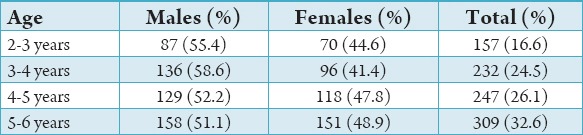
157 (16.6%) of them were 2-3 years
232 (24.5%) of them were 3-4 years
247 (26.1%) of them were 4-5 years
309 (32.6%) of them were 5-6 years.
About 82.1% of them have spaced dentition out of which 54% of males have spacing and 46% of females have spacing and 17.9% of them have non-spaced dentition out of which 53% of males and 47% of females of non-spaced dentition. The distribution of alignment was shown in Table 3.
Table 3.
Alignment distribution.

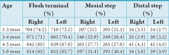
Of the 945 children examined, 75% had flush terminal plane, 4% distal step, and 21% mesial step molar relation. Percentage distribution among different age groups has shown flush terminal relation recorded highest among all the age groups followed by mesial step and distal step molar relation.
Among the age of 2-3 years, 75% flush terminal molar relation, 21% mesial step molar relation and 4% distal step molar relation. Among 3-4 years, 71% flush terminal molar relation, 25% mesial step molar relation, and 4% distal step molar relation. Among 4-5 years, 67% flush terminal molar relation, 28% mesial step molar relation, and 5% distal step molar relation.
Among 5-6 years, 65% flush terminal molar relation, 31% mesial step molar relation, and 4% distal step molar relation. No significant difference was noted between right and left side. Table 4 shows the percentage and correlation of the occlusal relationship of primary second molar for every age group.
Table 4.
Molar relation.
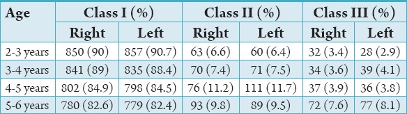
Of 945 children screened, 90% had Class I canine relation, 6% had Class II canine relation, and 4% had Class III canine relation. Percentage distribution among the different age groups has shown Class I relation recorded highest followed by Class II and Class III canine relation. Among 2-3 years, 90.7% of them have Class I canine relation, 6.4% of them have Class II, and 2.9% of them have Class III canine relation.
Among 3-4 years, 88.4% of them have Class I canine relation, 7.5% of them have Class II, and 4.1% of them have canine Class III relation. Among 4-5 years, 84.5% have Class I, 11.7% have Class II, and 4.8% of them have Class III canine relation. Among 5-6 years, 82.4% have Class I, 9.4% have Class II, and 8.1% have Class III canine relation. No significant difference was noted between right and left side. Table 5 shows the percentage and correlation of the occlusal relationship of primary second molar for every age group.
Table 5.
Canine relation.
Discussion
Early recognition of conditions predisposing young children to malocclusions is in the hands of primary care providers who for practical purposes are general practitioners and the pediatric dentist. It is important that conditions that predispose to develop a malocclusion of the permanent dentition be detected early in the primary dentition so the early interceptive procedure can be applied to prevent its further consequences.6
The presence of spacing or crowding and occlusion in the primary dentition and its relation to the development of malocclusion has been the long subject of discussion. There are many studies done regarding correlation of spacing or crowding with the malocclusion in pre-school children but still the knowledge of it is scares. The development of malocclusion starts from the primary dentition, so it is very important to know the occlusion in the primary dentition, as well as the changes of occlusal pattern, during the period of deciduous dentition. According to Bishara et al., a marked arch width (inter canine and intermolar width) expansion occurs significantly between 3 and 5 years of age in both maxilla and mandible. Therefore, the school children’s included in current study was limited to 6 years.7 Joshi and Makhija conducted study on 3-6 years school children in Gujarat and concluded that non-spaced dentition was less common in school children when compared to spaced dentition. An significant difference was noticed in which amount of spacing was found to be greater in males.8 Almost similar results were obtained in the present study. Im et al. concluded that maxilla showed more spacing when compared to mandible irrespective of sex. El-Nofely et al. assumption for spacing can be either due to small mesiodistal or wide inter canine width. Leighton’s hypothesis suggests that for the proper alignment of permanent teeth in mandibular arch, the amount of spacing in primary dentition should be of 6 mm or more.9
Traits of occlusal relationship in primary dentition can likely serve as a prognosticator for occlusal features in permanent dentition. The present study assessed for terminal molar and canine relation in 3-6 years school children. The study concluded that flush terminal molar relation recorded highest (74%) followed by mesial step (22%) and then distal step (4%) in all the age groups but with increase mesial step molar relation increased to 33% at the age of 6 years. Similar results were obtained by Baume showed that flush terminal plane (76%) is more commonly seen followed by mesial step (14%) and then distal step molar relation (10%). The study results shown highest bilateral flush terminal plane molar relation in children’s of Hyderabad (72.5%) and Chennai (74%) as concluded by Sriram et al.10 On contradictory, several other studies have shown that mesial step relation recorded highest followed by flush terminal plane and distal step. Bahadure et al. shown highest occurrence of mesial step molar relation (57.3%) followed by flush terminal plane (31.1%) and then the distal step molar relation (11.7%).3 Similar results were obtained by Ronald, Farsi, and Salama. The changes in the occlusion can be attributed to a combination of mesial migration of the lower arch and a mesial shift of the mandible, which is probably caused by growth.11 Considering canine relation, the most common relation found was Class I (90%) canine relation followed by Class II (6.4%) and Class III (3.6%) canine relation at the age of 2-3 years. At the age interval of 5-6 years Class I relation was found to be 82%, Class II relation was 9.8%, and Class III was 7.6%. Baidas et al. concluded that among the canine relation, Class I (90.1%) showed peak value succeeded by Class III (7.4%), and Class II (2.5%) relationship, which are similar to the present study. On the contradictory, Bahadure et al. concluded that among the canine relation, Class I (47.20%) showed highest value followed by Class II (42.83%) and Class III (9.97%). There were studies done on how spacing and occlusion is related to malocclusion and what were the possible reasons for the malocclusion. Howe et al. concluded that malocclusions were discripenancy in arch length and tooth size.12 The major reasons for the malocclusion may be due to the modern diet, which results in decreased inter-proximal wear and jaw growth. According to Profit et al. oral habits such as mouth breathing, thumb sucking might also be responsible for crowding in the arch.13
Conclusion
This study provides information of the prevalence of spacing and closed dentition, occlusion relation and its relation to a malocclusion. In the study, spaced dentition was more frequent the non-spaced dentition. Males have more percent of non-spaced dentition when compared to the females. The flush terminal plane molar relation was highest among all the age groups followed by mesial step and distal step molar relation with no significant difference was noted between right and left side among all the age groups. Hence, determining the malocclusion at its early age and early intervention procedure helps in preventing the problems at later stage.
Footnotes
Conflicts of Interest: None
Source of Support: Nil
References
- 1.Baidas L. Occlusion characteristics of the primary dentition by age in a sample of Saudi preschool children. Pak Oral Dent J. 2010;30(2):425–31. [Google Scholar]
- 2.Reddy BP, Rani MS, Santosh R, Shailaja AM. Incidence of malocclusion in deciduous dentition of Bangalore south population-India. Int J Contemp Dent. 2010;1(1):20–2. [Google Scholar]
- 3.Bahadure RN, Thosar N, Gaikwad R. Occlusal traits of deciduous dentition of preschool children of Indian children. Contemp Clin Dent. 2012;3(4):443–7. doi: 10.4103/0976-237X.107437. [DOI] [PMC free article] [PubMed] [Google Scholar]
- 4.Davies SJ, Gray RJ, Mackie IC. Good occlusal practice in children’s dentistry. Br Dent J. 2001;191(12):655–9. doi: 10.1038/sj.bdj.4801261a. [DOI] [PubMed] [Google Scholar]
- 5.Suma G, Das UM. Crowding, spacing and closed dentition and its relationship with malocclusion in primary dentition. Int J Clin Dent Sci. 2010;1(1):16–9. [Google Scholar]
- 6.Malandris M, Mahoney EK. Aetiology, diagnosis and treatment of posterior cross-bites in the primary dentition. Int J Paediatr Dent. 2004;14(3):155–66. doi: 10.1111/j.1365-263X.2004.00546.x. [DOI] [PubMed] [Google Scholar]
- 7.Bishara SE, Jakobsen JR, Treder J, Nowak A. Arch width changes from 6 weeks to 45 years of age. Am J Orthod Dentofacial Orthop. 1997;111(4):401–9. doi: 10.1016/s0889-5406(97)80022-4. [DOI] [PubMed] [Google Scholar]
- 8.Joshi MR, Makhija PG. Some observations on spacing in the normal deciduous dentition of 100 Indian children from Gujarat. Br J Orthod. 1984;11(2):75–9. doi: 10.1179/bjo.11.2.75. [DOI] [PubMed] [Google Scholar]
- 9.Vinay S, Keshav V, Sankalecha S. Prevalence of spaced and closed dentition and its relation to malocclusion in primary and permanent dentition. Int J Clin Pediatr Dent. 2012;5(2):98–100. doi: 10.5005/jp-journals-10005-1144. [DOI] [PMC free article] [PubMed] [Google Scholar]
- 10.Sriram CH, Priya VK, Sivakumar N, Reddy KR, Babu PJ, Reddy P. Occlusion of primary dentition in preschool children of Chennai and Hyderabad: A comparative study. Contemp Clin Dent. 2012;3(1):31–7. doi: 10.4103/0976-237X.94543. [DOI] [PMC free article] [PubMed] [Google Scholar]
- 11.Nanda RS, Khan I, Anand R. Age changes in the occlusal pattern of deciduous dentition. J Dent Res. 1973;52(2):221–4. doi: 10.1177/00220345730520020601. [DOI] [PubMed] [Google Scholar]
- 12.Howe RP, McNamara JA, Jr, O’Connor KA. An examination of dental crowding and its relationship to tooth size and arch dimension. Am J Orthod. 1983;83(5):363–73. doi: 10.1016/0002-9416(83)90320-2. [DOI] [PubMed] [Google Scholar]
- 13.Proffit WR, Fields HW, Ackerman JL, Sinclair PM, Thomas PM, Tulloch JF. 4th ed. St Louis: Mosby-Year Book, Early Stages of Development; 1993. Contemporary Orthodontics; pp. 72–106. [Google Scholar]