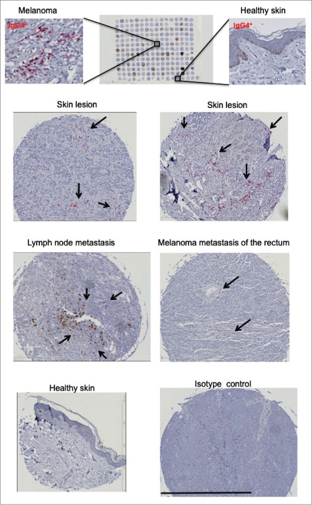
Figure 4.

IgG4+ cell infiltrates are detected in melanoma skin tumors. A tissue microarray (TMA; BioMax, n = 256) consisting of melanoma skin tumors (n = 108), melanoma lymph nodes metastases (n = 56), distant organ metastases (n = 60) and healthy skin (n = 32) specimens (top panel) was examined for the presence of IgG4 by immunohistochemistry. IgG4 positive infiltrates were detected by alkaline phosphatase (in red, selected areas shown by black arrows) and sections were counterstained in hematoxylin (in blue). Representative images of IgG4 immunohistochemical staining for IgG4+ infiltration revealed positive staining in skin lesions (top and second panels), lymph node and distant metastases (third panel), while staining was less frequent in healthy skin (bottom panel). Black bar represents 800 µm (third bottom right panel).
