Abstract
Background and aims
Ghrelin is a 28-amino-acid gut hormone that was first discovered as a potent growth hormone secretagogue. Recently, it has been shown to exert a strong anti-inflammatory effect. The purpose of the study reported here was to explore the effect and mechanism of ghrelin on concanavalin (Con) A-induced acute hepatitis.
Methods
Balb/C mice were divided into four groups: normal control (NC) (mice injected with vehicle [saline]); Con A (25 mg/kg); Con A + 10 μg/kg ghrelin; and Con A + 50 μg/kg ghrelin (1 hour before Con A injection). Pro-inflammatory cytokine levels were detected. Protein levels of phosphoinositide 3-kinase (PI3K); phosphorylated Akt (p-Akt); caspase 3, 8, and 9; and microtubule-associated protein 1 light chain 3 (LC3) were also detected. Perifosine (25 mM) (an Akt inhibitor) was used to investigate whether the protective effect of ghrelin was interrupted by an Akt inhibitor. Protein levels of p-AKT; Bcl-2; Bax; and caspase 3, 8, and 9 were also detected.
Results
Aspartate aminotransferase, alanine aminotransferase, and pathological damage were significantly ameliorated by ghrelin pretreatment in Con A-induced hepatitis. Inflammatory cytokines were significantly reduced by ghrelin pretreatment. Bcl-2; Bax; and caspase 3, 8, and 9 expression were also clearly affected by ghrelin pretreatment, compared with the Con A-treated group. However, the Akt kinase inhibitor reversed the decrease of Bax and caspase 3, 8, 9, and reduced the protein level of p-Akt and Bcl-2. Ghrelin activated the PI3K/Akt/Bcl-2 pathway and inhibited activation of autophagy.
Conclusion
Our results demonstrate that ghrelin attenuates Con A-induced acute immune hepatitis by activating the PI3K/Akt pathway and inhibiting the process of autophagy, which might be related to inhibition of inflammatory cytokine release, and prevention of hepatocyte apoptosis. These effects could be interrupted by an Akt kinase inhibitor.
Keywords: PI3K/Akt pathway, autophagy, inflammation, apoptosis
Introduction
Hepatitis is an increasingly serious human health problem worldwide, characterized by increased levels of aspartate aminotransferase (AST), alanine aminotransferase (ALT), and inflammatory cytokines, accompanied with hepatocyte apoptosis and necrosis. Hepatitis can be caused by various factors such as viruses, immune dysfunction, drugs, alcohol, lipids, genetics, and idiopathic factors. Although significant progress has been made in elucidating the mechanisms of hepatitis, effective therapeutic agents are still lacking.
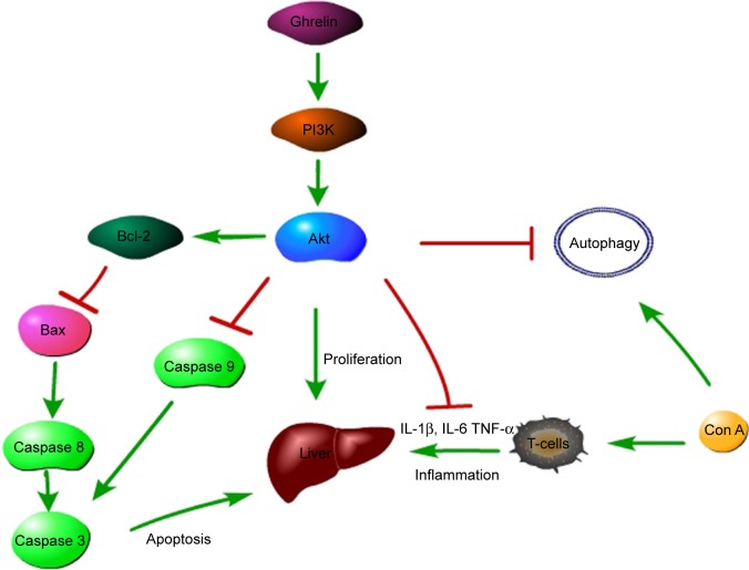
Concanavalin A (Con A) is a plant lectin and T-cell mitogen, which rapidly induces T-helper (Th) cell activation, mostly CD4+ T-cells, leading to inflammatory liver injury in mice.1–3 Natural killer (NK) T-cells and macrophages are also considered to play key roles in the development of liver damage induced by Con A.4 Pro-inflammatory cytokines – including interleukin (IL)-1, IL-2, IL-6, tumor necrosis factor (TNF)-α, and interferon-γ – are significantly increased in Con A-induced liver injury (Figure 1).5–7 Therefore, Con A-induced liver injury is an ideal model of T-cell-induced hepatitis, especially immune hepatitis.
Figure 1.
Mechanism of the protective effect of ghrelin on Con A-induced hepatitis.
Notes: Ghrelin activates PI3K and promotes the phosphorylation of its downstream Akt, leading to the proliferation of injured hepatocytes and the inhibition of pro-inflammatory cytokines such as IL-1β, IL-6, and TNF-α. Phosphorylated Akt promotes the expression of Bcl-2, which inhibits Bax and downregulates caspase 3, 8, and 9. PI3K/Akt may lead to the inhibition of the process of autophagy, which is activated by Con A treatment in uncertain mechanisms.
Abbreviations: Con, concanavalin; IL, interleukin; PI3K, phosphoinositide 3-kinase; TNF, tumor necrosis factor.
Several signaling pathways are involved in the mechanism of Con A-induced acute hepatitis. Recent studies demonstrated that activating phosphorylated Akt (p-Akt) significantly attenuated Con A-induced liver injury.8–11 Apoptosis, known as type I programmed cell death, is also involved in Con A-induced hepatitis.9 Members of the Bcl-2 family of proteins have an essential role in the apoptotic pathway: Bcl-2 and Bcl-xl as anti-apoptotic proteins, and Bax and Bad as pro-apoptotic proteins.12 Modulation of the balance between Bax and Bcl-2 proteins determines the survival status of cells challenged with various stimuli.
Autophagy is an intracellular process that targets defective organelles, surrounding them with autophagosomes, and delivering them to the lysosomes for degradation. Autophagy, usually activated by energy restriction during inflammation, plays crucial roles in development, differentiation, survival, and homeostasis, and has attracted considerable concern due to its implications in human disease.13 However, autophagy is characterized as type II programmed cell death, and can also cause cell death, therefore, we regard it as a double-edged sword.14 Microtubule-associated protein 1 light chain 3 (LC3), is widely used as a marker for monitoring autophagy.13
Ghrelin, a newly discovered 28-amino-acid gastric peptide hormone first purified from stomach of rats, is the endogenous ligand for growth hormone secretagogue receptor 1a.15 Originally regarded as a growth hormone, ghrelin has recently been shown to have various physiological effects, including on energy metabolism, glucose homeostasis, and food intake.16 Ghrelin is involved in the regulation of inflammation and shows strong anti-inflammatory effects.
The study reported here investigated whether ghrelin exerted any effect on Con A-induced immune hepatitis and its mechanism of action. We report that ghrelin significantly reduced liver injury, as measured by plasma hepatic enzymes, inflammatory cytokines, and histological changes, such as inflammatory cell infiltration and hepatocyte apoptosis and necrosis, which may be partly associated with PI3K/Akt/Bcl-2 pathway activation and autophagy inhibition.
Materials and methods
Reagents
Ghrelin was purchased from ProSpec (Ness-Ziona, Israel). Con A was purchased from Sigma-Aldrich (St Louis, MO, USA). Perifosine was provided by Keryx Biopharmaceuticals (New York, NY, USA). Antibodies against IL-1β, IL-6, TNF-α, total Akt, and p-Akt were purchased from Santa Cruz Biotechnology (Santa Cruz, CA, USA). LC3, Beclin 1, Bcl-2, and Bax were purchased from Cell Signaling Technology (Danvers, MA, USA). Antibodies against caspase 3, caspase 8, and caspase 9 were purchased from Proteintech (Chicago, IL, USA).
Animals and treatment
Balb/c mice (8 weeks old, 23±2 g) were purchased from Shanghai Laboratory Animal Co Ltd (Shanghai, People’s Republic of China). The mice were housed in a clean room maintained at 24°C±2°C with an alternating 12-hour light and dark cycle, with free access to food and water. The study was approved by the Animal Care and Use Committee and the Ethics Committee of Fudan University. Mice were randomly divided into four groups, with seven animals per group: (1) normal control (NC): mice injected with vehicle (saline), (2) Con A, (3) Con A + 10 μg/kg ghrelin, and (4) Con A + 50 μg/kg ghrelin. Ghrelin was diluted with normal saline and injected intraperitoneally 1 hour before injection of Con A. Con A was dissolved in normal saline solution at a concentration of 5 mg/mL and injected via the tail vein at a dose of 25 mg/kg to induce acute hepatic injury. Each group of mice was sacrificed randomly at 4, 8, and 24 hours.
To investigate whether the protective effect could be interrupted by an Akt kinase inhibitor, we created another four groups of mice: NC, Con A, Con A + 10 μg/kg ghrelin, and Con A + 10 μg/kg ghrelin + 25 mM perifosine. The choice and dose of Akt inhibitor was determined based on a high-quality study.17 Mice were killed at 8 hours. This was because the preliminary experiment showed that Con A-induced hepatitis was most serious at 8 hours, and ghrelin exerted its best therapeutic effect at that time. Liver samples and blood were acquired for histological and molecular analysis.
Biochemical assays
Serum levels of ALT and AST were measured with microplate test kits (Nanjing Jiancheng Bioengineering Institute, Nanjing, People’s Republic of China). Serum pro-inflammatory cytokines TNF-α and IL-6 were measured by enzyme-linked immunosorbent assay (R&D Systems, Minneapolis, MN, USA).
Histopathology
The middle portion of the left lobe of the liver was excised and sectioned and then perfused in 4% paraformaldehyde for at least 24 hours. After fixation, the tissues were embedded in paraffin, and 5 μm thick sections were stained with hematoxylin and eosin (H&E) to observe the tissue damage by light microscopy.
Immunohistochemistry
Liver sections (3 μm) were dewaxed, rehydrated regularly, and treated with 3% H2O2. The sections were pretreated with a microwave antigen-retrieval technique. The nonspecific sites were blocked with 10% goat serum for 30 minutes at room temperature. The liver slices were incubated overnight with antibody directed against p-Akt (1:500), IL-1β (1:500), IL-6 (1:500), TNF-α (1:500), LC3II (1:500), Bcl-2 (1:500), and Bax (1:500). On the second day after incubation with a secondary antibody, an antibody binding assay was performed using a diaminobenzidine kit. At last, the slides were counterstained with hematoxylin and observed under a light microscope. Three different fields of vision were randomly selected in one slide, and the ratios of brown staining areas and total areas were acquired with Image-Pro® Plus (v 6.0) software. We calculated the average of these three ratios. The described method was applied in all groups.
Western blotting
Total protein was extracted according to standard procedures. Equivalent amounts of total protein were separated by sodium dodecyl sulfate polyacrylamide gel electrophoresis and transferred to polyvinylidene difluoride membranes. Nonspecific binding was blocked with 5% nonfat milk (dissolved in phosphate-buffered saline) for 1 hour and then incubated overnight at 4°C with different primary antibodies. After incubation with peroxidase-conjugated secondary antibodies for 1 hour at 37°C, membranes were developed with the Odyssey two-color infrared laser imaging system (fluorescence detection).
Terminal deoxynucleotidyl transferase dUTP nick end labeling (TUNEL) assay
TUNEL assay was performed according to the manufacturer’s instructions (Roche, Mannheim, Germany). Liver sections were treated with 20 μg/mL proteinase K at room temperature for 15 minutes, followed by incubation with the TUNEL reaction mixture at 37°C in a humidified atmosphere for 1 hour. Three different fields of vision were randomly selected in one slide, the brown nucleus cells, regarded as positive cells, were visualized and counted, and the percentage of apoptotic cells to total cell numbers was calculated. We calculated the average of these three ratios.
RNA isolation and real-time quantitative reverse transcriptase polymerase chain reaction (qRT-PCR)
Liver tissue was analyzed with real-time qRT-PCR. Total RNA was extracted from frozen liver tissues or cells with TRIzol® reagent (Tiangen Biotech [Beijing] Co, Ltd, Beijing, People’s Republic of China). SYBR Green qRT-PCR was performed with an Applied Biosystems® 7900HT Fast Real-Time PCR [polymerase chain reaction] system (Thermo Fisher Scientific, Waltham, MA, USA) to determine expression of the target genes, according to the instructions for SYBR Premix EX Taq (TaKaRa Biotechnology [Dalian] Co, Ltd, Dalian, People’s Republic of China). The primers used in the PCR reactions are listed in Table 1.
Table 1.
Nucleotide sequences of primers used for PCR
| Gene | Primer sequence (5′-3′) | |
|---|---|---|
| IL-1β | Forward | ATAAGCCCACTCTACACCT |
| Reverse | ATTGGCCCTGAAAGGAGAGA | |
| IL-6 | Reverse | CTGCAAGAGACTTCCATCCAG |
| Forward | AGTGGTATAGACAGGTCTGTTGG | |
| TNF-α | Reverse | CAGGCGGTGCCTATGTCTC |
| Forward | CGATCACCCCGAAGTTCAGTAG | |
| Bcl-2 | Reverse | GCTACCGTCGTGACTTCGC |
| Forward | CCCCACCGAACTCAAAGAAGG | |
| Bax | Reverse | AGACAGGGGCCTTTTTGCTAC |
| Forward | AATTCGCCGGAGACACTCG | |
| β-actin | Reverse | GGCTGTATTCCCCTCCATCG |
| Forward | CCAGTTGGTAACAATGCCATGT |
Abbreviation: PCR, polymerase chain reaction.
Statistical analysis
All results are expressed as means ± standard deviation. Comparisons between two groups were made with Student’s t-test. Statistical differences in multiple groups were determined by multiple comparisons with analysis of variance, followed by Tukey’s post-hoc test. All statistical analyses were performed with Graphpad Prism (v 6.0) software. P-values <0.05 were considered statistically significant.
Results
Ghrelin pretreatment ameliorates Con A-induced hepatitis
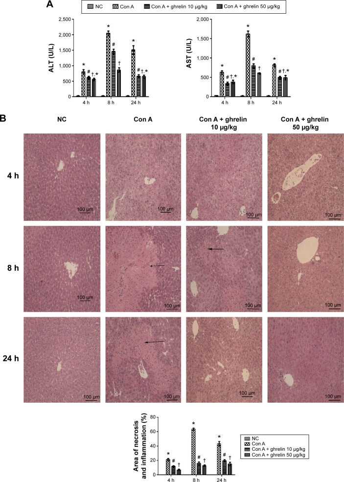
Con A significantly increased serum ALT and AST levels, with a peak at 8 hours after Con A injection, whereas both 10 and 50 μg/kg doses of ghrelin clearly reduced them, as shown in Figure 2A. Histopathological changes in the three groups were examined after H&E staining (Figure 2B). Massive areas of hepatic tissue inflammation and necrosis were observed in the mice treated with Con A alone, which was significantly ameliorated in the ghrelin and Con A co-treated group. Image-Pro Plus software showed that there were significant differences of hepatic injury between the Con A-treated and Con A and ghrelin co-treated groups.
Figure 2.
Ghrelin pretreatment ameliorates Con A-induced hepatitis.
Notes: (A) Serum ALT and AST levels are expressed as the mean ± SD of six animals per group at 4, 8, and 24 h after Con A injection in mice and the effects of both low (10 μg/kg) and high (50 μg/kg) dose ghrelin pretreatment at the same time. (B) Hematoxylin and eosin staining of liver sections. The cellular necrotic or edematous areas were analyzed with Image-Pro® Plus (v 6.0); (n=6). *P<0.05 for NC vs Con A, #P<0.05 for Con A vs Con A + 10 μg/kg ghrelin, †P<0.05 for Con A + 50 μg/kg ghrelin vs Con A + 10 μg/kg ghrelin, and †,*P>0.05 for Con A + 50 μg/kg ghrelin vs Con A + 10 μg/kg ghrelin.
Abbreviations: ALT, alanine aminotransferase; AST, aspartate aminotransferase; Con, concanavalin; h, hours; NC, normal control.
Ghrelin pretreatment inhibits release of inflammatory cytokines during Con A-induced hepatitis
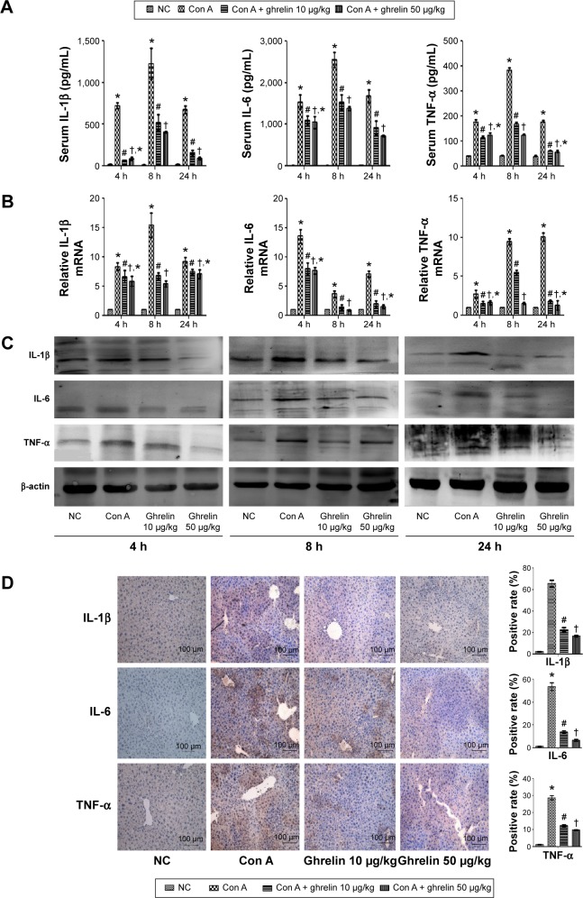
Hepatitis is associated with changes in the levels of inflammatory cytokines. Serum levels of IL-1β, IL-6, and TNF-α were significantly increased in the Con A-treated group and peaked at 8 hours after Con A injection. As expected, pretreatment with both doses of ghrelin significantly lowered the Con A-induced increase of serum levels of IL-1β, IL-6, and TNF-α, especially at 8 hours (Figure 3A). To confirm our observations, mRNA transcription and protein expression of the cytokines were also reduced by ghrelin pretreatment (Figures 3B and C). In addition, immunohistochemical staining was used to detect the expression of the inflammatory cytokines in liver tissues in the control, Con A, Con A + 10 μg/kg ghrelin, and Con A + 50 μg/kg ghrelin groups (Figure 3D). These results showed that ghrelin pretreatment reduced inflammatory cytokines in Con A-induced hepatitis.
Figure 3.
Ghrelin pretreatment inhibits release of IL-1β, IL-6, and TNF-α in Con A-induced hepatitis.
Notes: (A) The serum IL-1β, IL-6, and TNF-α levels are expressed as mean ± SD of six animals per group at 4, 8, and 24 h after Con A injection in mice and the effects of both low (10 μg/kg) and high (50 μg/kg) dose ghrelin pretreatment at the same time. (B) mRNA expression of IL-1β, IL-6, and TNF-α in the NC, Con A, Con A + 10 μg/kg ghrelin, and Con A + 50 μg/kg ghrelin groups was evaluated by real-time PCR. (C) Western blots showing the expression of IL-1β, IL-6, and TNF-α in liver tissues. (D) Immunohistochemistry staining (200×) showing the expression of IL-1β, IL-6, and TNF-α in liver tissue at 8 h. The ratio of brown area to total area was analyzed with Image-Pro® Plus (v 6.0); (n=6). *P<0.05 for NC vs Con A, #P<0.05 for Con A vs Con A + 10 μg/kg ghrelin, †P<0.05 for Con A + 50 μg/kg ghrelin vs Con A + 10 μg/kg ghrelin, and †,*P>0.05 for Con A + 50 μg/kg ghrelin vs Con A + 10 μg/kg ghrelin.
Abbreviations: Con, concanavalin; h, hours; IL, interleukin; NC, normal control; TNF, tumor necrosis factor; PCR, polymerase chain reaction.
Ghrelin attenuates hepatocyte apoptosis in Con A-induced hepatitis
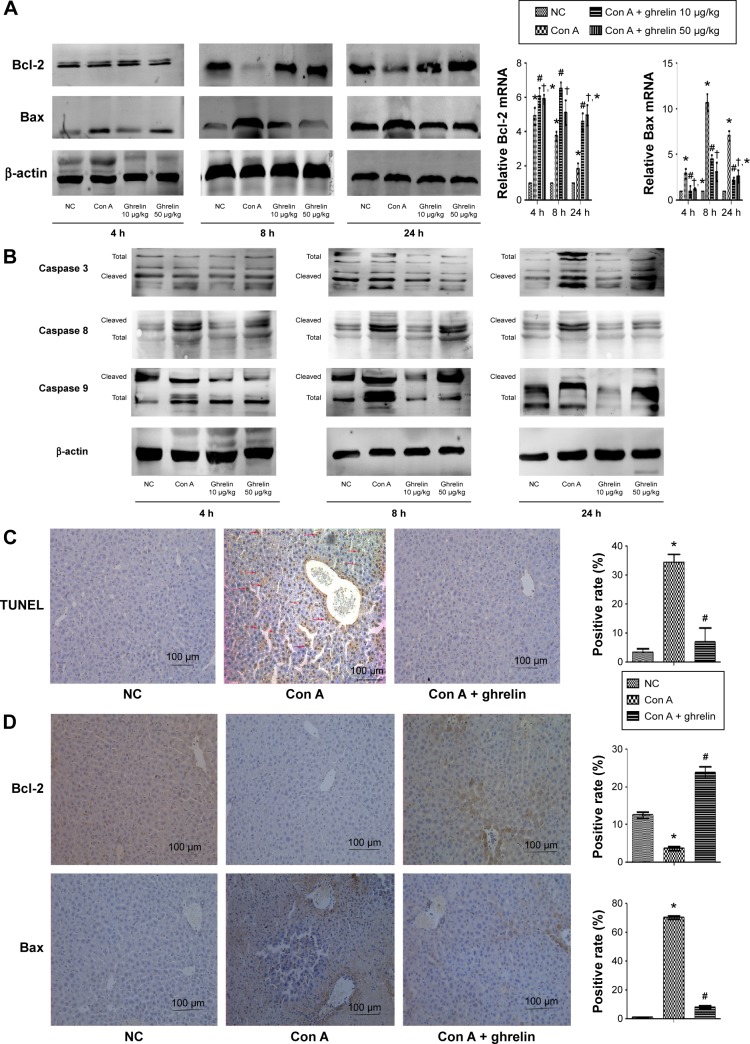
We investigated expression of caspase 3, 8, and 9. All three enzymes were activated by Con A injection and reduced by ghrelin treatment (Figure 4B). Likewise, as expected, TUNEL assay showed clear improvement of apoptosis in ghrelin-pretreated compared with Con A-treated mice (Figure 4C). Furthermore, ghrelin pretreatment significantly increased mRNA and protein expression of Bcl-2 and reduced expression of Bax at all three time points, especially at 8 hours (Figure 4A). In addition, the location and expression of Bcl-2 by immunohistochemical staining was reduced in the Con A group compared with in the normal group at 8 hours, while 10 μg/kg ghrelin significantly increased it. Immunohistochemical staining of Bax expression was stronger in the Con A-treated group, which was clearly reduced by 10 μg/kg ghrelin pretreatment at 8 hours (Figure 4D).
Figure 4.
Ghrelin attenuates hepatocyte apoptosis in Con A-induced hepatitis.
Notes: (A) Western blots and real-time PCR evaluation of expression of Bcl-2 and Bax in liver tissues. (B) Western blotting of expression of caspase 3, 8, and 9 in liver tissues. (C) TUNEL staining showed apoptotic cells in three groups at 8 h (200×). (D) Immunohistochemistry staining (200×) showed the expression of Bcl-2 and Bax protein in liver tissue at 8 h. *P<0.05 for NC vs Con A, #P<0.05 for Con A vs Con A + 10 μg/kg ghrelin, and †P<0.05 for Con A + 50 μg/kg ghrelin vs Con A + 10 μg/kg ghrelin.
Abbreviations: Con, concanavalin; h, hours; NC, normal control; PCR, polymerase chain reaction.
Ghrelin activates PI3K/Akt signaling in Con A-induced hepatitis
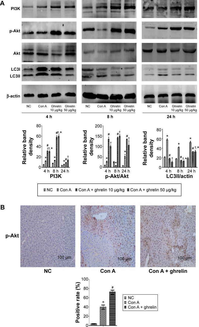
The protein level of PI3K was slightly elevated in the Con A-treated group but clearly increased in the ghrelin-pretreated group at three time points. Moreover, Con A impaired the expression of p-Akt at 8 hours and slightly increased the expression of p-Akt at 4 hours and 24 hours compared with in the normal group. Both doses of ghrelin significantly increased the expression of p-Akt (Figure 5A). Expression of p-Akt in the liver tissues of mice in the control, Con A, and Con A + 10 μg/kg ghrelin groups at 24 hours was detected using immunohistochemistry (Figure 5B).
Figure 5.
Ghrelin activates PI3K/Akt signaling and inhibits autophagy in Con A-induced hepatitis.
Notes: (A) Western blotting and quantitative evaluation of PI3K, p-Akt, and LC3 expression in liver tissues. (B) Immunohistochemistry staining (200×) showed the expression of p-Akt protein in liver tissue at 24 h. *P<0.05 for NC vs Con A, #P<0.05 for Con A vs Con A + 10 μg/kg ghrelin, †P<0.05 for Con A + 50 μg/kg ghrelin vs Con A + 10 μg/kg ghrelin, and †,*P>0.05 for Con A + 50 μg/kg ghrelin vs Con A + 10 μg/kg ghrelin.
Abbreviations: Con, concanavalin; h, hours; LC3, microtubule-associated protein 1 light chain 3; NC, normal control; p-Akt, phosphorylated Akt; PI3K, phosphoinositide 3-kinase.
We conclude that ghrelin treatment can ameliorate Con A- induced hepatitis at least in part through the PI3K/Akt pathway.
The protective effect of ghrelin could be interrupted by an Akt kinase inhibitor
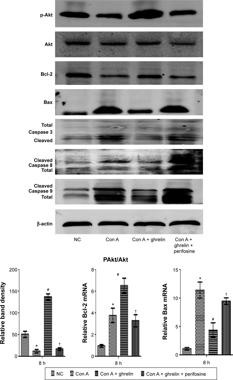
The protein levels of p-Akt; Bcl2; Bax; and caspase 3, 8, and 9 were detected. Perifosine, an Akt kinase inhibitor, reversed the decrease of Bax and caspase 3, 8, and 9, and reduced the protein level of p-Akt and Bcl2 in treated mice compared with in mice treated with Con A + ghrelin only (Figure 6).
Figure 6.
The protective effect of ghrelin could be interrupted by Akt kinase inhibitor.
Notes: Western blotting of expression of p-Akt; Bcl-2; Bax; and caspase 3, 8, and 9 in liver tissues. Real-time PCR evaluation of expression of Bcl-2 and Bax, and density quantification of p-Akt. *P<0.05 for NC vs Con A, #P<0.05 for Con A vs Con A + 10 μg/kg ghrelin, and †P<0.05 for Con A + 10 μg/kg ghrelin + perifosine vs Con A + 10 μg/kg ghrelin.
Abbreviations: Con, concanavalin; p-Akt, phosphorylated Akt; NC, normal control; PCR, polymerase chain reaction.
This result supports our speculation that ghrelin exerts its protective effect at least partly via PI3K/Akt/Bcl2 pathway.
Ghrelin attenuates autophagy in Con A- induced hepatitis
LC3 is an important marker of autophagy. We assessed the activation of autophagy in each group of mice. The protein level of LC3 was increased by Con A treatment but reduced by ghrelin, indicating that autophagy was activated by Con A but inhibited by ghrelin (Figure 5A).
Discussion
Hepatitis can be caused by virus infection, drugs, alcohol, fatty acids, and autoimmunity, and remains a global health problem for which effective drug therapies are still lacking. Hepatitis is critical in many liver diseases that progress to end-stage liver disease, even hepatocellular carcinoma.18 Therefore, more effective drugs are urgently needed for intervention at the hepatitis stage. Con A-induced hepatitis is a well-established experimental model of immune hepatitis,11 cell apoptosis, and necrosis, and possibly ultimately liver failure.
As we expected, our results show that ghrelin significantly reduced Con A-induced hepatocellular inflammation and apoptosis. Pretreatment with both doses of ghrelin markedly reduced Con A-induced elevation of serum ALT and AST levels, as well as levels of the pro-inflammatory cytokines IL-1β, IL-6, and TNF-α. Furthermore, qRT-PCR, Western blotting, and immunohistochemistry results demonstrate that ghrelin pretreatment significantly reduced expression of pro-inflammatory cytokines, especially at 8 hours (Figure 3). Hepatocyte inflammation and necrosis were also visualized by H&E staining in in the control, Con A, Con A + 10 μg/kg ghrelin, and Con A + 50 μg/kg ghrelin groups, which were quantified by calculating the area of hepatic inflammation and necrosis with Image-Pro Plus software (Figure 2). We found a significant difference between the ghrelin and Con A co-treated group and the Con A-treated group. To investigate whether ghrelin shows anti-apoptotic activity in hepatitis, we used the TUNEL essay and observed that the apoptotic nuclei were significantly reduced in the ghrelin pretreatment groups compared with in the Con A group. Likewise, the expression of apoptotic protein markers of caspase 3, 8, and 9 were increased in the Con A-treated group but attenuated by ghrelin administration in the ghrelin pretreatment groups. Therefore, we confirmed that ghrelin exhibits strong anti-inflammatory activity in Con A-induced immune hepatitis.
However, the mechanism of the anti-inflammatory and anti-apoptotic effects of ghrelin is not clear and requires further investigation. The PI3K/Akt pathway is a well-known cellular survival pathway that plays important roles in the regulation of cell proliferation and apoptosis, as well as inflammatory injury.19,20 Furthermore, PI3K/Akt exhibits a strong effect on apoptosis via promoting expression of Bcl2, an essential anti-apoptotic protein, and phosphorylating the pro-apoptotic protein Bad.21 A recent study showed that hydrogen sulfide attenuates Con A-induced hepatitis by activating the PI3K/Akt signaling pathway and promoting expression of Bcl2 and inhibiting Bax.9 As a newly discovered gut hormone with pleiotropic effects, ghrelin exhibits novel protective effects, such as anti-inflammatory, anti-apoptotic, and anti-fibrotic effects in the liver and other organs,22–25 in addition to increased food intake and maintenance of energy homeostasis.26 Some of the cytoprotective effects have been demonstrated via PI3K/Akt pathway activation.23,27,28 Therefore, we speculate that the PI3K/Akt pathway is associated with the anti-inflammatory and anti-apoptotic mechanism of ghrelin in Con A-induced hepatitis.
In our study, we found that Con A enhanced expression of PI3K and p-Akt which were further increased by ghrelin treatment. To investigate further the anti-apoptotic mechanism of ghrelin, we detected the expression of Bcl-2 and Bax. Con A increased Bax and decreased Bcl-2, resulting in cell death. However, ghrelin pretreatment significantly increased the expression Bcl-2, reduced the expression of Bax, and maintained the balance of Bcl-2 and Bax disrupted by Con A treatment (Figures 4A and D). The anti-apoptotic protein Bcl-2 inhibits apoptosis by preventing the release of cytochrome c,29 while Bax promotes mitochondrial membrane damage and cytochrome c release,30 leading to the activation of caspase 9 and 3 and cellular apoptosis (Figure 1). We supposed that ghrelin increases expression of Bcl-2 via the PI3K/Akt pathway, which may become a novel anti-inflammatory and anti-apoptotic agent for the treatment of liver injury. Moreover, we showed that the protective effect of ghrelin on hepatitis could be partly interrupted by an Akt kinase inhibitor, which supports our conclusion that ghrelin exerts its protective effect at least partly via the PI3K/Akt/Bcl-2 pathway (Figure 6). We observed a significant difference in effect between 10 and 50 μg/kg ghrelin-treated groups with reference to some but not all indicators and time points. We speculate that this effect is dose dependent, but the concentration gradient was not large enough; a greater concentration gradient is needed to investigate its dose-dependent effect.
The role of autophagy in hepatitis is worthy of further exploration. Autophagic cell death is involved in Con A-induced hepatitis.13 Several studies have reported that autophagy was activated in Con A-induced hepatitis, while autophagy inhibition was accompanied with hepatic injury attenuation.31,32 Therefore, we investigated whether ghrelin affected autophagy during Con A-induced hepatitis and whether it was mediated by the PI3K/Akt pathway. We showed that LC3II was inhibited in hepatocytes. This indicates that the PI3K/Akt pathway may be involved in the autophagic processes of Con A-induced hepatitis33 (Figure 1).
Conclusion
Our research demonstrates that ghrelin attenuated Con A-induced acute immune hepatitis, at least partly by activating the PI3K/Akt/Bcl-2 pathway and inhibiting autophagy. This is believed to be the first study to demonstrate that ghrelin attenuates Con A-induced liver injury and shows potential as a new therapeutic strategy for liver disease. However, the mechanism underlying ghrelin’s amelioration of liver injury is not clear and more research is needed.
Acknowledgments
This work was supported by a research grant for Key Clinical Discipline Construction of Shanghai Municipality, People’s Republic of China (number ZK2012B20).
Footnotes
Disclosure
The authors declare no conflicts of interest in this work.
References
- 1.Tiegs G, Hentschel J, Wendel A. A T cell-dependent experimental liver injury in mice inducible by concanavalin A. J Clin Invest. 1992;90(1):196–203. doi: 10.1172/JCI115836. [DOI] [PMC free article] [PubMed] [Google Scholar]
- 2.Carambia A, Herkel J. CD4 T cells in hepatic immune tolerance. J Autoimmun. 2010;34(1):23–28. doi: 10.1016/j.jaut.2009.08.006. [DOI] [PubMed] [Google Scholar]
- 3.Schümann J, Wolf D, Pahl A, et al. Importance of Kupffer cells for T-cell-dependent liver injury in mice. Am J Pathol. 2000;157(5):1671–1683. doi: 10.1016/S0002-9440(10)64804-3. [DOI] [PMC free article] [PubMed] [Google Scholar]
- 4.Kaneko Y, Harada M, Kawano T, et al. Augmentation of Valpha14 NKT cell-mediated cytotoxicity by interleukin 4 in an autocrine mechanism resulting in the development of concanavalin A-induced hepatitis. J Exp Med. 2000;191(1):105–114. doi: 10.1084/jem.191.1.105. [DOI] [PMC free article] [PubMed] [Google Scholar]
- 5.Trautwein C, Rakemann T, Brenner DA, et al. Concanavalin A-induced liver cell damage: activation of intracellular pathways triggered by tumor necrosis factor in mice. Gastroenterology. 1998;114(5):1035–1045. doi: 10.1016/s0016-5085(98)70324-5. [DOI] [PubMed] [Google Scholar]
- 6.Küsters S, Gantner F, Künstle G, Tiegs G. Interferon gamma plays a critical role in T cell-dependent liver injury in mice initiated by concanavalin A. Gastroenterology. 1996;111(2):462–471. doi: 10.1053/gast.1996.v111.pm8690213. [DOI] [PubMed] [Google Scholar]
- 7.Zhang X, Wei HX, Rui S, Wei H, Tian Z. Opposite effects of high and low doses of interleukin-2 on T cell-mediated hepatitis in mice (interleukin-2 on hepatitis) Hepatol Int. 2010;4(3):641–648. doi: 10.1007/s12072-010-9196-0. [DOI] [PMC free article] [PubMed] [Google Scholar]
- 8.Wang H, Hu B, Zou Y, et al. Dexmedetomidine premedication attenuates concanavalin A-induced hepatitis in mice. J Toxicol Sci. 2014;39(5):755–764. doi: 10.2131/jts.39.755. [DOI] [PubMed] [Google Scholar]
- 9.Cheng P, Chen K, Xia Y, et al. Hydrogen sulfide, a potential novel drug, attenuates concanavalin A-induced hepatitis. Drug Des Devel Ther. 2014;8:1277–1286. doi: 10.2147/DDDT.S66573. [DOI] [PMC free article] [PubMed] [Google Scholar]
- 10.Nakano T, Goto S, Lai CY, et al. Immunological aspects and therapeutic significance of an autoantibody against histone H1 in a rat model of concanavalin A-induced hepatitis. Immunology. 2010;129(4):547–555. doi: 10.1111/j.1365-2567.2009.03149.x. [DOI] [PMC free article] [PubMed] [Google Scholar]
- 11.Shao X, Qian Y, Xu C, et al. The protective effect of intrasplenic transplantation of Ad-IL-18BP/IL-4 gene-modified fetal hepatocytes on Con A-induced hepatitis in mice. PLoS One. 2013;8(3):e58836. doi: 10.1371/journal.pone.0058836. [DOI] [PMC free article] [PubMed] [Google Scholar]
- 12.Anilkumar U, Prehn JH. Anti-apoptotic BCL-2 family proteins in acute neural injury. Front Cell Neurosci. 2014;8:281. doi: 10.3389/fncel.2014.00281. [DOI] [PMC free article] [PubMed] [Google Scholar]
- 13.Yin XM, Ding WX, Gao W. Autophagy in the liver. Hepatology. 2008;47(5):1773–1785. doi: 10.1002/hep.22146. [DOI] [PubMed] [Google Scholar]
- 14.Shintani T, Klionsky DJ. Autophagy in health and disease: a double-edged sword. Science. 2004;306(5698):990–995. doi: 10.1126/science.1099993. [DOI] [PMC free article] [PubMed] [Google Scholar]
- 15.Kojima M, Hosoda H, Date Y, Nakazato M, Matsuo H, Kangawa K. Ghrelin is a growth-hormone-releasing acylated peptide from stomach. Nature. 1999;402(6762):656–660. doi: 10.1038/45230. [DOI] [PubMed] [Google Scholar]
- 16.Tacke F, Brabant G, Kruck E, et al. Ghrelin in chronic liver disease. J Hepatol. 2003;38(4):447–454. doi: 10.1016/s0168-8278(02)00438-5. [DOI] [PubMed] [Google Scholar]
- 17.Momota H, Nerio E, Holland EC. Perifosine inhibits multiple signaling pathways in glial progenitors and cooperates with temozolomide to arrest cell proliferation in gliomas in vivo. Cancer Res. 2005;65(16):7429–7435. doi: 10.1158/0008-5472.CAN-05-1042. [DOI] [PubMed] [Google Scholar]
- 18.Yang SW, Kim GH, Chung JW, et al. Prediction of risk for hepatocellular carcinoma by response of serum α-fetoprotein to entecavir therapy. J Gastroenterol Hepatol. 2015;30(7):1175–1182. doi: 10.1111/jgh.12921. [DOI] [PubMed] [Google Scholar]
- 19.Maddika S, Ande SR, Panigrahi S, et al. Cell survival, cell death and cell cycle pathways are interconnected: implications for cancer therapy. Drug Resist Updat. 2007;10(1–2):13–29. doi: 10.1016/j.drup.2007.01.003. [DOI] [PubMed] [Google Scholar]
- 20.Yang FJ, He YH, Zhou JH. Fenofibrate pre-treatment suppressed inflammation by activating phosphoinositide 3 kinase/protein kinase B (PI3K/Akt) signaling in renal ischemia-reperfusion injury. J Huazhong Univ Sci Technolog Med Sci. 2015;35(1):58–63. doi: 10.1007/s11596-015-1389-2. [DOI] [PubMed] [Google Scholar]
- 21.Drukker L, Margulis A, Chaouat M, Levitzki R, Maiorenko E, Ben Bassat H. Changes of PI3K/AKT/BCL2 signaling proteins in congenital Giant Nevi: melanocytes contribute to their increased survival and integrity. J Recept Signal Transduct Res. 2013;33(6):359–366. doi: 10.3109/10799893.2013.838785. [DOI] [PubMed] [Google Scholar]
- 22.Li Y, Hai J, Li L, et al. Administration of ghrelin improves inflammation, oxidative stress, and apoptosis during and after non-alcoholic fatty liver disease development. Endocrine. 2013;43(2):376–386. doi: 10.1007/s12020-012-9761-5. [DOI] [PubMed] [Google Scholar]
- 23.Hao XK, Wu W, Wang CX, et al. Ghrelin alleviates early brain injury after subarachnoid hemorrhage via the PI3K/Akt signaling pathway. Brain Res. 2014;1587:15–22. doi: 10.1016/j.brainres.2014.08.069. [DOI] [PubMed] [Google Scholar]
- 24.Moreno M, Chaves JF, Sancho-Bru P, et al. Ghrelin attenuates hepatocellular injury and liver fibrogenesis in rodents and influences fibrosis progression in humans. Hepatology. 2010;51(3):974–985. doi: 10.1002/hep.23421. [DOI] [PubMed] [Google Scholar]
- 25.Zhang D, Wang W, Zhou D, et al. Ghrelin inhibits apoptosis induced by palmitate in rat aortic endothelial cells. Med Sci Monit. 2010;16(12):BR396–BR403. [PubMed] [Google Scholar]
- 26.Muccioli G, Tschöp M, Papotti M, Deghenghi R, Heiman M, Ghigo E. Neuroendocrine and peripheral activities of ghrelin: implications in metabolism and obesity. Eur J Pharmacol. 2002;440(2–3):235–254. doi: 10.1016/s0014-2999(02)01432-2. [DOI] [PubMed] [Google Scholar]
- 27.Rossi F, Castelli A, Bianco MJ, Bertone C, Brama M, Santiemma V. Ghrelin induces proliferation in human aortic endothelial cells via ERK1/2 and PI3K/Akt activation. Peptides. 2008;29(11):2046–2051. doi: 10.1016/j.peptides.2008.07.002. [DOI] [PubMed] [Google Scholar]
- 28.Liang QH, Liu Y, Wu SS, Cui RR, Yuan LQ, Liao EY. Ghrelin inhibits the apoptosis of MC3T3-E1 cells through ERK and AKT signaling pathway. Toxicol Appl Pharmacol. 2013;272(3):591–597. doi: 10.1016/j.taap.2013.07.018. [DOI] [PubMed] [Google Scholar]
- 29.Zamzami N, Susin SA, Marchetti P, et al. Mitochondrial control of nuclear apoptosis. J Exp Med. 1996;183(4):1533–1544. doi: 10.1084/jem.183.4.1533. [DOI] [PMC free article] [PubMed] [Google Scholar]
- 30.Narita M, Shimizu S, Ito T, et al. Bax interacts with the permeability transition pore to induce permeability transition and cytochrome c release in isolated mitochondria. Proc Natl Acad Sci U S A. 1998;95(25):14681–14686. doi: 10.1073/pnas.95.25.14681. [DOI] [PMC free article] [PubMed] [Google Scholar]
- 31.Cheng P, Chen K, Xia Y, et al. Hydrogen sulfide, a potential novel drug, attenuates concanavalin A-induced hepatitis. Drug Des Devel Ther. 2014;8:1277–1286. doi: 10.2147/DDDT.S66573. [DOI] [PMC free article] [PubMed] [Google Scholar]
- 32.Shen M, Lu J, Dai W, et al. Ethyl pyruvate ameliorates hepatic ischemia-reperfusion injury by inhibiting intrinsic pathway of apoptosis and autophagy. 2013;2013:461536. doi: 10.1155/2013/461536. [DOI] [PMC free article] [PubMed] [Google Scholar]
- 33.Murrow L, Debnath J. Autophagy as a stress-response and quality-control mechanism: implications for cell injury and human disease. Annu Rev Pathol. 2013;8:105–137. doi: 10.1146/annurev-pathol-020712-163918. [DOI] [PMC free article] [PubMed] [Google Scholar]


