

Abstract
Ingestion of soapberry fruit toxins hypoglycin A and methylenecyclopropylglycine has been linked to public health challenges worldwide. In 1976, over 100 years after Jamaican Vomiting Sickness (JVS) was first reported, the cause of JVS was linked to the ingestion of the toxin hypoglycin A produced by ackee fruit. A structural analog of hypoglycin A, methylenecyclopropylglycine (MCPG), was implicated as the cause of an Acute Encephalitis Syndrome (AES). Much of the evidence linking hypoglycin A and MCPG to these diseases has been largely circumstantial due to the lack of an analytical method for specific metabolites. This study presents an analytical approach to identify and quantify specific urine metabolites for exposure to hypoglycin A and MCPG. The metabolites are excreted in urine as glycine adducts methylenecyclopropylacetyl-glycine (MCPA-Gly) and methylenecyclopropylformyl-glycine (MCPF-Gly). These metabolites were processed by isotope-dilution, separated by reverse-phase liquid chromatography, and monitored by electrospray-ionization tandem mass spectrometry. The analytical response ratio was linearly proportional to the concentration of MCPF-Gly and MCPA-Gly in urine from 0.10 to 20 μg/mL with a correlation coefficient of r > 0.99. The assay demonstrated accuracy ≥ 80 % and precision ≤ 20 % RSD across the calibration range. This method has been applied to assess exposure to hypoglycin A and MCPG as part of a larger public health initiative and was used to provide the first reported identification of MCPF-Gly and MCPA-Gly in human urine.
Keywords: Litchi, lychee, ackee, methylenecyclopropylglycine, hypoglycin A, soapberry, methylenecyclopropylalanine, Acer species, Aceraceae, cyclopropylamino acids, Sapindaceae, Aesculus
Graphical Abstract

INTRODUCTION
Hypoglycin A, the common name for methylenecyclopropylalanine (MCPA), is a toxin reported in members of the Soapberry family, including ackee fruit and some species of maple.1–7 The metabolite of hypoglycin A (Scheme 1), methylenecyclopropylacetyl coenzyme A ester (MCPA-CoA) is toxic and both induces hypoglycemia and depletes liver glycogen.8–13 In 1976, two fatal cases of Jamaican Vomiting Sickness (JVS) were investigated and conclusively linked to hypoglycin A in ackee fruit.14 Since 1976, over 500 additional poisonings, broadly referred to as “ackee poisoning”, have been linked to ackee fruit in Jamaica and other countries,4,15 including a non-fatal case in the United States.16 In the case of ackee fruit, the levels of the toxin hypoglycin A decrease significantly as the fruit ripens.17–18 Thus, it is believed that most cases of JVS are due to the ingestion of unripe ackee fruit. The FDA regulates the importation of ackee fruit to the United States, allowing only up to 100 ppm of hypoglycin A content in cans of ackee fruit.19
Scheme 1.

Literature-reported pathway of MCPG and Hypoglycin A distribution, metabolism and excretion. (asterisks represent label sites of internal standards)
Other fruits in the Soapberry family include longan and lychee fruit. Hypoglycin A has been isolated from the seeds of longan fruit while both hypoglycin A and methylenecyclopropylglycine (MCPG) have been recovered from lychee seeds.20–22 Similar to hypoglycin A, MCPG is metabolized to a toxic CoA ester (Scheme 1), methylenecyclopropylformyl-CoA (MCPF-CoA).22–28 MCPG has been implicated as a potential cause of AES,27–29 an ongoing public health problem in Asia,30–32 which bears a clinical resemblance to Reye’s syndrome33. Several species of maple have been reported to contain MCPG and/or hypoglycin A.7 Seasonal pasture myopathy (SPM) or equine atypical myopathy in horses has been linked to the ingestion of box elder seeds and sycamore maple seeds containing hypoglycin A.34–35 Both hypoglycin A and MCPG are documented compounds that can induce encephalopathy and hypoglycemia in animal studies.8–12,22,26
Once ingested, hypoglycin A is converted by an aminotransferase in the cytosol to methylenecyclopropylpyruvate (MCP-pyruvate) through α-oxidation.3,11,13 MCP-pyruvate is then decarboxylated and conjugated as a CoA ester (MCPA-CoA) in the liver by a mitochondrial branched-chain dehydrogenase. MCPA-CoA inactivates acyl-CoA dehydrogenases involved in the α–oxidation of fatty acids. The CoA ester is either reversibly converted to the free acid methylenecyclopropylacetic acid (MCPAA) via acyl-CoA hydrolase or conjugated to glycine via glycine-N-acylase to form methylenecyclopropylacetyl-glycine (MCPA-Gly).8–13 Studies in rats confirmed that the amount of MCPA-Gly excreted in urine accounted for 25–40% of the administered dose of hypoglycin A.8 During the investigation of fatal cases of JVS in 1976, MCPAA was not detected in the urine of pediatric patients.14 When the patients’ urine samples were treated with the hydrolyzing agent barium hydroxide, the samples tested positive for MCPAA, which suggests the majority of the metabolite had been conjugated prior to excretion.14 Previous studies suggest that MCPG undergoes a similar metabolic pathway to that of hypoglycin A.22–28 In the case of MCPG, α-oxidation forms MCP-glyoxalate, which is decarboxylated and conjugated as a CoA ester (MCPF-CoA). MCPF-CoA is also reported to inhibit α–oxidation of fatty acids but at a different stage than MCPA-CoA. Based on the accepted metabolism of hypoglycin A8–13, MCPF-CoA is expected to undergo hydrolysis to the free acid methylenecyclopropylformic acid (MCPFA) or conjugation to glycine as MCPF-Gly. The metabolism of hypoglycin A and MCPG, as compiled from previous literature, is provided in Scheme 1.3,6,8–14,22–27
Human exposure to hypoglycin A was evaluated previously by detecting the free acid MCPAA in urine treated with barium hydroxide.14 After hydrolysis with barium hydroxide, the samples were derivatized with diazomethane, and the methyl esters were analyzed by GC-MS.14 In another study, the toxin hypoglycin A was identified in the gastric juice of a deceased pediatric patient that suffered from major hypoglycemia and vomiting presumably due to ackee poisoning.4 Trimethylsilyl (TMS) derivatives of hypoglycin A were identified in the gastric juice using GC-MS.4 Methods for identifying hypoglycin A in the blood of patients with suspected ackee poisoning have been unsuccessful.15,34 In horses however, dansyl derivatives of hypoglycin A were identified in blood using UHPLC-MS/MS.35 Additionally, TMS derivatives of MCPA-carnitine in serum and MCPA-Gly were detected in the urine of horses exhibiting seasonal pasture myopathy caused by hypoglycin A ingestion.36
Given the breadth of Soapberry family members and the potential for acute toxicity, a reliable, high-throughput analytical method is necessary to rapidly confirm exposure. Current methods available to determine exposure to hypoglycin A are burdened by the need for analyte derivatization or samples of biological matrices not easily obtainable from patients, such as gastric juices.4 Thus far, no analytical methods using urine or blood to confirm exposure have been developed for clinical use. In this work we report an analytical method developed to specifically address these needs in assistance to public health investigators. This isotope-dilution HPLC-MS/MS method requires neither derivatization nor hydrolysis in order to monitor for MCPG and hypoglycin A urine metabolites, thus providing a straightforward, high-throughput method to evaluate exposure within one hour of sample receipt.
MATERIALS AND METHODS
Materials
Custom synthesis of isotopically-labeled and unlabeled MCPF-Gly and MCPA-Gly standards was contracted from IsoSciences, LLC (King of Prussia, PA). Label sites for isotopically-labeled standards are indicated by asterisks in Scheme 1. The purity of the unlabeled standards is ≥97%, and the isotopic incorporation of isotopically-labeled standards is ≥99.3%. HPLC grade solvents acetonitrile and water were obtained from Fisher Scientific (Pittsburgh, PA, USA). Formic Acid (98% purity) was obtained from Sigma Chemical Company (St. Louis, MO). Pooled human urine matrix used for calibrators and quality control (QC) materials was obtained commercially from Tennessee Blood Services (Memphis, TN, USA). The urine products used for calibrators, quality control materials, and the convenience set of 96 individual urine samples were acquired from commercial sources, and the work did not meet the definition of human subjects as specified in 45 CFR 46.102 (f).
Sample Preparation
For all calibrators, a 10 μL aliquot of sample matrix (pooled human urine) was added to a 96-well Eppendorf PCR plate. A 10 μL aliquot of stock isotopically-labeled calibrator solution (ISTD) at 10 μg/mL of the 15N13C2 isotope-labeled analogs MCPF-Gly and MCPA-Gly (written henceforth as MCPF-Gly* and MCPA-Gly*, respectively) was added to each well. A 10 μL aliquot of stock calibrator solution was added to the appropriate wells followed by a dilution with 170 μL of 0.1% formic acid in deionized water. QCs and clinical samples were processed similarly with 10 μL of QC or sample and 10 μL of isotopically labeled calibrator solution followed by dilution with 180 μL of 0.1% formic acid in deionized water.
Preparation of Stock Solutions and QC Materials
MCPF-Gly and MCPA-Gly were dissolved in deionized water to prepare a stock solution of 10 mg/mL. The stock solution was diluted with deionized water and calibrators 1–8 were dispensed in four-time-use aliquots and stored at working stock solutions of 0.100–20.0 μg/mL (0.645–129 μM MCPF-Gly and 0.592–118 μM MCPA-Gly) at −70 °C. Isotopically-labeled calibrator solutions were prepared in deionized water at 10 μg/mL (64.5 μM MCPF-Gly* and 59.2 μM MCPA-Gly*). QC-low, -mid and -high range samples were prepared in human urine at 0.300, 1.50 and 7.00 μg/mL (1.94, 9.68, and 45.2 μM MCPF-Gly and 1.78, 8.88, and 41.4 μM MCPA-Gly) and stored at −70 °C. Pooled human urine was purchased from Tennessee Blood Services (Memphis, TN, USA).
HPLC-MS/MS
MCPF-Gly and MCPA-Gly levels in human urine were determined on an AB Sciex 4000 triple quadrupole instrument (AB Sciex, Framingham, MA, USA). The instrument was tuned and calibrated monthly over a mass range of m/z 118–2720 using an Agilent ESI tuning mixture (P/N G2421A). Conventional HPLC elution was performed using an Agilent 1260 Infinity series HPLC system (Agilent, Santa Clara, CA, USA). Samples were injected at 5 μL volumes onto an Agilent Zorbax SB-C18 Rapid Resolution HT column (2.1 × 50 mm, 1.8 μm) equipped with an Agilent low-dispersion in-line filter (2 μm frit). Column and autosampler tray temperatures were 60 °C and 5 °C, respectively. Mobile phases consisted of 0.1% formic acid in (A) water and (B) acetonitrile. A gradient was delivered at 500 μL/min with an average back pressure of 250 bar, starting from 2% B for 0.25 min. From 0.25 to 1.50 min, mobile phase B was increased linearly from 2% to 80%, followed by an equilibration of the chromatography column at 2% B for 1.49 min. The following optimized instrument parameters were applied for the detection of the analytes: collision gas at 7 psig; curtain gas at 10 psig; ion source gas 1 at 60 psig; ion source gas 2 at 60 psig; ion spray voltage at 5500 V; temperature at 500 °C; collision exit potential at 10 V; declustering potential at 38 V; entrance potential at 10 V; dwell time at 75 ms; and a ‘unit’ resolution of 0.7 amu at full width half max. Quantitation was determined by MRM (MCPF-Gly quantitation ion m/z 156.1 → 81.0, collision energy of 16 V; MCPF-Gly confirmation ion m/z 156.1 → 53.0, collision energy of 32 V; 15N13C2-MCPF-Gly m/z 159.1 → 81.0, collision energy of 16 V; MCPA-Gly quantitation ion m/z 170.1 → 74.1, collision energy of 18 V; MCPA-Gly confirmation ion m/z 170.1 → 69.1, collision energy of 13 V; 15N13C2-MCPA-Gly m/z 173.1 → 76.1, collision energy of 18 V) in ESI positive ion mode (Figure 1).
Figure 1.

(A) MCPF-Gly chemical structure and product ion mass spectra at a CE of 16 V of the precursor ion, m/z 156.1. (B) MCPA-Gly chemical structure and product ion mass spectra at a CE of 18 V of the precursor ion, m/z 170.1.
Data Acquisition and Processing
Data acquisition and quantitative spectral analysis were carried out utilizing AB Sciex Analyst v.1.6 build 3773. Percent relative error was reported as %RE = [(Ce – Ct)/Ct)/]×100 where Ce is the experimental concentration determined from the calibration curve slope, and Ct is the theoretical concentration. The percent relative standard deviation %RSD= (SD/Cavg)× 100 was calculated as a measure of assay precision, where Cavg is the average concentration calculated, and SD is the standard deviation. Peak area ratios of MCPF-Gly/MCPF-Gly* and MCPA-Gly/MCPA-Gly* were plotted as a function of theoretical concentration to construct calibration curves of a series of eight calibrators in urine. Each calibrator was injected (n=20) and validated over the concentration range of 0.100–20 μg/mL (0.645–129 μM MCPF-Gly and 0.592–118 μM MCPA-Gly). Human urine QCs were made up at 0.300, 1.50 and 7.00 μg/mL (1.94, 9.68, and 45.2 μM for MCPF-Gly and 1.78, 8.88, and 41.4 μM for MCPA-Gly) and injected alongside calibrators. QC characterization (n=20) was completed over the course of two weeks, with three analysts participating and no more than two curves run per day.37
Application Sample Set
A convenience set of 96 individual human urine samples were commercially obtained from Tennessee Blood Services (Memphis, TN, USA). Samples for which laboratory analysis was requested included 73 potential exposures to MCPG and/or hypoglycin A and 34 unexposed samples. Potential exposure and non-exposure samples did not have identifying key information and therefore did not meet the criteria for IRB approval of research under 45-CFR 46.111.
RESULTS AND DISCUSSION
Previous Methodolog
The metabolism of MCPG has not been studied as extensively in the literature as hypoglycin A. In rats, MCPF-CoA was reported in liver mitochondrial fractions of rats treated with MCPG.22 To-date, neither methylenecyclopropylformic acid nor amino acid conjugates such as MCPF-Gly have been reported as a result of exposure to MCPG. In the case of the metabolism of hypoglycin A, MCPA-CoA is conjugated to glycine in the liver via the mitochondrial enzyme glycine-N-acylase to form the acylglycine MCPA-Gly.3 This enzyme is not specific to the MCPA-CoA ester, but rather conjugates glycine to a wide variety of acyl-CoA esters.38 In fact, some inborn errors of metabolism, such as organic acidemias are identified by elevated levels of acylglycines in urine, due to enzymatic failure to properly metabolize acyl-CoA esters.39–40 Due to the lack of specificity of glycine-N-acylase for acyl-CoA esters, MCPG’s metabolite MCPF-CoA would be expected to be cleared by renal excretion as the acylglycine MCPF-Gly. Neither MCPF-Gly nor MCPA-Gly has been reported previously in human urine, but the presence of MCPA-Gly in animal studies8,36 and previous knowledge of the metabolism of acyl-CoA esters suggests that these metabolites would be present in the event of human exposure to MCPG and hypoglycin A. Quantitation of the metabolites MCPF-Gly and MCPA-Gly in urine enables the evaluation of MCPG and hypoglycin A exposure in cases including Jamaican Vomiting Sickness, Acute Encephalitis Syndrome, and possibly Seasonal Pasture Myopathy.
Detection and Separation
The urine samples were processed by isotope-dilution with isotopically-labeled calibrators, MCPF-Gly* (15N13C2-MCPF-Gly) and MCPA-Gly* (15N13C2-MCPA-Gly), and a final dilution factor of 20 in 0.1% formic acid in deionized water. The CID fragmentation of the two analytes is shown in Figure 1 with structures of the protonated precursor ion, quantitation product ion, and confirmation product ion. Quantitation of MCPF-Gly was based on the transition m/z 156.1 → 81.0, and confirmation by m/z 156.1 → 53.0. MCPF-Gly* was analyzed by the transition m/z 159.1 → 81.0. Quantitation of MCPA-Gly was based on the transition m/z 170.1 → 74.1, and confirmation by m/z 170.1 → 69.0. MCPA-Gly* was analyzed by the transition m/z 173.1 → 76.1. Calibrators were processed in bulk pooled human urine obtained commercially. Matrix effects were not observed for either analyte (Figure 2). To quantitatively evaluate ion suppression, calibrators were prepared in both urine and water and evaluated as unknowns along with the standard curve (Table S1). The calculated % ion suppression for all three levels evaluated was greater than 100% confirming there is not significant ion suppression for either analyte. The peak signal intensity of the lowest calibrator (0.100 μg/mL) was at least 3-fold higher than the matrix blank (Figure 3). The theoretical LOD as determined by the Taylor method41 was 0.008 μg/mL for MCPF-Gly and 0.013 μg/mL MCPA-Gly. The lowest reportable limit (LRL) for the method is at the lowest calibrator, 0.100 μg/mL for both MCPF-Gly and MCPA-Gly. The LRL is dependent on the signal-to-noise of both the quantitation and confirmation ions for each analyte of interest.
Figure 2.

Evaluation of matrix effects. A 5 μL injection of 1:20 pooled urine: 0.1% formic acid in water was injected while infusing 1 μg/mL MCPF-Gly (gray) and MCPA-Gly (black). Matrix effects were not observed at the expected elution time (dashed vertical lines) of either analyte (0.93 min for MCPF-Gly and 1.43 min for MCPA-Gly) in the extracted ion chromatogram for the transitions m/z 156.1 → 81.0 and m/z 170.1 → 74.1, respectively.
Figure 3.

Extracted ion chromatograms of pooled human urine containing (A) no MCPF-Gly added, (B) 0.100 μg/mL MCPF-Gly, (C) 20 μg/mL MCPF-Gly, (D) no MCPA-Gly added, (E) 0.100 μg/mL MCPA-Gly, (F) 20 μg/mL MCPA-Gly. Detection of MCPF-Gly was based on the transition m/z 156.1 → 81.0 (A–C). MCPA-Gly used transition m/z 170.1 → 74.1 (D–F). The dashed lines indicate the chromatographic peak height.
Stability and Ruggedness
The stability of MCPF-Gly and MCPA-Gly in urine was evaluated by allowing the QC materials to stand for 4, 8 and 24 hours at 4, 22 and 37 °C prior to the addition of ISTD (Figure 4). The average response ratios (n=3) were within 15% of the initial value for up to 24 hours at 4 and 22 °C for all three QC levels. At 37 °C, all QCs were within 15% of the initial value, except for the low-level MCPA-Gly, which was within 17% of the initial value. The stability under storage conditions (4 °C) and bench-top conditions (22 °C) meets the FDA requirements for stored materials to remain within 15% of the initial concentration. The stability of QC materials at 37 °C demonstrates that the metabolites are stable within 17% of the initial concentration at body temperature for up to 24 hours. It is recommended that the standards and QCs be stored at −20 °C or less, but based on stability at 22 °C, the solutions may be left on the benchtop for several hours prior to sample preparation. The stability plot for the mid-level QC material is plotted in Figure 4. Storage effects were also assessed by measuring the peak area ratios after seven freeze-thaw cycles from −70 to 25 °C and were found to be stable within ± 10% of the theoretical value for both analytes. After dilution, the prepared plate may be left in the autosampler tray (5 °C) for up to at least 72 hours while maintaining QC values within 15% error (Table S2).
Figure 4.

Stability of QCs (A) MCPF-Gly and (B) MCPA-Gly in human urine at 4 °C, 22 °C and 37 °C. The stability of MCPF-Gly and MCPA-Gly in urine was evaluated by allowing the QC materials to stand for 0, 4, 8 and 24 hours at 4, 22 and 37 °C prior to the addition of ISTD. The average response ratios (n=3) were within 15% of the initial value for up to 24 hours at 4 and 22 °C, and within 17% of the initial value for up to 24 hours at 37 °C. The stability plot for the mid-level QC material is plotted above.
The ruggedness of the method was tested by assessing the changes in the following parameters: LC column temperature, injection volume, LC flow rate, LC gradient slope, and multiple column lots. The ruggedness of the method was evaluated by comparing the calculated quality control concentration at the adjusted parameter (± 10%) to its calculated concentration obtained from the optimized method parameters. Each parameter was evaluated at a higher level and a lower level than the final method. For example, the flow rate, 500 μL/min, was also evaluated at 450 and 550 μL/min. The calculated concentrations for the column temperature and LC flow rate were within 15% of the mean values obtained during QC characterization. The injection volume and LC gradient slope were found to be within 22% and 29% of the QC characterization values, respectively. Both the injection volume and LC gradient slope ruggedness testing fell outside of the established QC limits for the method. The use of LC columns from multiple product lots (n=2) showed negligible impact on method accuracy (less than 10% error).
Precision and Accuracy
The method accuracy and precision values shown in Table 1 for MCPF-Gly and MCPA-Gly were determined by calculating the % RE (% relative error) and % RSD of 20 separate measurements over a two week period. Three analysts participated in a two week method validation, analyzing no more than two calibration curves and corresponding QCs per day. A low-, mid-, and high-level QC was used for each analyte covering the calibration range. For MCPF-Gly, low-, mid- and high-level QCs demonstrated ≤ 0.85%, ≤ 7.7%, and ≤ 5.7% error, with corresponding % RSDs of ≤ 15%, ≤ 8.0%, and ≤ 7.5%, respectively. The % error observed for MCPA-Gly QCs was ≤ 0.77%, ≤ 7.0%, and ≤ 5.5%, with corresponding % RSDs of ≤ 11%, ≤ 6.0%, and ≤ 6.3%. The method’s accuracy and precision follow the guidelines in the FDA’s guidance for bioanalytical method validation and thus show applicability for the analysis of clinical samples.41–42
Table 1.
Precision (% RSD) and accuracy (% RE) data (n=20) for all calibrators and QCs for MCPF-Gly and MCPA-Gly in human urine.
| Conc. in Urine (μg/mL) | MCPF-Gly | MCPA-Gly | ||
|---|---|---|---|---|
| % RSD | % RE | % RSD | % RE | |
| 0.100 | 17 | −16 | 9.6 | −19 |
| 0.200 | 15 | −3.6 | 11 | −8.7 |
| 0.500 | 7.0 | 7.1 | 5.9 | 8.4 |
| 1.00 | 7.8 | 6.4 | 7.1 | 10 |
| 2.00 | 8.5 | 1.3 | 5.8 | 2.7 |
| 5.00 | 6.7 | 6.2 | 7.6 | 8.5 |
| 10.0 | 9.9 | −0.7 | 8.7 | 0.11 |
| 20.0 | 8.8 | −4.4 | 7.5 | −5.4 |
| QH | 7.5 | 5.7 | 6.3 | 5.5 |
| QM | 8.0 | 7.7 | 6.0 | 7.0 |
| QL | 15 | 0.85 | 11 | 0.77 |
Application of the Method
A convenience set of 96 individual urine samples were obtained commercially as samples without anticipated hypoglycin A or MCPG exposure levels. When analyzed, neither MCPF-Gly nor MCPA-Gly was observed in the samples. The method was further applied in a laboratory analysis of specimens collected from suspected AES cases. Of the samples (n=73) analyzed, 66% of the samples were found positive for MCPF-Gly and/or MCPA-Gly.44 Of the individual analytes, 64% were found positive for MCPA-Gly, 45% were found positive for MCPF-Gly and 34% were below the assay’s LRL (Figure 5). Both MCPA-Gly and MCPF-Gly were found in 44% of the samples. Samples (n=34) believed to not have been exposed were analyzed from the same location as the potential exposure samples, and all were found to be below the assay’s LRL for both analytes.
Figure 5.
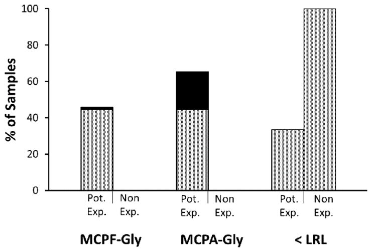
MCPF-Gly and MCPA-Gly metabolite presence in urine of potential exposures and non-exposures. Application of the method to potential exposure (Pot. Exp.) samples (n=73) of MCPG or hypoglycin A exposure yielded 45% MCPF-Gly, 64% MCPA-Gly and 34% beneath the method LRL. The black outline with gray fill represents both MCPF-Gly and MCPA-Gly (44%), while the additional portion with black fill represents the samples with only one of the metabolites, either MCPF-Gly (1%) or MCPA-Gly (21%). All non-exposure (Non. Exp.) samples (n=34) were found to be lower than the method’s LRL for both analytes.
For the 33 samples that were positive for the MCPF-Gly metabolite, the concentrations ranged from 0.105 to 2.62 μg/mL, with a median of 0.464 μg/mL. For the 47 samples that were positive for the MCPA-Gly metabolite, the concentrations ranged from 0.115 to 9.29 μg/mL, with a median of 0.569 μg/mL. In this application of the method, a wide range of concentrations were expected since urine samples were obtained when possible. Since the clearance rate of the metabolites is still unknown, the time lapse between collection and exposure could significantly affect the amount of metabolite remaining at the time of analysis.
CONCLUSION
The breadth of Soapberry family members and their possible toxicity constitutes a public health challenge for which laboratory support methods are needed. To date, fatalities linked to the ingestion of hypoglycin A have been reported in multiple countries, and of late, exposure to MCPG has been added to Soapberry compounds of concern. To address this public health need, we developed a quantitative method to evaluate human exposure to MCPG and hypoglycin A. The straightforward dilution of urine followed by HPLC-MS/MS analysis was developed for quantitation of the MCPG and hypoglycin A biomarkers MCPF-Gly and MCPA-Gly over a concentration range of 0.100 to 20.0 μg/mL. Evaluation of QC materials demonstrated ≤ 10% error with % RSDs of ≤ 15%. This method requires a urine sample volume of only 10 μL which is significant when sample volume is limited, such as in the analysis of pediatric samples. This work is the first quantitative HPLC-MS/MS assay reported for evaluating exposure to MCPG and hypoglycin A through quantitation of their respective metabolites MCPF-Gly and MCPA-Gly in human urine. Most importantly, the straightforward work up and ruggedness of this method will make it easily employable by public health laboratories. As a testament to its utility, this method has already been applied in a public health investigation, finding that 66% of the potential exposure samples were positive for MCPF-Gly and/or MCPA-Gly. This method is broadly applicable for investigating a number of public health challenges including Jamaican Vomiting Sickness and Acute Encephalitis Syndrome, both of which clinically resemble Reye’s syndrome.
Supplementary Material
Acknowledgments
Funding Sources
This work was supported by the Centers for Disease Control and Prevention and the Oak Ridge Institute for Science and Education.
The authors would like to thank Ms. Chariety Sapp of the CDC’s Incident Response Laboratory (IRL) for dispensing convenience set and public health investigation samples prior to analysis.
ABBREVIATIONS
- Cavg
average concentration
- Ce
experimental concentration
- Ct
theoretical concentration
- CoA
Coenzyme A
- ESI GC-MS
Gas chromatography-liquid chromatography
- HPLC-MS/MS
high-pressure liquid chromatography-tandem mass spectrometry
- IRB
Institutional Review Board
- ISTD
isotopically-labeled calibrator solution
- JVS
Jamaican Vomiting Sickness
- MCPA
methylenecyclopropylalanine
- MCPA-carnitine
methylenecyclopropylcarnitine
- MCPA-CoA
methylenecyclopropylacetyl-CoA
- MCPA-Gly
methylenecyclopropylacetyl-glycine
- MCPF-CoA
methylenecyclopropylformyl-Coenzyme A
- MCPF-Gly
methylenecyclopropylformyl-glycine
- MCPG
methylenecyclopropylglycine
- QC
quality control
- % RE
percent relative error
- % RSD
percent relative standard deviation
- SPM
Seasonal Pasture Myopathy
- UHPLC-MS/MS
ultra-high pressure liquid chromatography
Footnotes
Author Contributions
The manuscript was written through contributions of all authors. All authors have given approval to the final version of the manuscript.
DISCLAIMER
The findings and conclusions in this study are those of the authors and do not necessarily represent the views of the U.S. Department of Health and Human Services, or the U.S. Centers for Disease Control and Prevention. Use of trade names and commercial sources is for identification only and does not constitute endorsement by the U.S. Department of Health and Human Services, or the U.S. Centers for Disease Control and Prevention.
Evaluation of ion suppression and evaluation of post-preparative stability. This material is available free of charge via the Internet at http://pubs.acs.org.
References
- 1.Hassall CH, Reyle K, Feng P. Hypoglycin A, B: Biologically active polypeptides from Blighia sapida. Nature. 1954;173:356–357. doi: 10.1038/173356b0. [DOI] [PubMed] [Google Scholar]
- 2.Hassall CH, Reyle K. Hypoglycin A and B, two biologically active polypeptides from Blighia sapida. Biochem J. 1955;60:334–339. doi: 10.1042/bj0600334. [DOI] [PMC free article] [PubMed] [Google Scholar]
- 3.Sherratt HSA. Hypoglycin, the famous toxin of the unripe Jamaican ackee fruit. Trends Pharmacol Sci. 1986;7:186–191. [Google Scholar]
- 4.Gaillard Y, Carlier J, Berscht M, Mazoyer C, Bevalot F, Guitton J, Fanton L. Fatal intoxication due to ackee (Blighia sapida) in Suriname and French Guyana. GC-MS detection and quantification of hypoglycin-A. Forensic Sci Int. 2011;206:e103–e107. doi: 10.1016/j.forsciint.2011.01.018. [DOI] [PubMed] [Google Scholar]
- 5.Ware G. Method validation study of hypoglycin A determination in ackee fruit. J AOAC Int. 2002;85:933–937. [PubMed] [Google Scholar]
- 6.Henry SH, Page SW, Bolger PM. Hazard assessment of ackee fruit (Blighia sapida) Human Ecol Risk Assess. 1998;4:1175–1187. [Google Scholar]
- 7.Fowden L, Pratt HM. Cyclopropylamino acids of the genus Acer: Distribution and biosynthesis. Phytochemistry. 1973;12:1677–1681. [Google Scholar]
- 8.Tanaka K. On the mode of action of hypoglycin A. J Biol Chem. 1972;247:7465–7478. [PubMed] [Google Scholar]
- 9.Tanaka K, Ikeda Y. Hypoglycin and Jamaican Vomiting Sickness. Prog Clin Biol Res. 1990;321:167–184. [PubMed] [Google Scholar]
- 10.Al-Bassam SS, Sherratt HSA. The antagonism of the toxicity of hypoglycin by glycine. Biochem Pharmacol. 1981;30:2817–2824. doi: 10.1016/0006-2952(81)90420-2. [DOI] [PubMed] [Google Scholar]
- 11.von Holt C, Chang J, con Holt M, Bohm H. Metabolism and metabolic effects of hypoglycin. Biochim Biophys Acta. 1964;90:611–613. doi: 10.1016/0304-4165(64)90242-9. [DOI] [PubMed] [Google Scholar]
- 12.Blake OA, Bennink MR, Jackson JC. Ackee (Blighia sapida) hypoglycin A toxicity: Dose response assessment in laboratory rats. Food Chem Toxicol. 2006;44:207–213. doi: 10.1016/j.fct.2005.07.002. [DOI] [PubMed] [Google Scholar]
- 13.Billington D, Sherratt HSA. Hypoglycin and metabolically related inhibitors. Methods Ezymol. 1981;72:610–616. doi: 10.1016/s0076-6879(81)72051-2. [DOI] [PubMed] [Google Scholar]
- 14.Tanaka K, Kean EA, Johnson B. Jamaican Vomiting Sickness: Biochemical investigation of two cases. New Engl J Med. 1976;295:461–467. doi: 10.1056/NEJM197608262950901. [DOI] [PubMed] [Google Scholar]
- 15.Joskow R, Belson M, Vesper H, Backer L, Rubin C. Ackee fruit poisoning: An outbreak investigation in Haiti 2000–2001, and review of the literature. Clin Toxicol. 2006;44:267–273. doi: 10.1080/15563650600584410. [DOI] [PubMed] [Google Scholar]
- 16.McTague JA, Forney R., Jr Jamaican Vomiting Sickness in Toledo, Ohio. Ann Emerg Med. 1994;23:1116–1118. doi: 10.1016/s0196-0644(94)70112-1. [DOI] [PubMed] [Google Scholar]
- 17.Brown M, Bates RP, McGowan C, Cornell JA. Influence of fruit maturity on the hypoglycin A Level in ackee (Blighia Sapida) J Food Safety. 1991;12:167–177. [Google Scholar]
- 18.Bowen-Forbes CS, Minott DA. Tracking hypoglycins A and B over different maturity stages: Implications for detoxification of ackee (Blighia sapida K.D. Koenig Fruits) J Agr Food Chem. 59:3869–3875. doi: 10.1021/jf104623c. [DOI] [PubMed] [Google Scholar]
- 19.FDA. Guidance for FDA staff: Compliance policy guide sec. 550.050 canned ackee, frozen ackee, and other ackee products-hypoglycin A toxin. US Department of Health and Human Services. Food and Drug Administration, Center for Food Safety and Applied Nutrition, Office of Regulatory Affairs; 2014. [Google Scholar]
- 20.Minakata H, Komura H, Tamura SY, Ohfune Y, Nakanishi K, Kada T. Antimutagenic unusual amino acids from plants. Experientia. 1985;41:1622–1623. doi: 10.1007/BF01964840. [DOI] [PubMed] [Google Scholar]
- 21.Gray DO, Fowden L. α-(Methylenecyclopropyl)glycine from litchi seeds. Biochem J. 1962;82:385–389. doi: 10.1042/bj0820385. [DOI] [PMC free article] [PubMed] [Google Scholar]
- 22.Melde K, Jackson S, Bartlett K, Sherratt SA, Ghisla S. Metabolic consequences of methylenecyclopropylglycine poisoning in rats. Biochem J. 1991;274:395–400. doi: 10.1042/bj2740395. [DOI] [PMC free article] [PubMed] [Google Scholar]
- 23.Dakoji S, Ding L, Agnihotri G, Zhou H, Liu H. Studies on the inactivation of bovine liver enoyl-CoA hydratase by (methylenecyclopropyl)formyl-CoA: Elucidation of the inactivation mechanism and identification of cysteine-114 as the entrapped nucleophile. J Am Chem Soc. 2001;123:9749–9759. doi: 10.1021/ja011226k. [DOI] [PubMed] [Google Scholar]
- 24.Li D, Agnihotri G, Dakoji S, Oh E, Lantz M, Liu H. The toxicity of methylenecyclopropylglycine: Studies of the inhibitory effects of (methylenecyclopropyl)formyl-CoA on enzymes involved in fatty acid metabolism and the molecular basis of its inactivation of Enoyl-CoA hydratases. J Am Chem Soc. 1999;121:9034–3042. [Google Scholar]
- 25.Agnihotri G, He S, Hong L, Dakoji S, Withers SG, Liu H. A revised mechanism for the inactivation of bovine liver enoyl-CoA hydratase by (methylenecyclopropyl)formyl-CoA based on unexpected results with the C114A mutant. Biochemistry. 2002;41:1843–1852. doi: 10.1021/bi0119363. [DOI] [PubMed] [Google Scholar]
- 26.Melde K, Buettner H, Boschert W, Wolf HPO, Ghisla S. Mechanism of hypoglycaemic action of methylenecyclopropylglycine. Biochem J. 1989;259:921–924. doi: 10.1042/bj2590921. [DOI] [PMC free article] [PubMed] [Google Scholar]
- 27.John TJ, Das M. Acute encephalitis syndrome in children in Muzaffarpur: Hypothesis of toxic origin. Curr Sci India. 2014;106:1184–1185. [Google Scholar]
- 28.Shrivastava A, Srikantiah P, Kumar A, Bhushan G, Goel K, Kumar S, Kumar T, Mohankumar R, Pandey R, Pathan P, Tulsian Y, Pappanna M, Pasi A, Pradhan A, Singh P, Somashekar D, Velayudhan A, Yadav R, Chhabra M, Mittal V, Khare S, Sejvar JJ, Dwivedi M, Laserson K, Earhart KC, Sivaperumal P, Kumar AR, Chakrabarti A, Thomas J, Schier J, Signh R, Shankar RS, Dhariwal AC, Chauhan LS. Outbreaks of unexplained neurologic illness – Muzaffarpur, India, 2013–2014. MMWR. 2015;64:49–53. [PMC free article] [PubMed] [Google Scholar]
- 29.Pulla P. A child-killing toxin emerges from shadows: Scientists link mystery deaths in India to consumption of lychees. Science. 2015;348:15–16. doi: 10.1126/science.348.6230.15. [DOI] [PubMed] [Google Scholar]
- 30.Paireau J, Tuan NH, Lefrancois R, Buckwalter MR, Nghia ND, Hien NT, Lortholary O, Poiree S, Manuguerra J, Gessian A, Albert ML, Brey PT, Nga PT, Fontanet A. Litchi-associated acute encephalitis in children, Northern Vietnam, 2004–2009. Emerg Infect Dis. 2012;18:1817–1824. doi: 10.3201/eid1811.111761. [DOI] [PMC free article] [PubMed] [Google Scholar]
- 31.Biswas SK. Outbreak of illness and deaths among children living near lychee orchards in northern Bangladesh. ICDDRB Health Sci Bull. 2012;10:15–22. [Google Scholar]
- 32.Dinesh DS, Pandey K, Das VNR, Topno RK, Kesari S, Kumar V, Ranjan A, Sinha PK, Das P. Possible factors causing Acute Encephalitis Syndrome outbreak in Bihar, India. Int J Curr Microbiol App Sci. 2013;2:531–538. [Google Scholar]
- 33.Osterloh J, Cunningham W, Dixon A, Combest D. Biochemical relationships between Reye’s and Reye’s-like metabolic and toxicological syndromes. Med Toxicol Adverse Drug Exp. 1989;4:272–294. doi: 10.1007/BF03259913. [DOI] [PubMed] [Google Scholar]
- 34.Fincham AG. The determination of hypoglycin A in blood and plasma. West Indian Med J. 1977;26:62–65. [PubMed] [Google Scholar]
- 35.Carlier J, Guitton J, Moreau C, Boyer B, Bevalot F, Fanton L, Habyarimana J, Gault G, Gaillard Y. A validated method for quantifying hypoglycin A in whole blood by UHPLC-HRMS/MS. J Chromatogr B. 2015;978–979:70–77. doi: 10.1016/j.jchromb.2014.11.029. [DOI] [PubMed] [Google Scholar]
- 36.Valberg SJ, Sponseller BT, Hegeman AD, Earing J, Bender JB, Martinson KL, Patterson SE, Sweetman L. Seasonal pasture myopathy/atypical myopathy in North America associated with ingestion of hypoglycin A within seeds of the box elder tree. Equine Vet J. 2012:1–8. doi: 10.1111/j.2042-3306.2012.00684.x. [DOI] [PubMed] [Google Scholar]
- 37.Caudill SP, Schleicher RL, Pirkle JL. Multi-rule quality for the age-related eye disease study. Statist Med. 2008;27:4094–4106. doi: 10.1002/sim.3222. [DOI] [PubMed] [Google Scholar]
- 38.Gregersen N, Kolvraa S, Mortensen PB. Acyl-CoA: Glycine N-acyltransferase: In vitro studies on the glycine conjugation of straight- and branched-chained acyl-CoA esters in human liver. Biochem Med Metab B. 1986;35:210–218. doi: 10.1016/0885-4505(86)90076-9. [DOI] [PubMed] [Google Scholar]
- 39.Ombrone D, Salvatore F, Ruoppolo M. Quantitative liquid chromatography coupled with tandem mass spectrometry analysis of urinary acylglycines: Application to the diagnosis of inborn errors of metabolism. Anal Biochem. 2011;417:122–128. doi: 10.1016/j.ab.2011.05.042. [DOI] [PubMed] [Google Scholar]
- 40.Rinaldo P, O’Shea JJ, Coates PM, Hale DE, Stanley CA, Tanaka K. Medium-chain acyl-CoA dehydrogenase deficiency. New Engl J Med. 1988;319:1308–1313. doi: 10.1056/NEJM198811173192003. [DOI] [PubMed] [Google Scholar]
- 41.Taylor JK. Quality assurance of chemical measurements. CRC Press; Boca Raton, FL: 1987. pp. 79–82. [Google Scholar]
- 42.FDA. Guidance for industry, bioanalytical method validation. US Department of Health and Human Services. Food and Drug Administration, Center for Drug Evaluation and Research (CDER), Center for Veterinary Medicine (CV); 2001. [Google Scholar]
- 43.FDA. Draft Guidance. US Department of Health and Human Services. Food and Drug Administration, Center for Drug Evaluation and Research (CDER), Center for Veterinary Medicine (CV); 2013. Guidance for industry, bioanalytical method validation. [Google Scholar]
- 44.Yadav R. Outbreak of Acute Neurologic Illness Outbreak, Muzaffarpur, Bihar, India, 2014. 64th Annual Epidemic Intelligence Service Conference; April 20–23, 2015; Atlanta, GA: Centers for Disease Control and Prevention; 2015. [Google Scholar]
Associated Data
This section collects any data citations, data availability statements, or supplementary materials included in this article.